. 2011 Nov 15;83(22):8756-65.
doi: 10.1021/ac2022234. Epub 2011 Oct 17.

Two new "protected" oxyphors for biological oximetry: properties and application in tumor imaging

Affiliations

Two new "protected" oxyphors for biological oximetry: properties and application in tumor imaging

Tatiana V Esipova et al. Anal Chem. .

Abstract

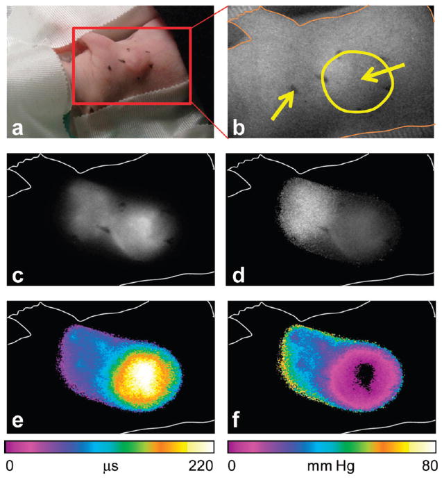
We report the synthesis, calibration, and examples of application of two new phosphorescent probes, Oxyphor R4 and Oxyphor G4, optimized specifically for in vivo oxygen imaging by phosphorescence quenching. These "protected" dendritic probes can operate in either albumin-rich (blood plasma) or albumin-free (interstitial space) environments at all physiological oxygen concentrations, from normoxic to deep hypoxic conditions. Oxyphors R4 and G4 are derived from phosphorescent Pd-meso-tetra-(3,5-dicarboxyphenyl)-porphyrin (PdP) or Pd-meso-tetra-(3,5-dicarboxyphenyl)-tetrabenzoporphyrin (PdTBP), respectively, and possess features common for protected dendritic probes, i.e., hydrophobic dendritic encapsulation of phosphorescent metalloporphyrins and hydrophilic PEGylated periphery. The new Oxyphors are highly soluble in aqueous environments and do not permeate biological membranes. The probes were calibrated under physiological conditions (pH 6.4-7.8) and temperatures (22-38 °C), showing high stability, reproducibility of signals, and lack of interactions with biological solutes. Oxyphor G4 was used to dynamically image intravascular and interstitial oxygenation in murine tumors in vivo. The physiological relevance of the measurements was demonstrated by dynamically recording changes in tissue oxygenation during application of anesthesia (isofluorane). These experiments revealed that changes in isofluorane concentration significantly affect tissue oxygenation.


Figures

Figure 1
Figure 1
Structures of Oxyphors R4 and G4 (PEG-amine av MW 1000).
Figure 2
Figure 2
Absorption (a) and emission (b) spectra of R4 (red) and G4 (green) in 50 mM phosphate buffer solutions (pH 7.2). The emission spectra are scaled by the brightness indexes (B) of the probes at zero-oxygen concentration. Brightness (B) is defined as a product of the molar extinction coefficient ε and the phosphorescence quantum yield φ (B = εφ).
Figure 3
Figure 3
Phosphorescence decays (a) of probe Oxyphor R4 recorded at three different oxygen concentrations (air saturation, intermediate pO2, deoxygenated) from 10 μM buffered solutions at 22.5 °C (phosphate buffer, 50 mM, pH 7.2). Phosphorescence was excited by 5 μs long LED pulses. Phosphorescence lifetime distributions (b) recovered by the maximum entropy method (MEM) (ref 76).
Figure 4
Figure 4
Temperature dependencies of oxygen quenching constants (kq) and lifetimes (τ0) for Oxyphors R4 (a and b) and G4 (c and d). The measurements were performed using 10 μM solutions of the probes in 50 mM phosphate buffer, pH 7.2.
Figure 5
Figure 5
Changes in interstitial (tissue) oxygenation in the leg muscle and in subcutaneous tumor (RIF) in a C3H mouse, as measured using Oxyphor G4 and a fiber-optic phosphorometer. Solution of G4 (20 μL, 10 μM) in physiological saline was injected directly into the tissue. Measurements were performed at 4 s intervals. The anesthetic gas mixture was delivered at the rate of 2 L/min, and the normal concentration of isofluorane in the mixture (1.75%) was changed briefly by higher (4%) or lower (1.25%) levels.
Figure 6
Figure 6
Imaging of intravascular oxygenation in a mouse using Oxyphor G4. The probe was injected via the tail vein to achieve the final concentration in the blood plasma of ca. 4 μM. (a) Photograph of the anesthetized animal on a heating pad and (b) the zoomed-in region, as imaged by the ICCD camera. The tumor is located on the right shoulder of the animal (pointed by yellow arrow). (c) Image (I1) of the phosphorescence intensity acquired after delay δt1 = 10 μs, following the excitation pulse. In total, 10 images at different delays, from 10 to 620 μs, were collected. Calculated images of (d) phosphorescence initial intensity (I0), (e) phosphorescence lifetimes (τ, color scale in μs), and (f) partial oxygen pressure (pO2, color scale in mm Hg).
Figure 7
Figure 7
Imaging of interstitial (pericellular space) oxygenation using Oxyphor G4. The probe (20 μM solution) was injected directly into the normal (40 μL) and tumor (20 μL) tissue. (a) Photograph of the anesthetized animal on a heating pad and (b) the zoomed-in region, as imaged by the ICCD camera. The tumor is located on the right shoulder of the animal (enclosed in a yellow circle), and the probe injection sites are marked by the arrows. (c) Image (I1) of the phosphorescence intensity acquired after delay of 10 μs, following the excitation pulse. Calculated images of (d) phosphorescence initial intensity (I0), (e) phosphorescence lifetimes (τ, color scale in μs), and (f) partial oxygen pressure (pO2, color scale in mm Hg). To simplify visualization, the contours of the animal body (as in image b) are shown in images c–f.

Similar articles

Cited by

References

    1. Yu DY, Cringle SJ. Exp Eye Res. 2005;80:745. - PubMed
    1. Tatum JL, Kelloff GJ, Gillies RJ, Arbeit JM, Brown JM, Chao KS, Chapman JD, Eckelman WC, Fyles AW, Giaccia AJ, Hill RP, Koch CJ, Krishna MC, Krohn KA, Lewis JS, Mason RP, Melillo G, Padhani AR, Powis G, Rajendran JG, Reba R, Robinson SP, Semenza GL, Swartz HM, Vaupel P, Yang D, Croft B, Hoffman J, Liu G, Stone H, Sullivan D. Int J Radiat Biol. 2006;82:699. - PubMed
    1. Blomgren K, Hagberg H. Free Radical Biol Med. 2006;40:388. - PubMed
    1. Schreml S, Szeimies RM, Prantl L, Karrer S, Landthaler M, Babilas P. Br J Dermatol. 2010;163:257. - PubMed
    1. Brown JM. J Natl Cancer Inst. 1990;82:338. - PubMed

Publication types