. 2011 Dec;176(6):842-8.
doi: 10.1667/rr2649.1. Epub 2011 Sep 30.

Cancer-prone mice expressing the Ki-rasG12C gene show increased lung carcinogenesis after CT screening exposures

Affiliations

Cancer-prone mice expressing the Ki-rasG12C gene show increased lung carcinogenesis after CT screening exposures

Michael T Munley et al. Radiat Res. 2011 Dec.

Abstract

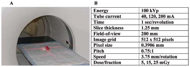
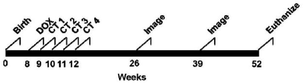
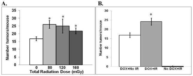
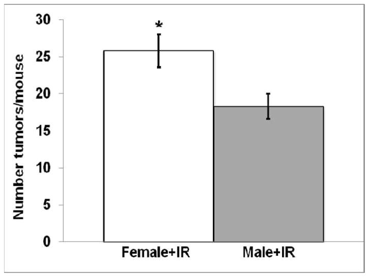
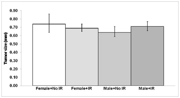
A >20-fold increase in X-ray computed tomography (CT) use during the last 30 years has caused considerable concern because of the potential carcinogenic risk from these CT exposures. Estimating the carcinogenic risk from high-energy, single high-dose exposures obtained from atomic bomb survivors and extrapolating these data to multiple low-energy, low-dose CT exposures using the Linear No-Threshold (LNT) model may not give an accurate assessment of actual cancer risk. Recently, the National Lung Cancer Screening Trial (NLST) reported that annual CT scans of current and former heavy smokers reduced lung cancer mortality by 20%, highlighting the need to better define the carcinogenic risk associated with these annual CT screening exposures. In this study, we used the bitransgenic CCSP-rtTA/Ki-ras mouse model that conditionally expresses the human mutant Ki-ras(G12C) gene in a doxycycline-inducible and lung-specific manner to measure the carcinogenic risk of exposure to multiple whole-body CT doses that approximate the annual NLST screening protocol. Irradiated mice expressing the Ki-ras(G12C) gene in their lungs had a significant (P = 0.01) 43% increase in the number of tumors/mouse (24.1 ± 1.9) compared to unirradiated mice (16.8 ± 1.3). Irradiated females had significantly (P < 0.005) more excess tumors than irradiated males. No tumor size difference or dose response was observed over the total dose range of 80-160 mGy for either sex. Irradiated bitransgenic mice that did not express the Ki-ras(G12C) gene had a low tumor incidence (≤ 0.1/mouse) that was not affected by exposure to CT radiation. These results suggest that (i) estimating the carcinogenic risk of multiple CT exposures from high-dose carcinogenesis data using the LNT model may be inappropriate for current and former smokers and (ii) any increased carcinogenic risk after exposure to fractionated low-dose CT-radiation may be restricted to only those individuals expressing cancer susceptibility genes in their tissues at the time of exposure.


Figures

FIG. 1
FIG. 1
Irradiation setup and parameters. Panel A: Position of one mouse and the phantom prior to starting irradiation. Panel B: CT irradiation parameters. The three doses were obtained by adjusting the tube current. The irradiation time was ~30 s for all doses.
FIG. 2
FIG. 2
Experimental timeline. At 8 weeks of age, groups of 12 bitransgenic CCSP-rtTA/Ki-ras mice were entered into the experiment. Half of the groups were given 500 μg/ml of doxycycline (DOX) in their drinking water; the other half were given normal drinking water. One week after initiating the DOX treatment, DOX or No DOX mice were lightly anesthetized and either sham-irradiated or irradiated once each week for 4 weeks with 5, 15 or 25 mGy of 100 kVp X rays from a clinical helical CT scanner. At 3, 6 and 9 months after the last weekly fraction, the lungs were imaged with CT (30 mGy/image) to noninvasively determine the size of the tumors. At 12 months of age the mice were euthanized, the lungs were excised, and the tumors were counted and sized.
FIG. 3
FIG. 3
Tumor formation with and without CT irradiation. Panel A: Mean number (± SEM) of lung tumors/mouse for each total dose level. There was a significant radiation effect that was independent of dose. *P < 0.05 compared to unirradiated mice expressing the Ki-rasG12C gene. Panel B: Comparison of the mean number of lung tumors/mouse for the combined data for irradiated mice. IR = ionizing radiation. White bar: unirradiated mice expressing the Ki-rasG12C gene; gray bar: irradiated mice expressing the Ki-rasG12C gene. *P = 0.01 compared to unirradiated mice expressing the Ki-rasG12C gene. Black bar: irradiated mice not expressing the Ki-rasG12C gene.
FIG. 4
FIG. 4
Comparison of the number of lung tumors/mouse (mean ± SEM) for irradiated female and male mice expressing the Ki-rasG12C gene after combining all of the irradiated groups (*P < 0.005).
FIG. 5
FIG. 5
Comparison of the mean tumor size (± 95% CI) for female and male mice expressing the Ki-rasG12C gene with and without exposure to CT radiation at 1 year of age (9 months postirradiation). P > 0.3 for all comparisons.
FIG. 6
FIG. 6
Panel A: Top left: DOX + No IR (ionizing radiation); top right: DOX + 80 mGy; bottom left: DOX + 120 mGy; bottom right: DOX + 160 mGy. At a magnification of 10×, after hematoxylin and eosin staining, all four lesions appear similar, being composed of a well-circumscribed, nonencapsulated, closely spaced collection of plump epithelial cells with little morphological variation. Panel B: Top left: DOX + No IR; top right: DOX + 80 mGy; bottom left: DOX + 120 mGy; bottom right: DOX + 160 mGy. At a magnification of 40×, similar cellular morphology for all four lesions can be seen. The lesions are composed of uniform cells with 10–12-μm-diameter round basophilic nuclei with coarsely clumped chromatin, often without nucleoli, and abundant eosinophilic cytoplasm. Mitoses are rare to absent in these lesions.

Similar articles

Cited by

References

    1. Brenner DJ, Hall EJ. Computed tomography—an increasing source of radiation exposure. N Engl J Med. 2007;357:2277–84. - PubMed
    1. Einstein AJ, Henzlova MJ, Rajagopalan S. Estimating risk of cancer associated with radiation exposure from 64-slice computed tomography coronary angiography. JAMA. 2007;298:317–23. - PubMed
    1. Dauer LT, Brooks AL, Hoel DG, Morgan WF, Stram D, Tran P. Review and evaluation of updated research on the health effects associated with low-dose ionising radiation. Radiat Prot Dosimetry. 2010;140:103–36. - PubMed
    1. Shuryak I, Sachs RK, Brenner DJ. Cancer risks after radiation exposure in middle age. J Natl Cancer Inst. 2010;102:1628–36. - PMC - PubMed
    1. Mossman KL. Economic and policy considerations drive the LNT debate. Radiat Res. 2008;169:245–7. - PubMed

Publication types

MeSH terms