. 2011 Sep 30;147(1):120-31.
doi: 10.1016/j.cell.2011.08.038.

A DNA repair complex functions as an Oct4/Sox2 coactivator in embryonic stem cells

Affiliations

A DNA repair complex functions as an Oct4/Sox2 coactivator in embryonic stem cells

Yick W Fong et al. Cell. .

Abstract

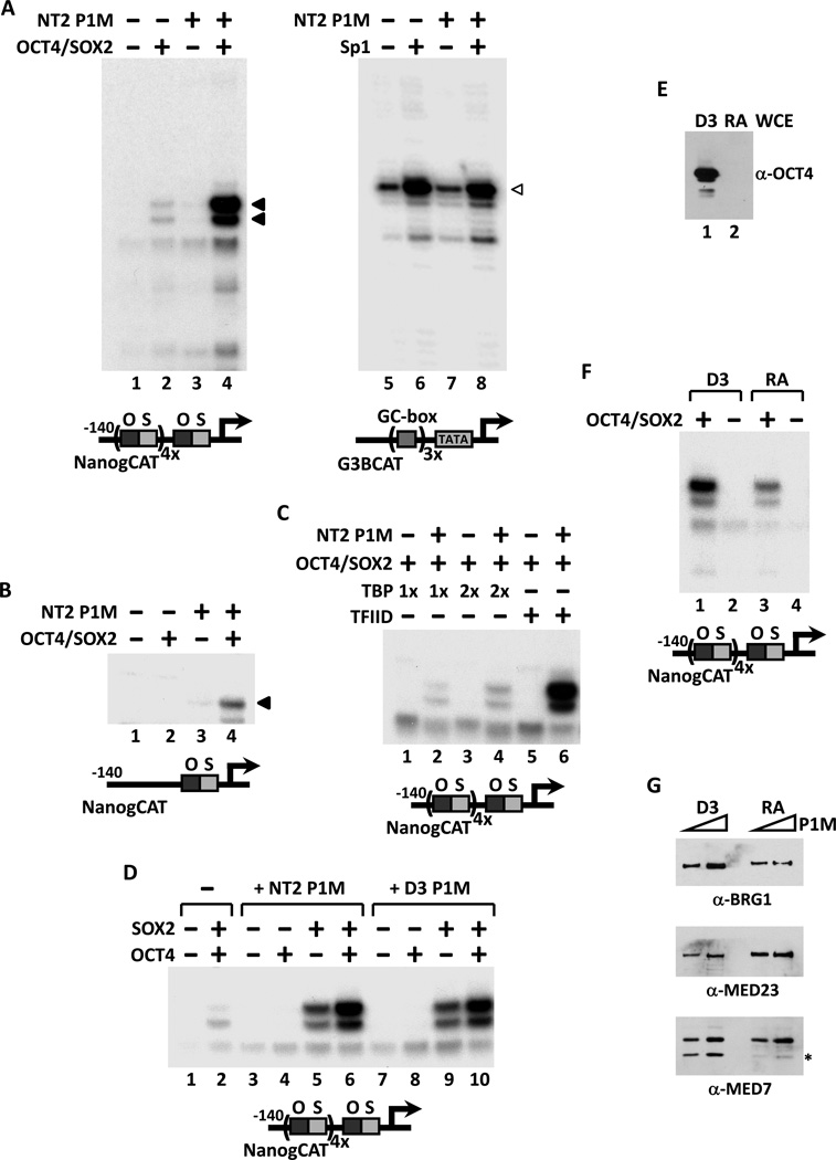
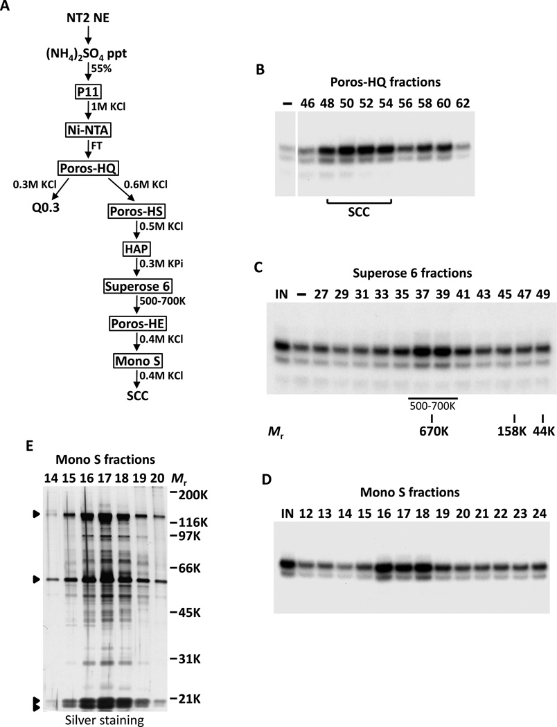
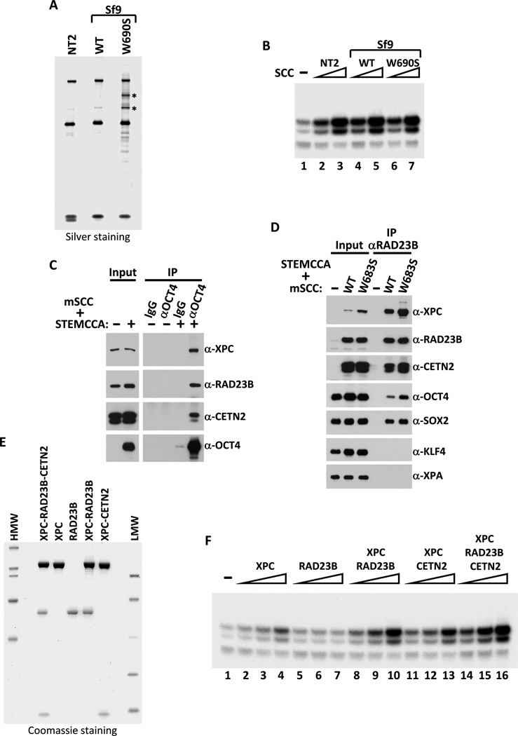
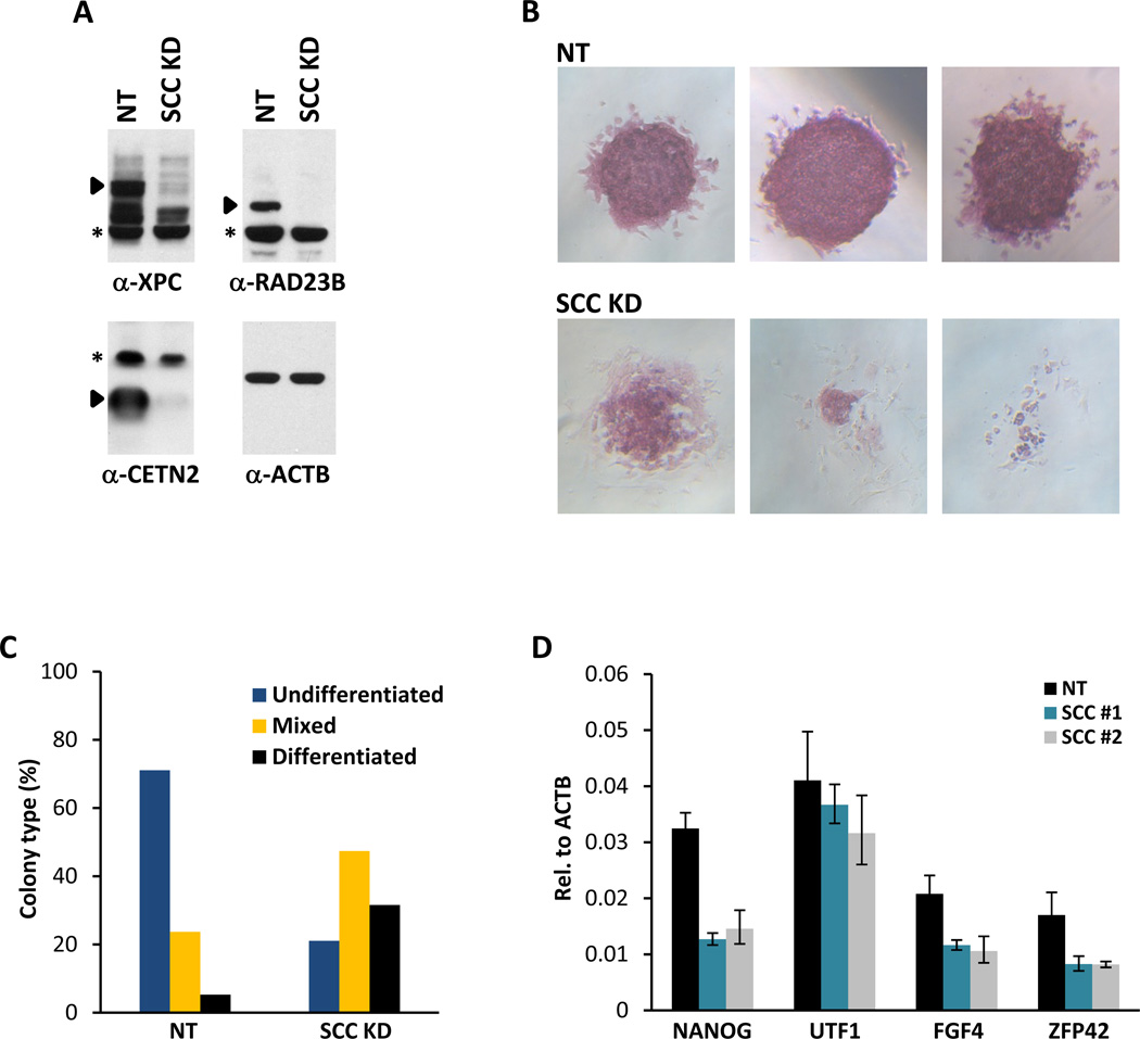
The transcriptional activators Oct4, Sox2, and Nanog cooperate with a wide array of cofactors to orchestrate an embryonic stem (ES) cell-specific gene expression program that forms the molecular basis of pluripotency. Here, we report using an unbiased in vitro transcription-biochemical complementation assay to discover a multisubunit stem cell coactivator complex (SCC) that is selectively required for the synergistic activation of the Nanog gene by Oct4 and Sox2. Purification, identification, and reconstitution of SCC revealed this coactivator to be the trimeric XPC-nucleotide excision repair complex. SCC interacts directly with Oct4 and Sox2 and is recruited to the Nanog and Oct4 promoters as well as a majority of genomic regions that are occupied by Oct4 and Sox2. Depletion of SCC/XPC compromised both pluripotency in ES cells and somatic cell reprogramming of fibroblasts to induced pluripotent stem (iPS) cells. This study identifies a transcriptional coactivator with diversified functions in maintaining ES cell pluripotency and safeguarding genome integrity.


Figures

Figure 1
Figure 1. Transcriptional Activation of Nanog by Oct4 and Sox2 Requires a Stem Cell-Specific Cofactor
(A) Reconstituted in vitro transcription reactions supplemented with Oct4 and Sox2 (lanes 2 and 4) or Sp1 (lanes 6 and 8), plus a phosphocellulose 1M KCl fraction derived from NT2 nuclear extracts (NT2 P1M, lanes 3, 4, 7 and 8) and programmed with either a Nanog template engineered with four extra copies of the oct-sox composite element (NanogCAT, lanes 1–4), or a GC box-containing template (G3BCAT, lanes 5–8). Oct4/Sox2, NT2 P1M-dependent transcripts are indicated by filled arrowheads and Sp1-dependent transcriptions by open arrowheads. (B) Transcription of the native Nanog promoter requires Oct4, Sox2 and NT2 P1M fraction (lane 4). (C) TFIID and NT2 P1M fraction are needed to potentiate Oct4/Sox2-dependent activation. Transcription reactions contain Oct4 and Sox2 (lanes 1–6), NT2 P1M fraction (lanes 2, 4 and 6), with increasing amounts of recombinant TBP (1x or 2x, lanes 1–4) or TFIID (lanes 5 and 6). (D) Synergistic activation of Nanog by Oct4 and Sox2 requires P1M fractions prepared from NT2 or mouse ES cell line D3 nuclear extracts. In vitro transcription reactions contain equal amounts (~0.7 µg) of NT2 (lanes 3–6) or D3 P1M fractions (lanes 7–10), with Oct4 alone (lanes 4 and 8), Sox2 alone (lanes 5 and 9), or both activators (lanes 2, 6 and 10). (E) Immunoblotting analysis of Oct4 levels in whole cell extracts (WCE) prepared from pluripotent D3 cells (D3, lane 1) and cells treated with retinoic acid for 6 days (RA, lane 2). (F) P1M fractions prepared from pluripotent (D3, lanes 1 and 2) and differentiated (RA, lanes 3 and 4) D3 nuclear extracts were added to transcription reactions with or without Oct4 and Sox2. (G) Western blots (2-fold titration) of P1M fractions prepared from pluripotent (D3) and differentiated (RA) D3 nuclear extracts using anti-BRG-1, anti-MED23 and anti-MED7 antibodies. Asterisk indicates a non-specific band or a breakdown product recognized by anti-MED7 antibody.
Figure 2
Figure 2. Purification of Stem Cell Coactivator (SCC)
(A) Chromatography scheme for partial purification of Q0.3 and purification of SCC from NT2 nuclear extracts (NT2 NE). NT2 NE is first subjected to ammonium sulfate precipitation (55% saturation) followed by a series of chromatographic columns as indicated. (B) Buffer (−) and fractions containing SCC eluted from a Poros-HQ anion exchanger (top) assayed in the presence of Oct4 and Sox2 in in vitro transcription assays. (C) Coactivator SCC migrates as a large complex. Input (IN), buffer (−), and Superose 6 fractions (top) assayed as in (B) except all reactions are supplemented with Q0.3 (Figure 2A). Mobilities of peak activity (500–700K) and gel filtration protein standards are shown (bottom). (D) Transcription profile of SCC activity after the final Mono S chromatography step. Reactions contain input (IN), Mono S fractions (top) and are assayed as in (C). (E) Silver-stained SDS-PAGE gel of the active Mono S fractions. Filled arrowheads indicate polypeptides that co-migrate with SCC activity.
Figure 3
Figure 3. SCC is the XPC-RAD23B-CETN2 Nucleotide Excision Repair Complex
(A) Mass spectrometry analysis of Mono S peak activity fractions (16–18) in Figure 2E with protein identities indicated. (B) SCC is highly enriched in NT2 P1M fraction. Comparative western blot analysis of HeLa and NT2 P1M fractions (1.5 µg each), and purified Mono S SCC fraction (Purif, ~30 ng) using anti-XPC, anti-RAD23B and anti-CETN2 antibodies. (C) Down-regulation of XPC and RAD23B upon RA-induced differentiation of mouse D3 ES cells. Western blot analysis of whole cell extracts prepared from D3 cells (D3 WCE) collected at indicated days post RA treatment using antibodies against XPC, RAD23B, CETN2, OCT4, XPB, XPA, TFIIEβ, TBP and loading control β-actin (ACTB).
Figure 4
Figure 4. Reconstitution of Recombinant SCC Complexes
(A) Silver-stained SDS-PAGE gel of purified NT2 SCC (NT2), recombinant wild-type (WT) and DNA-binding defective mutant (W690S) XPC-containing SCC complexes reconstituted in insect Sf9 cells by co-infection with baculoviruses expressing His-tagged XPC, FLAG-tagged RAD23B and untagged-CETN2. Major proteolytic fragments of mutant XPC are indicated by asterisks. (B) Recombinant SCC complex enhances Oct4/Sox2-activated transcription of Nanog independent of DNA binding. Buffer (−), NT2 (Mono S peak activity fractions; lanes 2 and 3), recombinant WT (lanes 4 and 5) and W690S mutant (lanes 6 and 7) SCC complexes are assayed (over a three-fold concentration range). All transcription reactions contain Oct4, Sox2 and Q0.3 (lanes 1–7). (C) Oct4 interacts with SCC. Western blot analysis of input lysates (2%) and co-immunoprecipitated proteins from extracts of 293T cells transfected with a polycistronic expression plasmid encoding all three subunits of mouse SCC (mSCC) with or without a polycistronic plasmid expressing mouse Oct4, Sox2, Klf4 and c-Myc (STEMCCA) using normal IgG or anti-Oct4 antibody. (D) SCC-B interacts directly with Oct4 and Sox2 independent of DNA binding. Control vector (−), plasmids expressing wild-type (WT) or mutant (W683S) XPC-containing mSCC complexes were co-transfected with STEMCCA into 293T cells and immunoprecipitated with anti-RAD23B antibody. Input lysates (2%) and RAD23B-bound proteins were detected by immunoblotting. (E) Coomassie-stained SDS-PAGE gel of purified recombinant XPC, RAD23B, dimeric (XPC-RAD23B and XPC-CETN2) and holo-SCC (XPC-RAD23B-CETN2) complexes. (F) Titrations (over a four-fold concentration range) of XPC (lanes 2–4), RAD23B (lanes 5–7), XPC-RAD23B (lanes 8–10), XPC-CETN2 (lanes 11–13) and XPC-RAD23B-CETN2 (lanes 14–16) in in vitro transcription reactions supplemented with Q0.3 (lanes 1–16) and assayed as in (B).
Figure 5
Figure 5. SCC is Required for ES Cell Maintenance
(A) Efficiency of shRNA-mediated depletion of SCC in mouse ES cell line D3. Whole cell extracts of mouse D3 cells infected with non-target (NT) lentiviruses (MOI of 300) or with an equal mixture of three lentiviruses (MOI of 100 each) targeting XPC, RAD23B and CETN2 (SCC KD) are analyzed by western blotting. Specific bands recognized by their respective antibodies are indicated by filled arrowheads. Asterisks denote non-specific signals. (B) ES cell colony morphology and alkaline phosphatase (AP) activity (red) are maintained in control D3 cells (NT, top panels) but are compromised in SCC-depleted D3 cells (SCC KD, bottom panels) (See also Figure S4C). (C) Clonal assays on SCC-depleted D3 ES cells. Stable non-target (NT) and SCC-depleted (SCC KD) D3 cell pools were plated at 300 cells per well in 6-well plates, emerging colonies were stained for AP activity and scored according to differentiation status after 6 days. (D) Two non-overlapping sets of shRNAs targeting SCC (SCC #1 and SCC #2) are used to deplete SCC. Quantification of Nanog, Utf1, Fgf4 and Zfp42 mRNA levels are analyzed by real time quantitative PCR (qPCR) and normalized to Actb. Data from representative experiments are shown; error bars represent standard deviations (n=3).
Figure 6
Figure 6. SCC is Recruited to the Nanog and Oct4 Promoters and Genomic Regions Occupied by Oct4 and Sox2
(A) Co-occupancy of SCC, Oct4 and Sox2 on the promoters of Nanog and Oct4. ChIP analysis of RAD23B occupancy on distal enhancers (enh), proximal promoter (transcription start site, TSS), and upstream (positions indicated by numbers) and downstream intronic regions of the Nanog (left), Oct4 (middle), and Actb (right) gene loci. Representative data (n>5) showing the enrichment of RAD23B (black bars) compared to normal IgGs (white bars) are analyzed by qPCR and expressed as percentage of input chromatin. Schematic diagrams of Oct4 and Sox2 binding sites on the Nanog and Oct4 regulatory regions (TSS and enhancers; see also Figure S5A) are indicated at the bottom. Error bars represent standard deviations (n=3). (B) Percent peak overlap between RAD23B and control IgG ChIP-seq data relative to published Oct4/Sox2 and Nanog/Tcf3 peak data. (C) Percent base-pair overlap between RAD23B and control IgG ChIP-seq data relative to Oct4/Sox2 and Nanog/Tcf3 ChIP-seq data sets. (D) The distribution of distance (in base pair) of RAD23B and control IgG peaks from Oct4/Sox2 and Nanog/Tcf3 peaks.
Figure 7
Figure 7. SCC is Required for Efficient Somatic Cell Reprogramming
(A) Depletion of SCC blocks somatic cell reprogramming. Oct4-GFP mouse embryonic fibroblasts infected with lentiviruses expressing STEMCCA and rtTA together with non-target shRNA (NT), shRNAs against Oct4, individual subunits of SCC, or all three subunits simultaneously at low or high multiplicity of infection (SCC LO or HI) are plated in 6 well plates for colony counting and FACS, or in 24 well plates for AP staining. AP-positive (red) cells are stained and counted 17 days (14 days + dox, 3 days − dox) post induction (dpi). Results from two separate experiments are shown. (B) Single cell suspensions of 17 dpi Oct4-GFP MEFs as described in (A) are stained with anti-mouse SSEA-1 antibodies and analyzed by FACS. (C) Wild-type (WT), RAD23A and B double-heterozygous (23A/B d-Het) MEFs, together with XPC, RAD23A and RAD23B knockout (KO) MEFs, are induced with STEMCCA. AP-positive colonies are stained and counted as in (A).

Comment in

References

    1. Araki M, Masutani C, Takemura M, Uchida A, Sugasawa K, Kondoh J, Ohkuma Y, Hanaoka F. Centrosome protein centrin 2/caltractin 1 is part of the xeroderma pigmentosum group C complex that initiates global genome nucleotide excision repair. J. Biol. Chem. 2001;276:18665–18672. - PubMed
    1. Bernardes de Jesus BM, Bjoras M, Coin F, Egly JM. Dissection of the molecular defects caused by pathogenic mutations in the DNA repair factor XPC. Mol. Cell. Biol. 2008;28:7225–7235. - PMC - PubMed
    1. Boyer LA, Lee TI, Cole MF, Johnstone SE, Levine SS, Zucker JP, Guenther MG, Kumar RM, Murray HL, Jenner RG, et al. Core transcriptional regulatory circuitry in human embryonic stem cells. Cell. 2005;122:947–956. - PMC - PubMed
    1. Camenisch U, Trautlein D, Clement FC, Fei J, Leitenstorfer A, Ferrando-May E, Naegeli H. Two-stage dynamic DNA quality check by xeroderma pigmentosum group C protein. EMBO J. 2009;28:2387–2399. - PMC - PubMed
    1. Cervantes RB, Stringer JR, Shao C, Tischfield JA, Stambrook PJ. Embryonic stem cells and somatic cells differ in mutation frequency and type. Proc. Natl. Acad. Sci. USA. 2002;99:3586–3590. - PMC - PubMed

Publication types

Substances