

Structural plasticity of dendritic spines
- PMID: 21963169
- PMCID: PMC4281347
- DOI: 10.1016/j.conb.2011.09.002
Structural plasticity of dendritic spines
Abstract
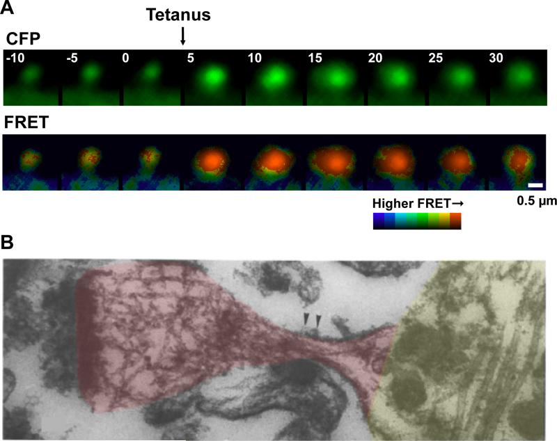
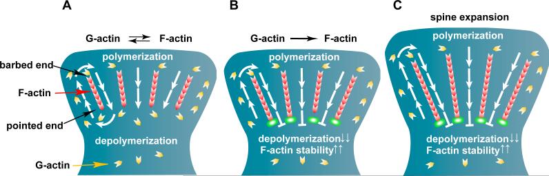
Dendritic spines are small mushroom-like protrusions arising from neurons where most excitatory synapses reside. Their peculiar shape suggests that spines can serve as an autonomous postsynaptic compartment that isolates chemical and electrical signaling. How neuronal activity modifies the morphology of the spine and how these modifications affect synaptic transmission and plasticity are intriguing issues. Indeed, the induction of long-term potentiation (LTP) or depression (LTD) is associated with the enlargement or shrinkage of the spine, respectively. This structural plasticity is mainly controlled by actin filaments, the principal cytoskeletal component of the spine. Here we review the pioneering microscopic studies examining the structural plasticity of spines and propose how changes in actin treadmilling might regulate spine morphology.
Copyright © 2011 Elsevier Ltd. All rights reserved.
Figures
References
-
- Yuste R. Dendritic spines. The MIT Press; Cambridge: 2010.
-
- Hayashi Y, Majewska AK. Dendritic spine geometry: functional implication and regulation. Neuron. 2005;46:529–532. - PubMed
-
- Knott GW, Quairiaux C, Genoud C, Welker E. Formation of dendritic spines with GABAergic synapses induced by whisker stimulation in adult mice. Neuron. 2002;34:265–273. - PubMed
Publication types
MeSH terms
Grants and funding
LinkOut - more resources
Full Text Sources
