. 2012 Feb;24(2):435-442.
doi: 10.1016/j.cellsig.2011.09.016. Epub 2011 Sep 22.

Angiotensin II mediates cell survival through upregulation and activation of the serum and glucocorticoid inducible kinase 1

Affiliations

Angiotensin II mediates cell survival through upregulation and activation of the serum and glucocorticoid inducible kinase 1

Rebekah Baskin et al. Cell Signal. 2012 Feb.

Abstract

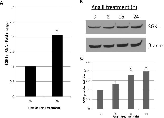
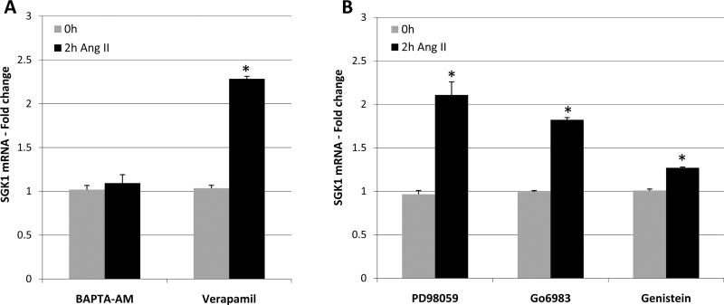
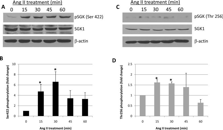
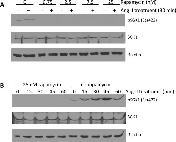
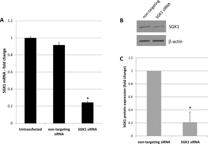
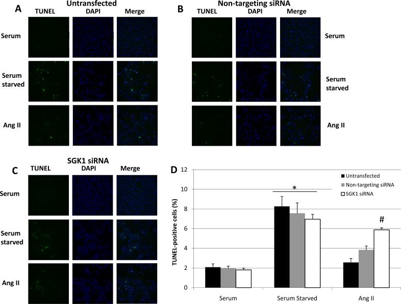
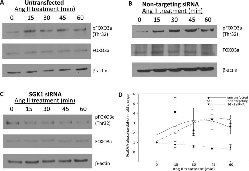
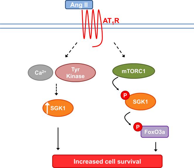
The serum- and glucocorticoid-inducible kinase 1 (SGK1) is known to regulate a wide variety of cellular processes, including renal sodium retention and cell survival. Angiotensin II (Ang II) is one of the many signaling molecules capable of regulating SGK1 expression, and is also known to impact cell survival. Here, we examined the role of SGK1 in Ang II-mediated cell survival. We hypothesized that Ang II protects cells from apoptosis by upregulating and activating SGK1. To test this, we examined the effects of Ang II stimulation on SGK1 expression and downstream signaling. We also examined the effects of Ang II treatment and siRNA-mediated SGK1 knockdown on apoptosis after serum starvation. We found that after 2h of Ang II treatment, SGK1 mRNA expression was increased approximately 2-fold. This induction was sensitive to reductions in intracellular calcium levels after pretreatment with BAPTA-AM, but insensitive to the L-type calcium channel blocker verapamil. SGK1 induction was also sensitive to the tyrosine kinase inhibitor genistein. Ang II treatment also caused a rapid increase in the level of phosphorylation of SGK1 at Ser422 and Thr256, and Ser422 phosphorylation was rapamycin-sensitive. We found that Ang II treatment was protective against serum starvation-induced apoptosis, and this protective effect was significantly blunted when SGK1 was silenced via siRNA. Lastly, Ang II induced FOXO3A phosphorylation in an SGK1-dependent manner, thereby reducing the pro-apoptotic actions of FOXO3A. Overall, these results indicate that Ang II upregulates and activates SGK1, leading to increased cell survival via multiple, non-redundant mechanisms.


Figures

Figure 1
Figure 1
Increase in SGK1 expression in response to Ang II. A, Cells were serum starved overnight and treated with Ang II for 0 or 2h and SGK1 mRNA was quantified by qRT-PCR. n=6. *, p<0.05 vs. 0 h control. B, Cells were serum starved overnight and treated with Ang II for 0, 8, 16, or 24 h and SGK1 protein levels were analyzed by Western blot. C, SGK1 protein levels were quantified using densitometry and normalized to β-actin. n=3. *, p<0.05 vs. 0 h control.
Figure 2
Figure 2
Ang II increases SGK1 mRNA levels via mobilization of intracellular calcium and tyrosine kinase activation. A, Cells were serum starved overnight and pre-treated with BAPTA-AM (50 μM, 10 min) or verapamil (10 μM, 5 min) prior to treatment with Ang II for 0 or 2 h. SGK1 mRNA was quantified via qRT-PCR. n=3. *, p<0.05 vs. 0 h control. B, Cells were serum starved overnight and pre-treated with PD98059 (50 μM, 1 h), Go6983 (10 μM, 1 h), or genistein (100 μM, 1 h) prior to treatment with Ang II for 0 or 2 h. SGK1 mRNA was quantified using qRT-PCR. n=3. *, p<0.05 vs. 0 h control.
Figure 3
Figure 3
Ang II increases the phosphorylation of SGK1 at Ser422 and Thr256. Cells were serum starved overnight and then treated with Ang II for the indicated times and SGK1 phosphorylation at Ser422 (A and B) and Thr256 (C and D) was examined by Western blot. Phosphorylation was quantified using densitometry and normalized to total SGK1 protein levels. n=3. *, p<0.05 vs. 0 min control.
Figure 4
Figure 4
The Ang II-mediated phosphorylation of SGK1 at Ser422 is mTOR-dependent. A, Cells were serum starved overnight and then pre-treated with rapamycin (0 – 25 nM, 1 h) prior to treatment with Ang II for 0 or 30 min. n=3. B, Cells were serum starved overnight and then pre-treated with rapamycin (25 nM, 1 h) prior to treatment with Ang II for 0–60 min. n=3.
Figure 5
Figure 5
Confirmation of SGK1 knockdown. A, Cells were left untransfected or transfected with non-targeting or SGK1-targeting siRNA and SGK1 mRNA levels were then quantified by qRT-PCR. n=4. *, p<0.05 vs. untransfected and non-targeting siRNA. B, Cells were transfected with non-targeting or SGK1-targeting siRNA and SGK1 protein levels were examined by Western blot. C, SGK1 protein levels were quantified using densitometry and normalized to β-actin. n=2. *, p<0.05 vs. non-targeting siRNA.
Figure 6
Figure 6
Ang II protects cells from starvation-induced apoptosis and this is SGK1-dependent. Cells were left untransfected (A), transfected with non-targeting siRNA (B), or transfected with SGK1-targeting siRNA (C) and then serum starved for 48 h in the presence or absence of 100 nM Ang II. Cells were analyzed for apoptosis by TUNEL assay. TUNEL-positive cells are green and the DAPI nuclear counter stain is blue. D, The percentage of TUNEL-positive cells was quantified and plotted as a function of condition. n=4. *, p<0.05 vs. serum containing groups. #, p<0.05 vs. untransfected Ang II-treated group.
Figure 7
Figure 7
Ang II induces FOXO3A phosphorylation in an SGK1-dependent manner. Cells were left untransfected (A), transfected with non-targeting siRNA (B), or transfected with SGK1-targeting siRNA (C) and then serum starved overnight prior to Ang II treatment for 0–60 min. FOXO3A phosphorylation at Thr32 was examined by Western blot. D, FOXO3A phosphorylation was quantified using densitometry and normalized to total FOXO3A protein. n=3 for untransfected and n=2 for non-targeting and SGK1-targeting siRNA.
Figure 8
Figure 8
Proposed signaling pathway for the role of SGK1 in Ang II-mediated cell survival. Ang II acts via the AT1 receptor to 1) increase SGK1 mRNA and SGK1 protein levels via calcium and tyrosine kinase-dependent mechanisms and 2) increase SGK1 and FOXO3A phosphorylation levels via an mTORC1-dependent mechanism. The combined effect of increased SGK1 mRNA levels, increased SGK1 protein levels, and activation of existing pools of SGK1 protein via post translational phosphorylation, is an increase in overall cell survival.

References

    1. Lang F, Bohmer C, Palmada M, Seebohm G, Strutz-Seebohm N, Vallon V. Physiol Rev. 2006;86:1151–1178. - PubMed
    1. Brunet A, Park J, Tran H, Hu LS, Hemmings BA, Greenberg ME. Mol Cell Biol. 2001;21:952–965. - PMC - PubMed
    1. Zhang L, Cui R, Cheng X, Du J. Cancer Res. 2005;65:457–464. - PubMed
    1. Wu W, Chaudhuri S, Brickley DR, Pang D, Karrison T, Conzen SD. Cancer Res. 2004;64:1757–1764. - PubMed
    1. Mehta PK, Griendling KK. Am J Physiol Cell Physiol. 2007;292:C82–97. - PubMed

Publication types

MeSH terms