. 2012 Jan;181(1):7-16.
doi: 10.1016/j.molbiopara.2011.09.006. Epub 2011 Sep 22.

Nuclear actin-related protein is required for chromosome segregation in Toxoplasma gondii

Affiliations

Nuclear actin-related protein is required for chromosome segregation in Toxoplasma gondii

Elena S Suvorova et al. Mol Biochem Parasitol. 2012 Jan.

Abstract

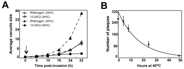
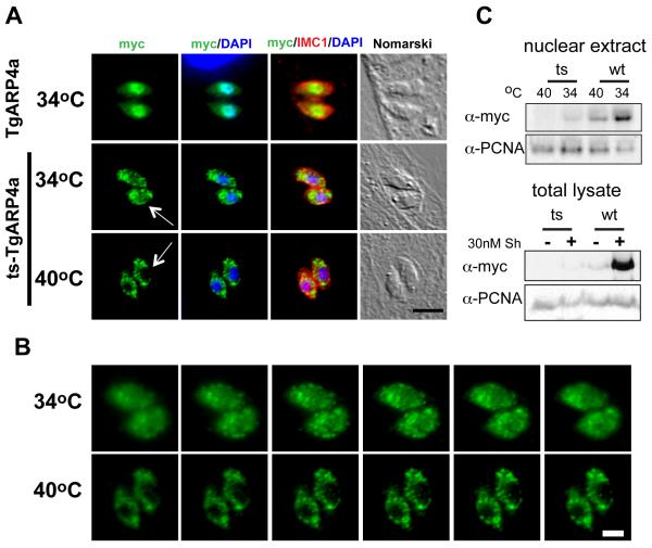
Apicomplexa parasites use complex cell cycles to replicate that are not well understood mechanistically. We have established a robust forward genetic strategy to identify the essential components of parasite cell division. Here we describe a novel temperature sensitive Toxoplasma strain, mutant 13-20C2, which growth arrests due to a defect in mitosis. The primary phenotype is the mis-segregation of duplicated chromosomes with chromosome loss during nuclear division. This defect is conditional-lethal with respect to temperature, although relatively mild in regard to the preservation of the major microtubule organizing centers. Despite severe DNA loss many of the physical structures associated with daughter budding and the assembly of invasion structures formed and operated normally at the non-permissive temperature before completely arresting. These results suggest there are coordinating mechanisms that govern the timing of these events in the parasite cell cycle. The defect in mutant 13-20C2 was mapped by genetic complementation to Toxoplasma chromosome III and to a specific mutation in the gene encoding an ortholog of nuclear actin-related protein 4. A change in a conserved isoleucine to threonine in the helical structure of this nuclear actin related protein leads to protein instability and cellular mis-localization at the higher temperature. Given the age of this protist family, the results indicate a key role for nuclear actin-related proteins in chromosome segregation was established very early in the evolution of eukaryotes.


Figures

Figure 1
Figure 1. Tachyzoite strain 13-20C2 carries a lethal mutation that is conditional with respect to temperature
(A) The growth of parental RHΔhxgprt and mutant 13-20C2 parasites were monitored in populations partially synchronized by limited invasion over a 32 h period at 34°C and 40°C. The arrow indicates a post-invasion time when cultures were shifted to 40°C. To determine average vacuole sizes, randomly selected vacuoles (>50) in three independent cultures per strain and temperature condition were directly monitored by light microscopy. (B) The lethality of the temperature defect was determined by plaque assay. Freshly invaded mutant parasites were incubated at 40°C for the indicated times before the cultures were shifted to the permissive temperature (34°C) to allow for plaque development. Plaque numbers represent the average of three independent flasks.
Figure 2
Figure 2. Immunofluorescent analysis of mutant 13-20C2 reveals severe defects in chromosome segregation and nuclear division
Immunofluorescent microscopy of parental strain RHΔhxgprt (a, b, c) and mutant 13-20C2 was performed 16 h post-infection at 34°C (d, e, f) and 40°C (g, h, I, k, l, m) to determine the primary temperature defect. Representative images showing the co-staining with anti-IMC1 (green) antibodies and DAPI (blue) revealed the primary retention of genomic DNA in the mother residual mass (dashed circled) occurred in parasites grown at the non-permissive temperature (40°C). The loss of chromosome material was variable with some parasites retaining significant DNA (middle panels), while other newly formed parasites lacked detectable DAPI staining (lower panels). Magnification bar (5 μm) is indicated in the lower right hand Nomarski image.
Figure. 3
Figure. 3. The mitotic defect in mutant 13-20C2 does not prevent daughter formation or organellar duplication and targeting
Cell biologic analyses of mutant 13-20C2 parasites cultured for 24 h at 34°C (A) or 40°C (B). Co-stains: Green stains are anti-centrin (centrosome), anti-MORN1 (centrocone and basal complex), anti-Atrx1 (apicoplast) or anti-ROP7 (Rhoptries). Red stain is anti-IMC1. Blue stain is DAPI. Marker guides are decolorized/inverted green/red merged images. Note the consistent posterior location of centrosomes in daughter parasites formed at 40°C (d image panel). Different MORN1 patterns (see guides in h image panels) mark the sequence of cell cycles with the larger posterior body (arrow) retained from the previous basal complex, while the duplicated MORN1 centrocone with ring (star) was formed in the current cell cycle. It is interesting to note that in this example of parasites grown at high temperature (a four vacuole trying to make eight parasites) showed evidence of all three cell cycles with two residual basal complexes evident (total of six basal MORN1 bodies). The two basal MORN1 bodies (arrows) from parasites originally dividing 1-to-2 is still present along with the four MORN1 basal bodies from the 2-to-4 division and the newly formed centrocone/ring structure (star). Marker guides for apicoplast/daughters are indicated in m-panels, and for rhoptries/daughters in q-panels. Dashed circles denote the chromosome material lost in the residual mass. Magnification is indicated in the lower marker guide panels with a 5 μm black bar. (C) Comparison of two chromosome mis-segregation mutants showing distinct severity of defect. Mutant 12-42D6 has an uncoupling phenotype leading to relaxed chromosomes (expanded blue DAPI staining) and severely degenerated bud structures (anti-IMC1, red). By contrast, mutant 13-20C2 has compact, if fragmented nuclei, and well developed buds.
Figure 4
Figure 4. Identification of the defective gene in mutant 13-20C2 by genetic complementation
(A) Results of mutant complementation with cosmid libraries and individual cosmids are diagrammed. Mutant 13-20C2 was complemented with cosmid genomic library DNA. Sequence tags (black arrows) were recovered from temperature and drug resistant populations and mapped to a single locus on chromosome III (665,366bp to 711,113bp). To confirm the complementing locus, the original mutant 13-20C2 was complemented with DNA from two cosmid clones, PSBMM94 (no growth at 40°C) that spans the right side of the locus and cosmid PSBLQ51 (100% growth at 40°C compared to growth at 34°C), which spans the center of the locus and includes genes TGGT1_002010 (Gene #2, TgG2a), TGGT1_002020 (Gene #3, TgARP4a), and TGGT1_002030 (Gene #4, hypothetical). To narrow the gene list of cosmid PSBLQ51 further, mutant 13-20C2 was complemented with cosmid DNA bearing either a deletion in Gene #2 (TgG2a) or Gene #3 (TgARP4a). Mutant parasites transfected with individual cosmids were first selected in bleomycin and the drug resistant populations tested for temperature resistant growth by plaque assay. Ability to form plaques is expressed as a percentage of the plaques formed at 40°C versus parallel controls grown at 34°C. To test the ability of genomic fragments to complement, DNA fragments spanning the TgARP4a locus including promoter and 3′-UTR sequences were amplified from mutant 13-20C2 or parental genomic DNA. (B) Sequencing of TgARP4a cDNA from mutant 13-20C2 and parental RHΔhxgprt parasites identified a single transition mutation at 1,862bp (T/C) in the coding sequence of the tsTgAPR4a resulting in a change of isoleucine to threonine at residue 621 of the predicted TgARP4a coding sequence. Mutated codon is shown boxed. (C) Alignment of the helical region that contains the I621T mutation of tsTgARP4a with selected orthologs (Plasmodium falciparum PF14_0218; Saccharomyces cereviseae SacArp4p; Homo sapiens HmALP6B). Hydrophobic residue corresponding to isoleucine 621 (starred column) in TgARP4a is conserved in the analyzed orthologs. Note that the 621-residue in the helix is preserved by conservative substitutions isoleucine and valine. Identical residues as compared to TgARP4a are indicated in the ortholog sequences as a period. (D, E) Predicted folding of the polypeptides I383-L432 (Sc_Arp4) and F614-L665 (TgARP4a). Crystal structure of yeast protein (pdb:3QB0) was used to model a part of the Subdomain 3 of the actin fold of TgARP4a contain the temperature sensitive mutation. The alpha helix is shown in blue and the corresponding residue I621 in TgARP4a to the V385 in yeast Arp4p are shown in red. Bulky side chains of the residues of three α-helixes and one β-sheet that form a hydrophobic pocket are shown and corresponding residues are labeled on the outside. Note the position of I621 in TgARP4a may interact with a hydrophobic pocket that would like be altered by the mutation to threonine in the ts-TgARP4a protein. (F) Ribbon drawing of the helix I617-D626 showing the precise location of the I621T change.
Figure 5
Figure 5. The I621T mutation in tsTgARP4a results in mis-localization
(A) In order to better understand the temperature defect caused by tsTgARP4a, we compared the subcellular localization of this mutant protein to the wild type isoform over expressed in the parental strain. Transgenic clones expressing DDmyc-wtTgARP4a showed tight nuclear localization at either temperature (34°C expression shown only in top panels); green=anti-myc stain. By contrast, DDmyc-tsTgARP4a was primarily cytoplasmic with some nuclear localization at the non-permissive temperature of 34°C (middle panels). Fluorescent micrographs of DDmyc-tsTgARP4a transgenic parasites were taken using >10-fold longer time exposures. Magnification bar (5 μm) is indicated. (B) Image series scanning through representative 34°C and 40°C vacuoles demonstrated the absence of detectable nuclear DDmyc-tsTgARP4a at the non-permissive temperature. Magnification bar (2 μm) is indicated. (C) Upper panels: To assess the affect of temperature on the levels of nuclear ts or wtTgARP4a, the transgenic clones were induced with 30nM Shield1 at 34°C or 40°C and nuclear extracts prepared as described in the Material and Methods. Nuclear levels of the fusion proteins were detected by Western blots stained with anti-myc antibody. Note the levels of either TgARP4a isoform are affected by higher temperature with the tsTgARP4a undetectable in the parasite nuclear extracts at 40°C similar to the IFA results above. Lower panels: Protein levels in total lysates of transgenic parasites expressing DDmyc-wtTgARP4a or DDmyc- tsTgARP4a fusion proteins grown in the presence or absence of 30 nM Shield1 (Sh) at 34°C were subjected to the western blot analysis using anti-myc antibody. The Western blot membrane was secondarily probed with anti-PCNA1 antibody to ensure equal loading.

References

    1. Dubey JP, Carpenter JL. Toxoplasma gondii-like schizonts in the tracheal epithelium of a cat. J Parasitol. 1991;77:792–6. - PubMed
    1. Canning EU, Sinden RE. The organization of the ookinete and observations on nuclear division in oocysts of Plasmodium berghei. Parasitology. 1973;67:29–40. - PubMed
    1. Speer CA, Dubey JP. An ultrastructural study of first- and second-generation merogony in the coccidian Sarcocystis tenella. J Protozool. 1981;28:424–31. - PubMed
    1. Striepen B, Jordan CN, Reiff S, van Dooren GG. Building the perfect parasite: Cell division in apicomplexa. PLoS Pathog. 2007;3:e78. - PMC - PubMed
    1. Sheffield HG, Melton ML. The fine structure and reproduction of Toxoplasma gondii. J Parasitol. 1968;54:209–26. - PubMed

Publication types

LinkOut - more resources