. 2011 Dec 5;671(1-3):18-25.
doi: 10.1016/j.ejphar.2011.09.033. Epub 2011 Sep 19.

The effect of dehydroleucodine in adipocyte differentiation

Affiliations

The effect of dehydroleucodine in adipocyte differentiation

Adriana Galvis et al. Eur J Pharmacol. .

Abstract

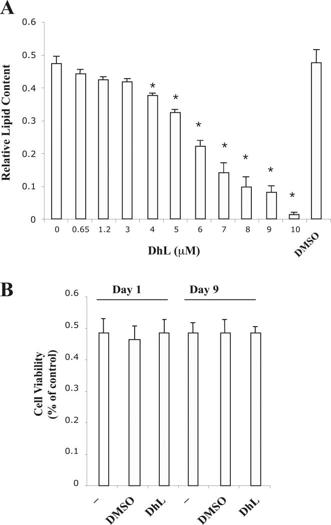
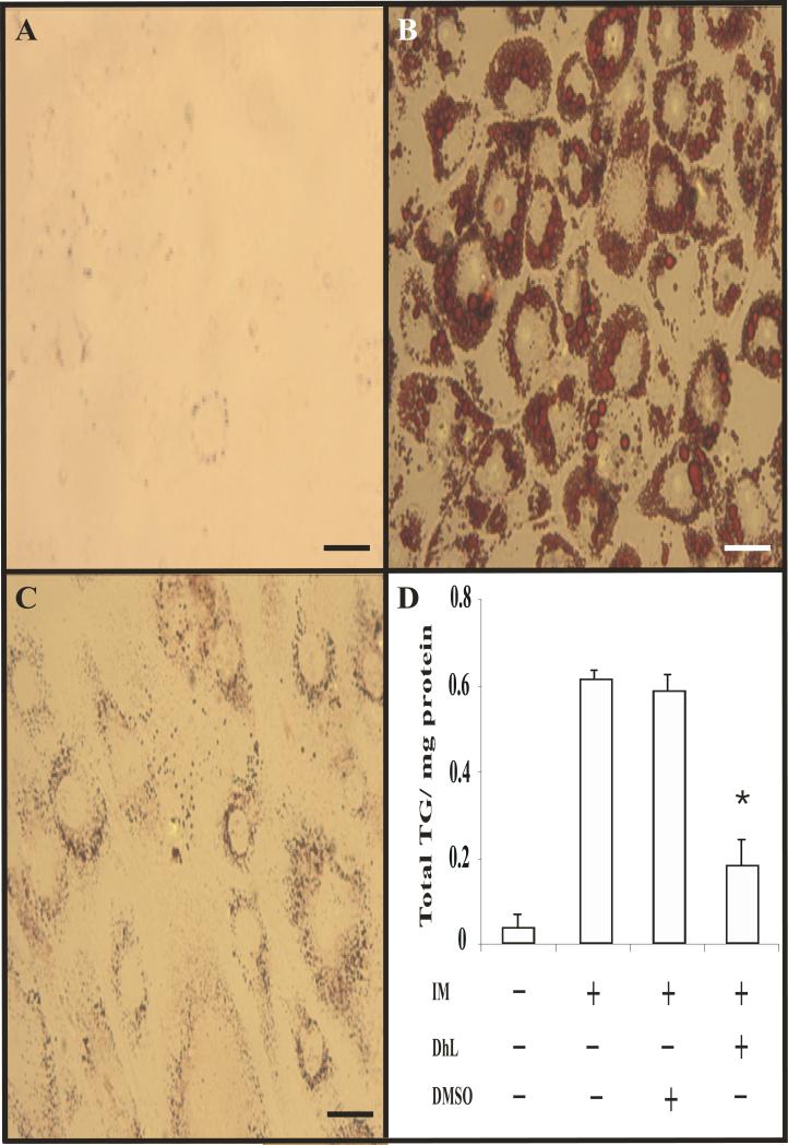
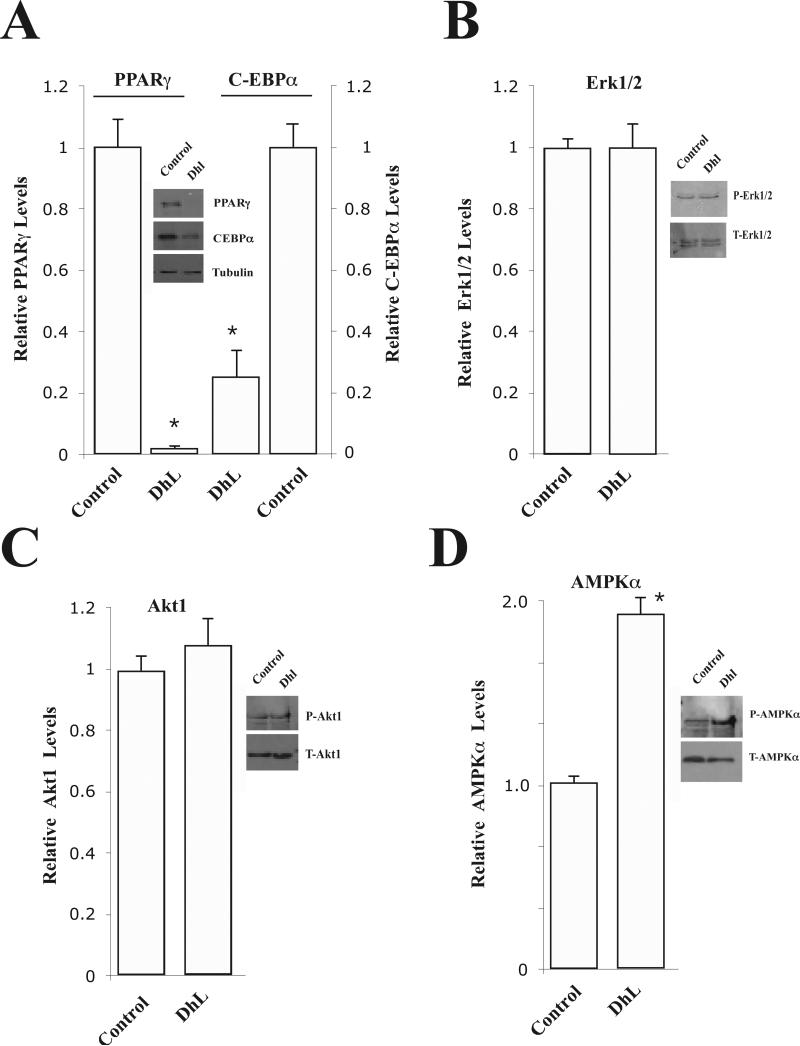
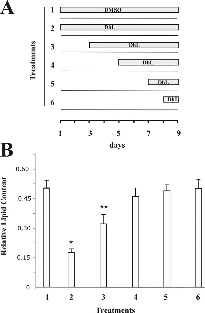
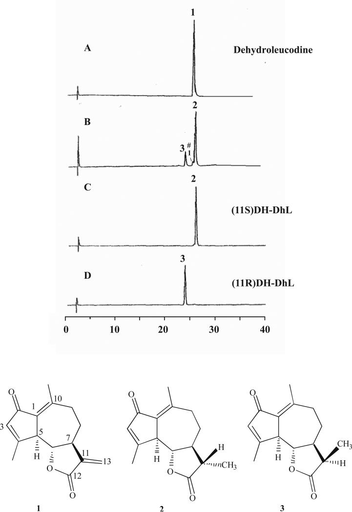
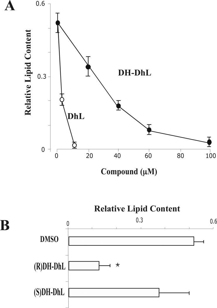
Dehydroleucodine (DhL) is a sesquiterpene lactone of the guaianolide group with gastric cytoprotective activity. Recent studies have also demonstrated that DhL inhibits the proliferation of vascular smooth muscle cells. In this study we examined the effect of DhL in the differentiation of 3T3-L1 preadipocytes. The addition of DhL significantly inhibited the differentiation of 3T3-L1 preadipocytes along with a significant decrease in the accumulation of lipid content by a dramatic downregulation of the expression of adipogenic-specific transcriptional factors PPARγ and C-EBPα. However, phosphorylation of AMPKα, Erk1/2 and Akt1 was not inhibited by DhL treatment. Interestingly, we also found that 11,13-dihydrodehydroleucodine, a derivative of DhL with inactivated α-methylene-γ-lactone function, also inhibited the differentiation of 3T3-L1 preadipocytes. Taken together, these data suggest that DhL has an important inhibitory effect in cellular pathways regulating adipocyte differentiation by modulating the PPARγ expression, which is known to play a pivotal role during adipogenesis.


Figures

Fig. 1
Fig. 1. Dehydroleucodine inhibited adipogenesis of 3T3-L1 preadipocytes without reducing cell viability
(A) 3T3-L1 preadipocytes were differentiated into adipocytes in the absence or in the presence of various amounts of DhL (0.65 to 10 μM) as described in Material and Methods. Results were represented as relative lipid contents. Data represent the mean ± S.E.M. of three independent experiments. *P < 0.05 by Student's t-test compared to DMSO and only induction media-treated cells. (B) Cells were treated with 8 μM DhL for either 24hrs (Day 1) or for 9 days (Day 9). Cell viability was measured using the MTT assay as described in Material and Methods. Data represent the mean ± S.E.M. of three independent experiments.
Fig. 2
Fig. 2. Dehydroleucodine blocked the formation of lipid droplet by induction media in 3T3-L1 cells
Adipocyte differentiation was induced by treating confluent 3T3-L1 preadipocytes with induction media in the absence or presence of 8 μM DhL. Morphological changes of 3T3-L1 preadipocytes were monitored by microscope and photographed after 9 days from the onset of differentiation. (A) Vehicle only, (B) cells treated induction media in the presence DMSO, or (C) in the presence of DhL. (D) Nine days after induction of differentiation, cells were lysed for triglyceride and protein assays as described in Material and Methods. Vehicle only (line 1), cells treated with induction media alone (line 2), cells treated with induction media in the presence DMSO (line 3), and cells treated with induction media in the presence of 8 μM DhL (line 4). Bars=10μm. Data represent the mean ± S.E.M. of three independent experiments. *P < 0.05 by Student's t-test compared to DMSO-treated cells and only induction media-treated cells.
Fig. 3
Fig. 3. Dehydroleucodine attenuated the expression of PPARγ during 3T3-L1 preadipocyte differentiation
3T3-L1 preadipocytes were induced to differentiate by induction media into adipocytes in the absence (insert: –DhL, control) or in the presence (insert: +DhL) of 8 μM DhL. Total protein extracts were prepared at day 9 from each sample. The proteins were subset to 12% SDS-PAGE electrophoresis, blotted to a nitrocellulose membrane, and probed with anti-bodies specific to (A) PPARγ, C-EBPα and tubulin, (B) P-Erk1/2, T-Erk1/2, (C) P-Akt1, T-Akt1, and (D) P-AMPKα and T-AMPKα respectively. Relative levels of proteins were determined by densitometry as describe in Material and Methods. Data represent the mean ± S.E.M. of three independent experiments. *P < 0.05 by Student's t-test compared to DMSO induction media-treated cells (control).
Fig. 4
Fig. 4. Dehydroleucodine blocked adipocyte differentiation in a time-dependent manner
(A) 3T3-L1 preadipocyte cells were treated with 8 μM DhL for the time indicated in the schematic representation of the experiment. (B) In each treatment (1-6), the accumulation of lipid droplets was measured by the incorporation of Oil red O as described in Material and Methods. Data represent the mean ± S.E.M. of three independent experiments. *P < 0.05 and **P < 0.01 by Student's t-test compared to DMSO induction media-treated cells (control).
Fig. 5
Fig. 5. GC analysis of dehydroleucodine and 11,13-dihydro-dehydroleucodine epimers
A) DhL isolated from Artemisia douglassiana; (B) Mixture DH-DhL epimers as obtained by reduction of DhL, (#) denotes small amount of DhL after the reduction reaction; (C) DH-DhL epimer S, and, (D) DH-DhL epimer R after separation from the mixture. Chemical structures of DhL (compound 1), DH-DhL epimer 11S (compound 2) and DH-DhL epimer 11R (compound 3) (numbering according to classical nomenclature).
Fig. 6
Fig. 6. 11,13-dihydro-dehydroleucodine inhibited 3T3-L1 preadipocyte differentiation
(A) 3T3-L1 preadipocytes were differentiated into adipocytes in the absence or in the presence of either DHL or DH-DhL. Results were represented as relative lipid content. Data represent the mean ± S.E.M. of three independent experiments. (B) 3T3-L1 preadipocyte cells were incubated with induction media supplemented with either DMSO, 80 μM DH-DhL epimer S or 80 μM DH-DhL epimer R and the incorporation of Oil Red O was measured by as described in Material and Methods. Results were represented as relative lipid content. Data represent the mean ± S.E.M. of three independent experiments. **P < 0.01 by Student's t-test compared to DMSO-treated cells.

References

    1. Auld CA, Hopkins RG, Fernandes KM, Morrison RF. Novel effect of helenalin on Akt signaling and Skp2 expression in 3T3-L1 preadipocytes. Biochem. Biophys. Res. Commun. 2006;346:314–320. - PubMed
    1. Beekman AC, Woerdenbag HJ, van Uden W, Pras N, Konings AW, Wikstrom HV, Schmidt TJ. Structure-cytotoxicity relationships of some helenanolide-type sesquiterpene lactones. J. Nat. Prod. 1997;60:252–257. - PubMed
    1. Blanco JG, Gil RR, Alvarez CI, Patrito LC, Genti-Raimondi S, Flury A. A novel activity for a group of sesquiterpene lactones: inhibition of aromatase. FEBS. Lett. 1997;409:396–400. - PubMed
    1. Bohlmann F, Zdero C. Zwei neue Sesquiterpen-lactone aus Lidbeckia pectinata Berg. und Pentzia elegans DC. Tetrahedron Lett. 1972;13:621–624.
    1. Bost F, Aouadi M, Caron L, Binetruy B. The role of MAPKs in adipocyte differentiation and obesity. Biochimie. 2005;87:51–56. - PubMed

Publication types