. 2011 Dec 1;360(1):132-42.
doi: 10.1016/j.ydbio.2011.09.017. Epub 2011 Sep 22.

Functional dissection of phosphorylation of Disheveled in Drosophila

Affiliations

Functional dissection of phosphorylation of Disheveled in Drosophila

Wang A Yanfeng et al. Dev Biol. .

Abstract

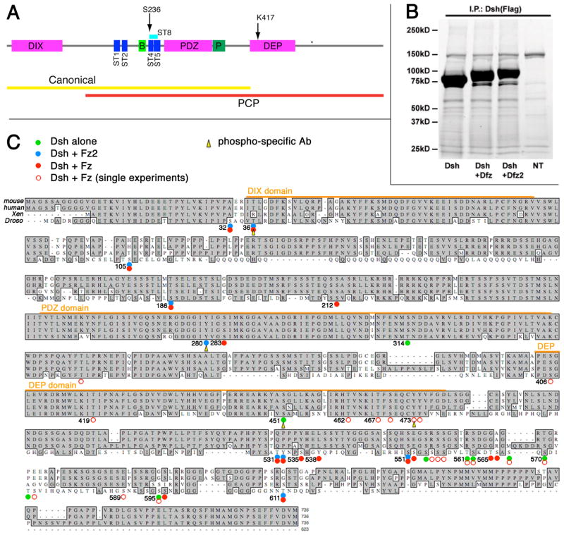
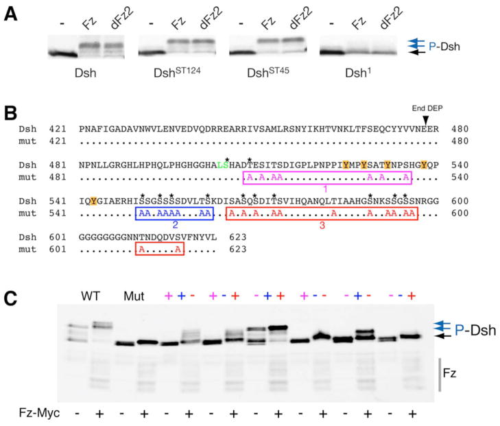
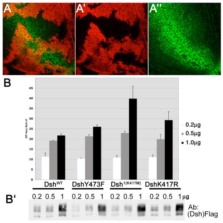
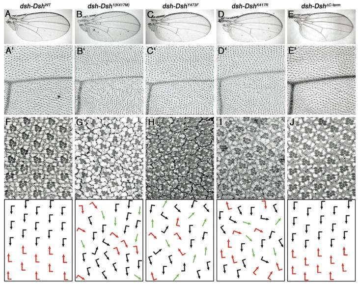
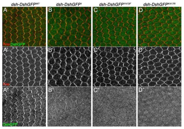
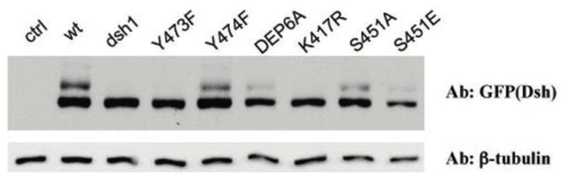
Disheveled/Dsh proteins (Dvl in mammals) are core components of both Wnt/Wg-signaling pathways: canonical β-catenin signaling and Frizzled (Fz)-planar cell polarity (PCP) signaling. Although Dsh is a key cytoplasmic component of both Wnt/Fz-pathways, regulation of its signaling specificity is not well understood. Dsh is phosphorylated, but the functional significance of its phosphorylation remains unclear. We have systematically investigated the phosphorylation of Dsh by combining mass-spectrometry analyses, biochemical studies, and in vivo genetic methods in Drosophila. Our approaches identified multiple phospho-residues of Dsh in vivo. Our data define three novel and unexpected conclusions: (1) strikingly and in contrast to common assumptions, all conserved serines/threonines are non-essential for Dsh function in either pathway; (2) phosphorylation of conserved Tyrosine473 in the DEP domain is critical for PCP-signaling - Dsh(Y473F) behaves like a PCP-specific allele; and (3) defects associated with the PCP specific dsh(1) allele, Dsh(K417M), located within a putative Protein Kinase C consensus site, are likely due to a post-translational modification requirement of Lys417, rather than phosphorylation nearby. In summary, our combined data indicate that while many Ser/Thr and Tyr residues are indeed phosphorylated in vivo, strikingly most of these phosphorylation events are not critical for Dsh function with the exception of DshY473.


Figures

Figure 1
Figure 1. Identification of phosphorylated residues of Dsh
(A) Schematic of Dsh with the DIX, PDZ, and DEP domains indicated in purple. Light and dark green domain correspond to the Basic and Proline rich regions, respectively. Dark blue boxes represent the Serine/Threonine clusters that were mutated in Penton et al. (2002). ST8 (light blue box) corresponds to the 8 S/T residues that were mutated in Strutt et al. 2006 and have been identified as potential Par1 sites (Ossipova et al 2005). S236 corresponds to the CK1ε site in Klein et al. 2006. K417 and (*) depict the position of the dsh1 mutation and the BlpI site used to engineer the C-terminal mutations, respectively. Note that re-sequencing of the ST5 region of the mutant constructs from Penton et al. (2002) showed that T252 is not mutated in the ST5 containing constructs. The defined functional requirements for the two Wnt-signaling pathways (Boutros et al. 1998; 1999; Wallingford and Habas, 2005) are indicated as yellow (canonical pathway) and red (PCP signaling) bars below the scheme. (B) Purification of transfected Dsh protein from HEK293T cells. Protein staining (Simply Blue, Invitrogen) of SDS-PAGE gel is shown. 3xFlag-tagged Drosophila Dsh was transfected alone or co-transfected with either Dfz or Dfz2 as indicated. Cell lysates were immunoprecipitated with anti-Flag antibodies. The respective products were analyzed in 4–15% gradient SDS-PAGE gel (Biorad; BSA was used to estimate protein concentration). NT lane: untransfected control. The upper bands/region (hyperphosphorylated Dsh) were cut out, eluted and subjected to mass-spectrometry (MS) studies (see Methods). (C) Phosphorylated residues of Drosophila Dsh as identified by mass-spectrometry, Drosophila Dsh sequence is aligned with mouse Dvl2, human Dvl2, and Xenopus Dsh. Marked residues were detected as phosphorylated in independent transfections and MS experiments. The results shown reflect three independent experimental analyses; phosphorylation events detected only in one of the three analyses are shown with open circles instead of dots. Green dots: Dsh transfected alone (naïve state); blue dots: Dsh co-tranfected with Fz2 (“canonical” signaling state); red dots: Dsh co-transfected with Fz (“PCP” state); the respective signaling states were assigned based on observations of Dsh membrane recruitment (T.J. Klein and MM, unpublished) and activation of the Top-flash Wnt-signaling reporter assay. Orange color bars indicate the extent of the three conserved domains: DIX (residue 35–83), PDZ (252–338) and DEP (404–478). Yellow arrowheads indicate residues that were chosen for anti-phospho-residue antiserum generation. All four phospho-residues were confirmed by in vivo analyses.
Figure 2
Figure 2. Dsh mobility shift assays
(A) Gel-shift assays show that DshST124 and DshST45 phosphorylation (blue arrows on right) can be induced by co-transfections of Fz or dFz2 in S2 cells. As previously shown in vivo (Axelrod 2001), dsh1 barely shows a gel-shift under these conditions. (C, D) The C-terminal S/T residues affect the Dsh gel-shift. (B) Sequence of the C-terminus of Dsh with the end of the DEP domain indicated. To assess the contribution of the 29 S/T residues of the C-tail to the phosphorylation induced gel-shift, we mutated 28 of them in three clusters (#1: purple, #2: blue, and #3: red in color code; note that the first serine is part of the BlpI site (green) and was not mutated for cloning reasons). Asterisks indicate conserved S/Ts. The region around cluster #1 also contained 5 Tyrosines (marked by orange boxes). (C) Myc-Fz induced Dsh gel-shift assay in 293T cells. In contrast to wild-type Dsh, a Dsh with a fully mutant C-terminus is not shifted (Mut; blue arrows indicate phosphorylated forms of Dsh; note the strong shift due to endogenous Dsh activation of WT-Dsh in the 293T cells used). Analysis of mutations of individual clusters shows that the second and third clusters have bigger effects on the gel-shift than the first cluster. Combinations of the clusters, however, indicate that the first cluster also contributes to Dsh phosphorylation as assessed by gel-shifts. Grey bar on the right shows migration of Myc-Fz.
Figure 3
Figure 3. Phosphorylation of Dsh protein in vivo
Immunoprecipitated Dsh protein from late larval and pupal stages was analyzed with phospho-residue specific antibodies (see Methods for details). Purified Dsh protein from dsh-Dsh3xFlag, dshV26 flies were probed with the following antibodies (all control lanes are BSA): 1: anti-Dsh; 2: anti-phosphoThr36; 3: anti-phosphoTyr280; 4: anti-phosphoSer451; 5–7: anti-phosphoTyr473 with phosphorylated antigen peptide as competitor (lane 6) and non-phosphorylated antigen peptide as competitor (lane 7).
Figure 4
Figure 4. Canonical Wg-signaling activity of Dsh1, DshY473F, and DshK417R mutants
(A) dsh-DshY473F (in a dsh- background) fully rescues the expression of Wg-signaling target genes during wing disc development. Example shown is Distalless (Dll). Senseless/Sens was also tested and rescued (not shown). 3rd instar larval wing disc stained for the clonal marker lacZ (red, marks wild-type regions) and Distalless (Dll; green), expressed along the dorso-ventral margin flanking the Wg expression stripe and weaker throught the wing blade anlage. Dll is a direct target of Wg. dshV26 clones loose Dll expression (not shown). The presence of DshY473F fully rescued the mutant dshV26 effect. Dll expression showed no difference between mutant and wild-type tissue. (A′) and (A″) show single channels for lacZ and Dll, respectively. (B-B′) Dsh mutants do not affect expression levels of Top-flash reporter. Dsh mutant expression constructs were transfected with Top-flash luciferase reporter (and SV40-renilla luciferase reporter as control) in HEK293T cells. The ratios of Top-flash vs. renilla were normalized against empty vector control at three doses of transfection as indicated. Note that DshY473 showed no significant reduction as compared to wild-type Dsh (DshWT); the same was the case with Dsh1(K417M) and DshK417R mutants. (B″): Western-blot against Dsh to visualize similar protein levels.
Figure 5
Figure 5. Functional requirements of specific Dsh residues in vivo
All rescue constructs were expressed under the control of the endogenous dsh promoter in a dshV26 (null), f background; the only Dsh protein present is provided by the transgenic lines (note that all genotypes are forked [f] to control for dshV26, giving the cellular hairs a wavy appearance). For all constructs, several independent lines behaved identically and displayed very similar expression levels, as expected (see Suppl. Fig. S5). (A–E′) Wings of rescued flies of indicated genotype are shown; low magnification (A–E) highlight normal Wg-signaling: development with of normal sized wing and normal wing margin development demonstrating correct canonical Wnt signaling. High magnification (panels A′-E′) show PCP arrangements of the respective wings. The high magnification pictures show a region near the posterior cross vein and L4. Note PCP defects in the genotypes dsh-Dsh1, dsh-DshY473F, dsh-DshK417R. (F–J) Adult eye sections of the respective genotypes with schematic presentation in lower panels; black and red arrows represent ommatidia with chiral arrangements, which are normally arranged in a mirror-image across the dorso-ventral midline (see panel F for wild-type); green arrows represent symmetric ommatidia. Note that in G–I, the mirror-image arrangement is lost and a random distribution of the two chiral forms and symmetrical ommatidia is detected, reflecting typical PCP defects.
Figure 6
Figure 6. Localization of mutated Dsh isoforms in pupal wings during PCP signaling. Confocal images of DshGFP (green) and Fmi (red) staining in pupal wings at the 30 to 34hrs APF stage are shown. The respective genotypes are indicated above
A′-D′ display anti-Fmi monochrome images (highlighting membranes and the PCP complexes localized there), and A″-D″ show monochromes of DshGFP (A–D are the merged channels). Note that in B/B″, C/C″, and D/D″ the membrane levels of DshGFP are reduced or even absent, although the overall signal and protein levels are not affected (see also Suppl. Figure S5).
Figure 7
Figure 7
Migration behavior and phophorylation status of mutant DshGFP isoforms as detected by Western blots of late 3rd instar/early pupal tissues. The respective Dsh isoforms are indicated above the gel and were detected with an anti-GFP antibody (“ctrl” lane has no DshGFP protein present as control). All lines are expressed from the endogenous dsh-expression cassette and exhibit very similar protein levels (also Suppl. Figure S5). Note that Dsh1, DshY473F and DshK417R show markedly reduced gel shift, indicative of reduced Dsh phosphoprylation and correlating with their defective behavior in PCP signaling.

Similar articles

Cited by

References

    1. Adler PN. Planar signaling and morphogenesis in Drosophila. Dev Cell. 2002;2:525–535. - PubMed
    1. Axelrod JD. Unipolar membrane association of Dishevelled mediates Frizzled planar cell polarity signaling. Genes Dev. 2001;15:1182–1187. - PMC - PubMed
    1. Axelrod JD, Miller JR, Shulman JM, Moon RT, Perrimon N. Differential requirement of Dishevelled provides signaling specificity in the Wingless and planar cell polarity signaling pathways. Genes & Dev. 1998;12:2610–2622. - PMC - PubMed
    1. Bhanot P, Fish M, Jemison JA, Nusse R, Nathans J, Cadigan KM. Frizzled and DFrizzled-2 function as redundant receptors for wingless during drosophila embryonic development [In Process Citation] Development. 1999;126:4175–86. - PubMed
    1. Bhat KM. Frizzled and Frizzled2 play a partially redundant role in Wingless signaling and have similar requirements to Wingless in neurogenesis. Cell. 1998;95:1027–1036. - PubMed

Publication types

MeSH terms