

Niaspan enhances vascular remodeling after stroke in type 1 diabetic rats
- PMID: 21963653
- PMCID: PMC3265018
- DOI: 10.1016/j.expneurol.2011.09.022
Niaspan enhances vascular remodeling after stroke in type 1 diabetic rats
Abstract
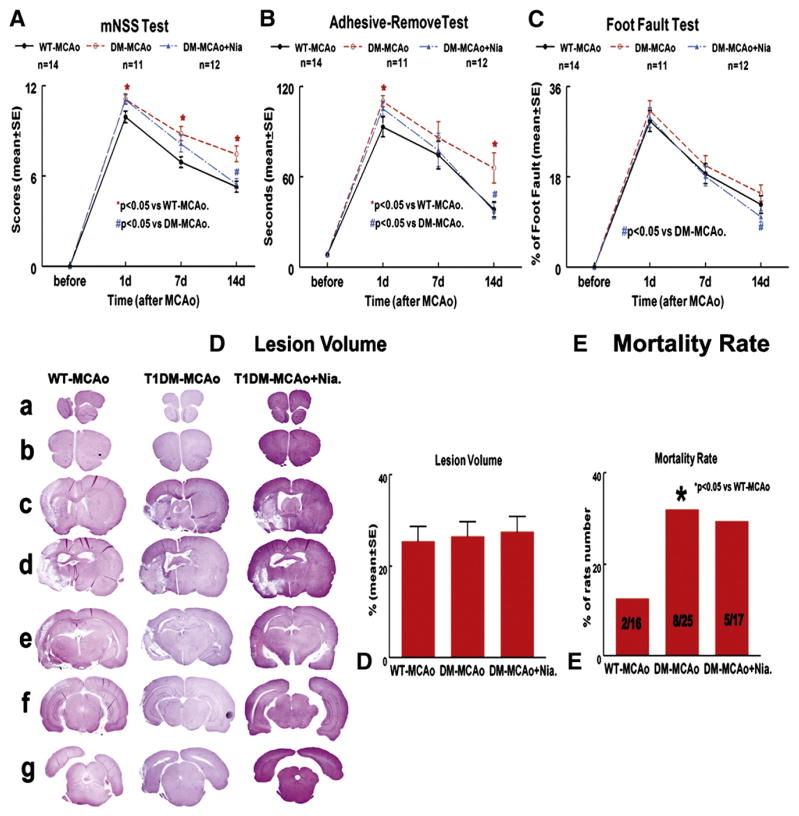
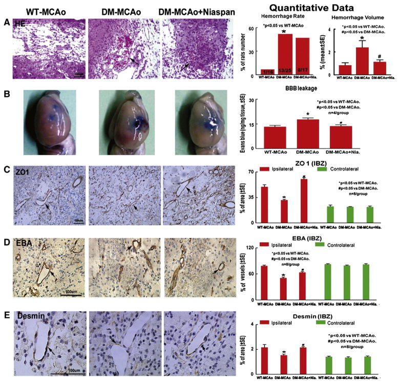
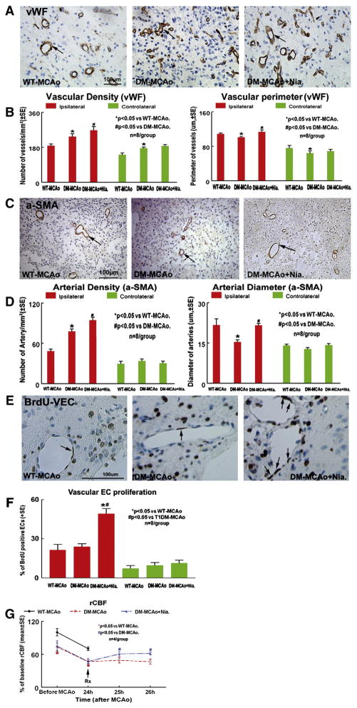
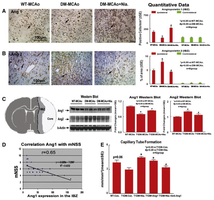
We investigated the changes and the molecular mechanisms of cerebral vascular damage and tested the therapeutic effects of Niaspan in type-1 streptozotocin induced diabetic (T1DM) rats after stroke. T1DM-rats were subjected to transient middle cerebral artery occlusion (MCAo) and treated without or with Niaspan. Non-streptozotocin rats (WT) were also subjected to MCAo. Functional outcome, blood-brain-barrier (BBB) leakage, brain hemorrhage, immunostaining, and rat brain microvascular endothelial cell (RBEC) culture were performed. Compared to WT-MCAo-rats, T1DM-MCAo-rats did not show an increase lesion volume, but exhibited significantly increased brain hemorrhage, BBB leakage and vascular damage as well as decreased functional outcome after stroke. Niaspan treatment of stroke in T1DM-MCAo-rats significantly attenuated BBB damage, promoted vascular remodeling and improved functional outcome after stroke. T1DM-MCAo-rats exhibited significantly increased Angiopoietin 2 (Ang2) expression, but decreased Ang1 expression in the ischemic brain compared to WT-MCAo-rats. Niaspan treatment attenuated Ang2, but increased Ang1 expression in the ischemic brain in T1DM-MCAo-rats. In vitro data show that the capillary-like tube formation in the WT-RBECs marginally increased compared to T1DM-RBEC. Niaspan and Ang1 treatment significantly increased tube formation compared to non-treatment control. Inhibition of Ang1 attenuated Niacin-induced tube formation in T1DM-RBECs. Niaspan treatment of stroke in T1DM-rats promotes vascular remodeling and improves functional outcome. The Ang1/Ang2 pathway may contribute to Niaspan induced brain plasticity. Niaspan warrants further investigation as a therapeutic agent for the treatment of stroke in diabetics.
Copyright © 2011 Elsevier Inc. All rights reserved.
Figures
References
-
- Adams HP, Jr, Brott TG, Furlan AJ, Gomez CR, Grotta J, Helgason CM, Kwiatkowski T, Lyden PD, Marler JR, Torner J, Feinberg W, Mayberg M, Thies W. Guidelines for thrombolytic therapy for acute stroke: a supplement to the guidelines for the management of patients with acute ischemic stroke. A statement for health-care professionals from a Special Writing Group of the Stroke Council, American Heart Association. Circulation. 1996;94:1167–1174. - PubMed
-
- Alvarez-Sabin J, Molina CA, Montaner J, Arenillas JF, Huertas R, Ribo M, Codina A, Quintana M. Effects of admission hyperglycemia on stroke outcome in reperfused tissue plasminogen activator-treated patients. Stroke. 2003;34:1235–1241. - PubMed
-
- Arora S, Pomposelli F, LoGerfo FW, Veves A. Cutaneous microcirculation in the neuropathic diabetic foot improves significantly but not completely after successful lower extremity revascularization. J Vasc Surg. 2002;35:501–505. - PubMed
-
- Ben-nun J, Alder VA, Constable IJ. Retinal microvascular patency in the diabetic rat. Int Ophthalmol. 2004;25:187–192. - PubMed
Publication types
MeSH terms
Substances
Grants and funding
LinkOut - more resources
Full Text Sources
Medical
Miscellaneous
