Review
. 2011 Dec;162(1-2):80-99.
doi: 10.1016/j.virusres.2011.09.020. Epub 2011 Sep 22.

Progress in understanding and controlling respiratory syncytial virus: still crazy after all these years

Affiliations
Review

Progress in understanding and controlling respiratory syncytial virus: still crazy after all these years

Peter L Collins et al. Virus Res. 2011 Dec.

Abstract

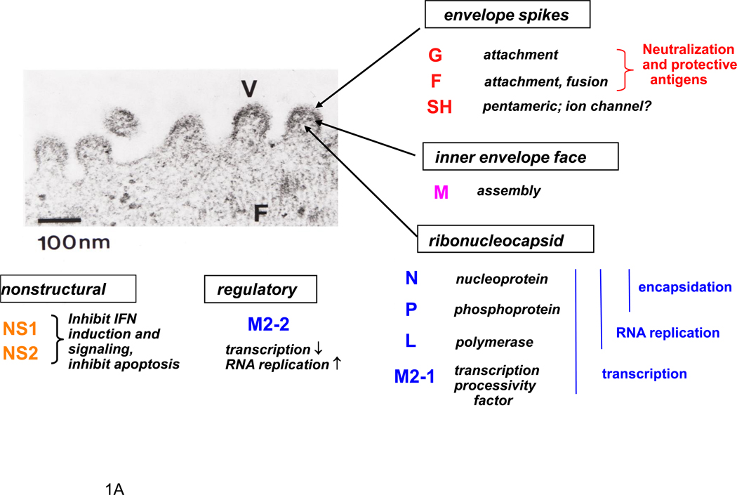
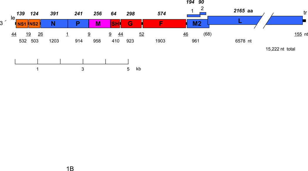
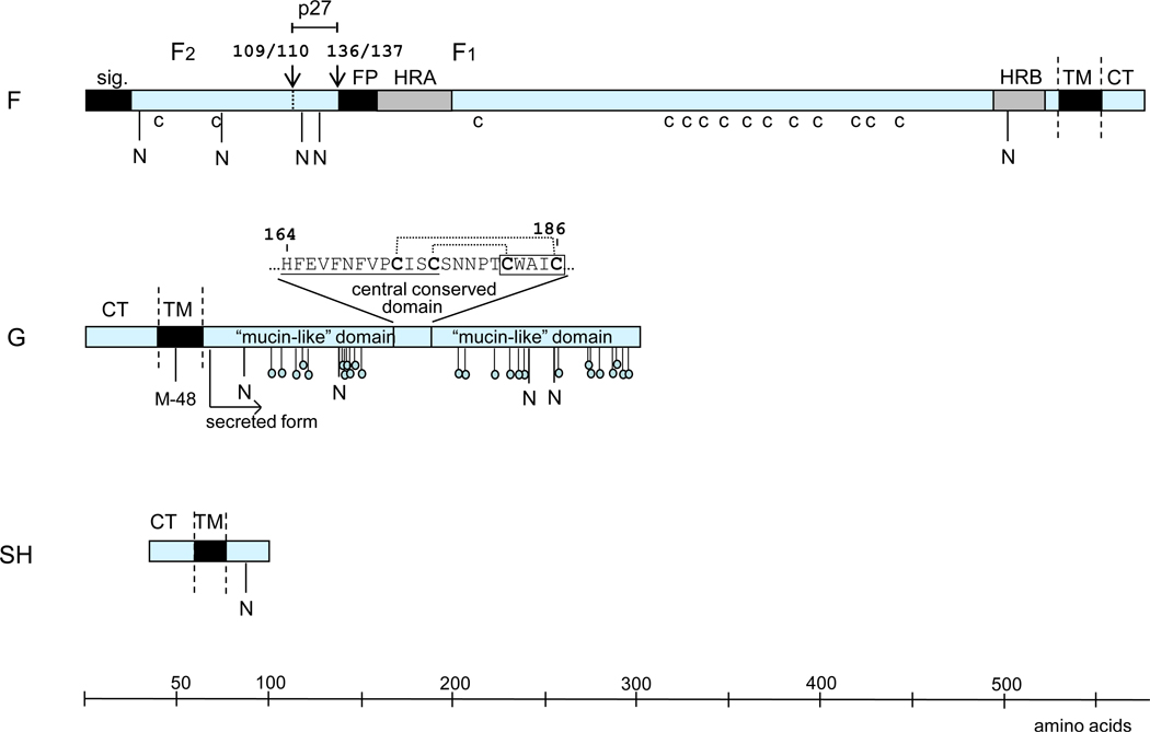
Human respiratory syncytial virus (RSV) is a ubiquitous pathogen that infects everyone worldwide early in life and is a leading cause of severe lower respiratory tract disease in the pediatric population as well as in the elderly and in profoundly immunosuppressed individuals. RSV is an enveloped, nonsegmented negative-sense RNA virus that is classified in Family Paramyxoviridae and is one of its more complex members. Although the replicative cycle of RSV follows the general pattern of the Paramyxoviridae, it encodes additional proteins. Two of these (NS1 and NS2) inhibit the host type I and type III interferon (IFN) responses, among other functions, and another gene encodes two novel RNA synthesis factors (M2-1 and M2-2). The attachment (G) glycoprotein also exhibits unusual features, such as high sequence variability, extensive glycosylation, cytokine mimicry, and a shed form that helps the virus evade neutralizing antibodies. RSV is notable for being able to efficiently infect early in life, with the peak of hospitalization at 2-3 months of age. It also is notable for the ability to reinfect symptomatically throughout life without need for significant antigenic change, although immunity from prior infection reduces disease. It is widely thought that re-infection is due to an ability of RSV to inhibit or subvert the host immune response. Mechanisms of viral pathogenesis remain controversial. RSV is notable for a historic, tragic pediatric vaccine failure involving a formalin-inactivated virus preparation that was evaluated in the 1960s and that was poorly protective and paradoxically primed for enhanced RSV disease. RSV also is notable for the development of a successful strategy for passive immunoprophylaxis of high-risk infants using RSV-neutralizing antibodies. Vaccines and new antiviral drugs are in pre-clinical and clinical development, but controlling RSV remains a formidable challenge.


Figures

Figure 1
Figure 1
RSV proteins (A) and gene map (B). Panel A shows a negatively-stained electron micrograph of budding RSV virions: V indicates a budding virion and F indicates filamentous cytoplasmic structures thought to be nucleocapsids (courtesy of Dr. Robert M. Chanock) (Kalica et al., 1973). The locations of viral proteins in the virion, and their functions when known, are indicated. Panel B shows a map of the negative sense genome (RSV strain A2), approximately to scale. The overlapping M2-1 and M2-2 ORFs are shown over the gene. Numbers below the map indicate nucleotide (nt) lengths: those of the 3´ leader (le) and 5´ trailer (tr) and intergenic regions are underlined, and the length of the gene overlap in parentheses. Italicized, bold numbers over the map indicate the amino acid (aa) lengths of the unmodified proteins. The viral proteins are as follows: G, attachment glycoprotein; F, fusion glycoprotein; SH, small hydrophobic glycoprotein; M, matrix protein; N, nucleoprotein; P, phosphoprotein; L, large polymerase protein; M2-1, product of the first ORF in the M2 mRNA; M2-2; product of the second ORF in the M2 RNA.
Figure 1
Figure 1
RSV proteins (A) and gene map (B). Panel A shows a negatively-stained electron micrograph of budding RSV virions: V indicates a budding virion and F indicates filamentous cytoplasmic structures thought to be nucleocapsids (courtesy of Dr. Robert M. Chanock) (Kalica et al., 1973). The locations of viral proteins in the virion, and their functions when known, are indicated. Panel B shows a map of the negative sense genome (RSV strain A2), approximately to scale. The overlapping M2-1 and M2-2 ORFs are shown over the gene. Numbers below the map indicate nucleotide (nt) lengths: those of the 3´ leader (le) and 5´ trailer (tr) and intergenic regions are underlined, and the length of the gene overlap in parentheses. Italicized, bold numbers over the map indicate the amino acid (aa) lengths of the unmodified proteins. The viral proteins are as follows: G, attachment glycoprotein; F, fusion glycoprotein; SH, small hydrophobic glycoprotein; M, matrix protein; N, nucleoprotein; P, phosphoprotein; L, large polymerase protein; M2-1, product of the first ORF in the M2 mRNA; M2-2; product of the second ORF in the M2 RNA.
Figure 2
Figure 2
The surface glycoproteins of RSV (strain A2), drawn approximately to scale. Hydrophobic domains are shown as black boxes (sig., signal peptide; FP, fusion peptide; TM, transmembrane anchor). Heptad repeats (HRA and HRB) in F are in grey. CT: cytoplasmic tail. The cleavage sites in F are indicated with downward facing arrows and identified by amino acid position. Cysteine residues in F that are conserved among human pneumoviruses are indicated (c). Potential acceptor sites for N-linked carbohydrate are indicated as downward facing stalks with N. The 25 potential acceptor sites for O-linked sugars in G that are predicted by NetOGlyc 2.0 to be the most likely to be utilized are indicated as downward facing stalks with small circles. The expanded sequence above G protein shows the conserved 13-amino acid segment (underlined) and cystine noose; cysteine residues are bold; the disulfide bonding pattern is indicated by dotted lines (Gorman et al., 1997); and the fractalkine CX3C motif is boxed. M-48 in the HRSV G protein is the translational start site for the secreted form, and the mature secreted form is indicated (Roberts et al., 1994).

Similar articles

Cited by

References

    1. Reduction of respiratory syncytial virus hospitalization among premature infants and infants with bronchopulmonary dysplasia using respiratory syncytial virus immune globulin prophylaxis. The PREVENT Study Group. Pediatrics. 1997;99(1):93–99. - PubMed
    1. Palivizumab, a Humanized Respiratory Syncytial Virus Monoclonal Antibody, Reduces Hospitalization From Respiratory Syncytial Virus Infection in High-risk Infants. The IMpact-RSV study group. Pediatrics. 1998;102(3):531–537. - PubMed
    1. Diagnosis and management of bronchiolitis. Pediatrics. 2006;118(4):1774–1793. - PubMed
    1. From the American Academy of Pediatrics: Policy statements--Modified recommendations for use of palivizumab for prevention of respiratory syncytial virus infections. Pediatrics. 2009;124(6):1694–1701. - PubMed
    1. Abu-Harb M, Bell F, Finn A, Rao WH, Nixon L, Shale D, Everard ML. IL-8 and neutrophil elastase levels in the respiratory tract of infants with RSV bronchiolitis. Eur Respir J. 1999;14(1):139–143. - PubMed

Publication types

MeSH terms