. 2011 Dec;301(6):H2422-32.
doi: 10.1152/ajpheart.00343.2011. Epub 2011 Sep 30.

Intramuscular VEGF activates an SDF1-dependent progenitor cell cascade and an SDF1-independent muscle paracrine cascade for cardiac repair

Affiliations

Intramuscular VEGF activates an SDF1-dependent progenitor cell cascade and an SDF1-independent muscle paracrine cascade for cardiac repair

David Zisa et al. Am J Physiol Heart Circ Physiol. 2011 Dec.

Abstract

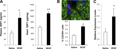
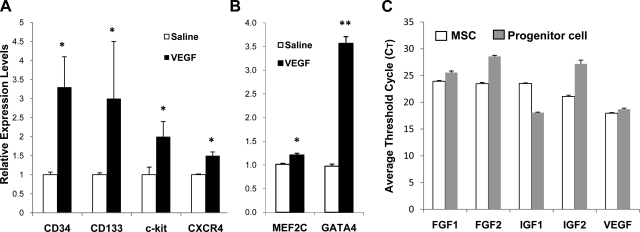
The skeletal muscle is endowed with an impressive ability to regenerate after injury, and this ability is coupled to paracrine production of many trophic factors possessing cardiovascular benefits. Taking advantage of this humoral capacity of the muscle, we recently demonstrated an extracardiac therapeutic regimen based on intramuscular delivery of VEGF-A(165) for repair of the failing hamster heart. This distal organ repair mechanism activates production from the injected hamstring of many trophic factors, among which stromal-derived factor-1 (SDF1) prominently mobilized multi-lineage progenitor cells expressing CXCR4 and their recruitment to the heart. The mobilized bone marrow progenitor cells express the cardiac transcription factors myocyte enhancer factor 2c and GATA4 and several major trophic factors, most notably IGF1 and VEGF. SDF1 blockade abrogated myocardial recruitment of CXCR4(+) and c-kit(+) progenitor cells with an insignificant effect on the hematopoietic progenitor lineage. The knockdown of cardiac progenitor cells led to deprivation of myocardial trophic factors, resulting in compromised cardiomyogenesis and angiogenesis. However, the VEGF-injected hamstring continued to synthesize cardioprotective factors, contributing to moderate myocardial tissue viability and function even in the presence of SDF1 blockade. These findings thus uncover two distinct but synergistic cardiac therapeutic mechanisms activated by intramuscular VEGF. Whereas the SDF1/CXCR4 axis activates the progenitor cell cascade and its trophic support of cardiomyogenesis intramuscularly, VEGF amplifies the skeletal muscle paracrine cascade capable of directly promoting myocardial survival independent of SDF1. Given that recent clinical trials of cardiac repair based on the use of marrow-mobilizing agents have been disappointing, the proposed dual therapeutic modality warrants further investigation.


Figures

Fig. 1.
Fig. 1.
Intramuscular VEGF increases circulating and myocardial stromal-derived factor-1 (SDF1). A: hamster plasma and heart tissue protein extracts were analyzed by SDF1 ELISA. Plasma and heart SDF1 were expressed as picograms per milliliter and picograms per milligrams soluble proteins, respectively (n = 4). B: immunostaining of CXCR4+ (pink) cells in the heart. Cardiomyocytes were stained by a troponin-T antibody (green). Nuclei were stained by diamidino-2-phenylindole (DAPI) (blue). Computer quantification of CXCR4+ cells in the heart after intramuscular VEGF is presented below the image (n = 4). C: myocardial CXCR4 gene expression after intramuscular saline or VEGF injections were analyzed by quantitative PCR (qPCR). *P < 0.05 vs. saline; **P < 0.01 vs. saline.
Fig. 2.
Fig. 2.
VEGF-mobilized bone marrow progenitor cells express cardiac transcription factors and major trophic factors. A: qPCR analysis of expression of stem cell surface markers by peripheral blood mononuclear cells isolated from saline- and VEGF-injected animals. B: qPCR analysis of expression of cardiac transcription factors by peripheral blood mononuclear cells. C: comparison of TO2 hamster bone marrow mesenchymal stem cells (MSC) and VEGF-mobilized progenitor cells by qPCR analysis. RNA was isolated from culture-expanded MSC and progenitor cells. *P < 0.05 vs. saline; **P < 0.01 vs. saline. MEF, myocyte enhancer factor.
Fig. 3.
Fig. 3.
SDF1 blockade reduces circulating SDF1 after intramuscular VEGF. TO2 hamsters were divided into 4 treatment groups as indicated in the graph. Two groups were injected twice per week for 4 wk with an SDF1 blocking antibody (Ab) by intraperitoneal injection. Plasma samples were collected after 4 wk. *P < 0.05 vs. saline; #P < 0.05 vs. VEGF + SDF1 blockade (n = 3–8).
Fig. 4.
Fig. 4.
Mobilization of bone marrow progenitor cells after VEGF treatment and SDF1 blockade. The 3 animal groups (n = 5 per group) are saline control, intramuscular VEGF, and intramuscular VEGF plus SDF1 blockade. Peripheral blood samples were collected 1 mo after the treatments. Flow cytometry quantification of CXCR4+, c-kit+, and CD31+ cells per 106 peripheral blood mononuclear cells was performed, and data were graphed as fold change compared with saline control. *P < 0.05 vs. saline; #P < 0.05 vs. VEGF.
Fig. 5.
Fig. 5.
Correlation between recruitment of cardiac progenitor cells and myocardial expression of trophic factors. qPCR analysis of progenitor cell surface markers (A) and expression of trophic factors (B) in the TO2 hamster heart was performed 1 mo after the VEGF and SDF1 blocking antibody treatments. *P < 0.05 vs. saline; #P < 0.05 vs. VEGF + SDF1 blocking antibody (n = 4).
Fig. 6.
Fig. 6.
Regeneration of cardiomyocytes depends on the trophic action of cardiac progenitor cells. Hematoxylin-eosin-stained ventricular heart tissue sections were prepared from the saline group (A), VEGF group (B), and VEGF plus SDF1 blocking antibody group (C) 1 mo after therapy. Representative images from each group were presented. D: cross-sectional myocyte areas were measured for each group, and data were expressed as squared micrometers. E: analysis cross-sectional myocyte area in the TO2 hamster heart from ∼4 mo of age (pretreatment) to ∼10 mo of age. *P < 0.05 comparing A and B; #P < 0.005 comparing B and C (n = 4 per group).
Fig. 7.
Fig. 7.
SDF1 blockade abrogates angiogenesis and cardiomyogenesis mediated by intramuscular VEGF. A: representative images (200×) of capillary and cardiomyocyte staining of heart sections from the saline control, VEGF, and VEGF + SDF1 blocking antibody groups. Cardiomyocytes and capillaries were stained using a troponin-T antibody (red) and FITC-labeled GSL-IB4 lectin (green), respectively. Nuclei were stained by DAPI (blue). B: capillary density per squared millimeters 4 wk after treatments. C: cardiomyocyte nuclear density per squared millimeters 1 mo after treatments. D: analysis myocyte nuclear density in the TO2 hamster heart from ∼4 mo of age (pretreatment) to ∼10 mo of age. *P < 0.05 vs. saline; #P < 0.05 vs. VEGF (n = 5).
Fig. 8.
Fig. 8.
Analysis of myocardial apoptosis and injury reveals an SDF1-independent cardioprotective mechanism. A: analysis of cardiomyocyte apoptosis by TUNEL staining. Data were presented as percent TUNEL+ nuclei. B: TO2 hamster plasma cardiac troponin-I levels were determined by ELISA after treatments. *P < 0.05 vs. saline; #P < 0.05 vs. VEGF (n = 5).
Fig. 9.
Fig. 9.
SDF1 blockade does not inhibit expression of cardioprotective trophic factors in the VEGF-injected hamstring. qPCR analysis of major trophic factor gene expression in the hamstring 1 mo post-treatments is shown. The 3 animal groups are saline control, VEGF, and VEGF + SDF1 blocking antibody as described above. *P < 0.05 vs. saline (n = 4).
Fig. 10.
Fig. 10.
Echocardiography reveals the SDF1-independent cardioprotective mechanism mediated by the VEGF-injected hamstring. Left ventricular ejection fraction (LVEF), left ventricular diastolic dimension (LVDd), and left ventricular systolic dimension (LVDs) preinjection and 1 mo post-treatments are presented. *P < 0.05 vs. saline; †P < 0.05 vs. VEGF + SDF1 blocking antibody; #P < 0.05 vs. preinjection (n = 5); ##P < 0.001 vs. preinjection.
Fig. 11.
Fig. 11.
A model of cardiac repair mediated by extracardiac VEGF. The diagram illustrates an extracardiac VEGF protein administration strategy based on intramuscular (i.m.) injection into the hamstring muscle. Three major tissues illustrated are the skeletal muscle, bone marrow, and the diseased heart. The intramuscular injected VEGF activates local expression of trophic factors such as SDF1, FGF, hepatocyte growth factor, IGF, and VEGF as documented previously (61). The 2 dashed lines indicate the 2 major therapeutic mechanisms activated by intramuscular VEGF: 1) SDF1 serves to mobilize CXCR4-expressing c-kit+, CD133+, and CD34+ bone marrow progenitor cells. The CXCR4-containing c-kit+ progenitor cells are preferentially recruited to the injured myocardium for growth factor production necessary for angiogenesis and cardiomyogenesis; and 2) skeletal muscle-derived paracrine factors serve to attenuate myocardial apoptosis and promote cell survival, and this pathway is independent of SDF1.

References

    1. Abbott JD, Huang Y, Liu D, Hickey R, Krause DS, Giordano FJ. Stromal cell-derived factor-1alpha plays a critical role in stem cell recruitment to the heart after myocardial infarction but is not sufficient to induce homing in the absence of injury. Circulation 110: 3300–3305, 2004 - PubMed
    1. Abdel-Latif A, Bolli R, Zuba-Surma EK, Tleyjeh IM, Hornung CA, Dawn B. Granulocyte colony-stimulating factor therapy for cardiac repair after acute myocardial infarction: a systematic review and meta-analysis of randomized controlled trials. Am Heart J 156: 216–226, 2008 - PMC - PubMed
    1. Arsic N, Zacchigna S, Zentilin L, Ramirez-Correa G, Pattarini L, Salvi A, Sinagra G, Giacca M. Vascular endothelial growth factor stimulates skeletal muscle regeneration in vivo. Mol Ther 10: 844–854, 2004 - PubMed
    1. Asahara T, Takahashi T, Masuda H, Kalka C, Chen D, Iwaguro H, Inai Y, Silver M, Isner JM. VEGF contributes to postnatal neovascularization by mobilizing bone marrow-derived endothelial progenitor cells. EMBO J 18: 3964–3972, 1999 - PMC - PubMed
    1. Bearzi C, Rota M, Hosoda T, Tillmanns J, Nascimbene A, De Angelis A, Yasuzawa-Amano S, Trofimova I, Siggins RW, Lecapitaine N, Cascapera S, Beltrami AP, D'Alessandro DA, Zias E, Quaini F, Urbanek K, Michler RE, Bolli R, Kajstura J, Leri A, Anversa P. Human cardiac stem cells. Proc Natl Acad Sci USA 104: 14068–14073, 2007 - PMC - PubMed

Publication types

MeSH terms