. 2012 May 31;31(22):2783-93.
doi: 10.1038/onc.2011.450. Epub 2011 Oct 3.

EGFR-dependent pancreatic carcinoma cell metastasis through Rap1 activation

Affiliations

EGFR-dependent pancreatic carcinoma cell metastasis through Rap1 activation

M Huang et al. Oncogene. .

Abstract

Tyrosine kinase receptors have an essential role in various aspects of tumor progression. In particular, epidermal growth factor receptor (EGFR) and its ligands have been implicated in the growth and dissemination of a wide array of human carcinomas. Here, we describe an EGFR-mediated signaling pathway that regulates human pancreatic carcinoma cell invasion and metastasis, yet does not influence the growth of primary tumors. In fact, ligation/activation of EGFR induces Src-dependent phosphorylation of two critical tyrosine residues of p130CAS, leading to the assembly of a Crk-associated substrate (CAS)/Nck1 complex that promotes Ras-associated protein-1 (Rap1) signaling. Importantly, GTP loading of Rap1 is specifically required for pancreatic carcinoma cell migration on vitronectin but not on collagen. Furthermore, Rap1 activation is required for EGFR-mediated metastasis in vivo without impacting primary tumor growth. These findings identify a molecular pathway that promotes the invasive/metastatic properties of human pancreatic carcinomas driven by EGFR.


Conflict of interest statement

Conflict of Interest: The authors declare no conflict of interest.

Figures

Figure 1
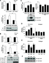
Figure 1. Rap1 activation is required for EGFR-mediated metastasis
(a, Upper) FG human pancreatic carcinoma cells stably expressing non-silencing or Rap1 shRNA were treated with or without EGF for 10 minutes and subsequently implanted on the chorioallantoic membrane (CAM) of 10-day old chick embryos. After 10 days, primary tumors were weighed and pulmonary metastasis was quantified by qPCR for human Alu sequence (see Materials and Methods) n ≥ 15 in each group. (Lower) Cells were lysed to confirm Rap1 knockdown. (b, Upper) FG cells stably expressing empty vector or Rap1GAP were treated with or without EGF for 10 minutes and subsequently implanted on the chick CAM to assess the effect on primary tumor growth and metastasis. (Lower) Cells were lysed to analyze for Rap1GAP expression and Rap1 activation status. (c) FG cells stably expressing the empty vector, FLAG-tagged WT Rap1, Rap1 G12V (active) or Rap1 S17N (inactive) mutants were implanted on the chorioallantoic membrane (CAM) of 10-day old chick embryos. After 10 days, primary tumors were weighed and pulmonary metastasis was quantified by qPCR for human Alu sequence (see Materials and Methods) n ≥ 10 in each group. (Lower) Cell lysates from WT Rap1, Rap1 G12V, Rap1 S17N expressing FG cells were analyzed for Rap1 activation status (d) Migration assay with FG cells expressing empty vector, WT Rap1, Rap1 G12V or Rap1 S17N on vitronectin or collagen in a Boyden chamber. (e, f) FG cells transiently stimulated with EGF (50ng/mL, 10min), MSP (10ng/mL, 10min), or IGF (100ng/mL, 30 min) were analyzed for their (e) migration on vitronectin or collagen, and for (f) Rap1-GTP levels. *p < 0.05, **p < 0.01. Data in d, e represent the mean±s.e.m. of triplicates and are representative of three independent experiments.
Figure 2
Figure 2. Src and CAS are required for EGFR-dependent migration and Rap1 activation
(a) Lysates from FG cells transiently stimulated with EGF, MSP or IGF were immunoblotted for pSrc Y416 and subsequently re-probed for total Src (loading control). (b) FG cells were transiently transfected with control siRNA or Src siRNA. After 24h, the cells were serum starved overnight, followed by treatment with or without EGF and analyzed for Rap1-GTP, (c) (top) migration on vitronectin or collagen, (bottom) Src knockdown, and (d) CAS phosphorylation. (e) FG cells were transiently transfected with control siRNA or CAS siRNA. After 24h, they were serum starved overnight, treated with or without EGF and analyzed for Rap1-GTP, (f) (upper) migration on vitronectin or collagen, (bottom) and CAS knockdown. *p < 0.01. Data in c, f represent the mean±s.e.m. of triplicates and are representative of three independent experiments. Quantification of Rap1-GTP levels in b, e was performed by normalizing total Rap1 levels by densitometric evaluation (ImageJ)
Figure 3
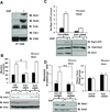
Figure 3. Nck1, a CAS binding partner, is necessary for EGFR-mediated migration, metastasis and Rap1-GTP loading
(a) Lysates from serum-starved FG cells treated with or without EGF were immunoprecipitated with anti-CAS. Eluted proteins were analyzed by immunoblotting with anti-CAS, anti-CrkI, anti-CrkL, anti-Nck1 or anti-Nck2 antibody, as indicated. (b) FG cells were transiently transfected with control siRNA, Nck1 siRNA, or Nck2 siRNA. After 24h, cells were serum starved overnight, subsequently treated with or without EGF and analyzed for migration on vitronectin (upper) and for confirmation of Nck1 and Nck2 knockdown (lower). (c) FG cells were transiently transfected with control siRNA or Nck1 siRNA. After 24h, they were serum starved overnight, subsequently treated with or without EGF and analyzed for Rap1-GTP levels. (d) (upper) FG cells stably expressing control shRNA or Nck1 shRNA were serum starved, treated with or without EGF and implanted on the CAM of 10-day old chick embryos. After 10 days, primary tumors were weighed and pulmonary metastasis was quantified by qPCR for human Alu sequence and chicken GAPDH and normalized to a standard curve. n ≥ 10 in each group. (lower) Nck1 knockdown was confirmed by western blot analysis using indicated antibodies. *p < 0.01, **p < 0.05. Data in b represent the mean±s.e.m. of triplicates and are representative of three independent experiments. Quantification of Rap1-GTP levels in c was performed by normalizing total Rap1 levels by densitometric evaluation (ImageJ)
Figure 4
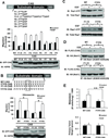
Figure 4. EGFR-induced metastasis, CAS/Nck1 association and Rap1-GTP loading require CAS Y253/Y271
(a) (upper) Schematic of the tyrosine residues of the first 9 YxxP motifs in the substrate domain of CAS. (lower) FG cells stably expressing CAS mutants with individual or a subset of Y/F mutations in the first 9 YxxP motifs were serum starved, treated with or without EGF and analyzed for their migratory capacity on vitronectin. (b, c) (upper b) Schematic of the tyrosine residues of the YxxP motifs in the substrate domain of WT CAS, F1–15 CAS or Y7/Y8 CAS. Box represents the tyrosine residues of the 7th and 8th YxxP motifs. (lower b) FG cells stably expressing WT CAS, F7/F8 CAS, F1–15 CAS or Y7/Y8 CAS were serum starved, treated with or without EGF and assessed for (b) their migration phenotype on vitronectin, and (c) Rap1-GTP statuses. (d) FG cells stably expressing HA-tagged Nck1 (W38K, W143K, W229K) were transiently transfected with WT CAS, F7/F8 CAS, F1–15 CAS or Y7/Y8 CAS. After 24h, cells were serum starved overnight, followed by treatment with or without EGF and lysed. Lysates were immunoprecipitated with anti-HA and analyzed by immunoblotting for FLAG and HA (loading control). (e) FG cells stably expressing WT CAS or F7/F8 CAS were serum starved, treated with or without EGF and implanted on the CAM of 10-day old chick embryos. After 10 days, primary tumors were weighed and pulmonary metastasis was quantified by qPCR for human Alu sequence and chicken GAPDH and normalized to a standard curve. n ≥ 10 in each group. *p < 0.01, **p < 0.05. Data in b represent the mean±s.e.m. of triplicates and are representative of three independent experiments. Quantification of Rap1-GTP levels in c was performed by normalizing total Rap1 levels by densitometric evaluation (ImageJ)
Figure 5
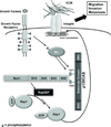
Figure 5. EGFR activates migration and metastasis via a CAS/Nck1/Rap1 signaling module
EGF stimulation of EGFR activates Src kinase, leading to phosphorylation of CAS Y253 and Y271, which binds to the SH2 domain of Nck1. The assembly of the CAS/Nck1 complex along with RapGEFs leads to GTP loading of Rap1. This facilitates actin remodeling, cell migration, invasion and metastasis.

References

    1. Bailey CL, Kelly P, Casey PJ. Activation of Rap1 promotes prostate cancer metastasis. Cancer Res. 2009;69:4962–4968. - PMC - PubMed
    1. Bos JL. Linking Rap to cell adhesion. Current Opinion in Cell Biology. 2005;17:123–128. - PubMed
    1. Brabek J, Constancio SS, Shin NY, Pozzi A, Weaver AM, Hanks SK. CAS promotes invasiveness of Src-transformed cells. Oncogene. 2004;23:7406–7415. - PubMed
    1. Brabek J, Constancio SS, Siesser PF, Shin NY, Pozzi A, Hanks SK. Crk-Associated Substrate Tyrosine Phosphorylation Sites Are Critical for Invasion and Metastasis of Src-Transformed Cells. Mol Cancer Res. 2005;3:307–315. - PubMed
    1. Brooks PC, Klemke RL, Schon S, Lewis JM, Schwartz MA, Cheresh DA. Insulin-like growth factor receptor cooperates with integrin alpha v beta 5 to promote tumor cell dissemination in vivo. J Clin Invest. 1997;99:1390–1398. - PMC - PubMed

Publication types

MeSH terms