. 2012 May 31;31(22):2761-72.
doi: 10.1038/onc.2011.452. Epub 2011 Oct 3.

BAG-1 interacts with the p50-p50 homodimeric NF-κB complex: implications for colorectal carcinogenesis

Affiliations

BAG-1 interacts with the p50-p50 homodimeric NF-κB complex: implications for colorectal carcinogenesis

S L Southern et al. Oncogene. .

Abstract

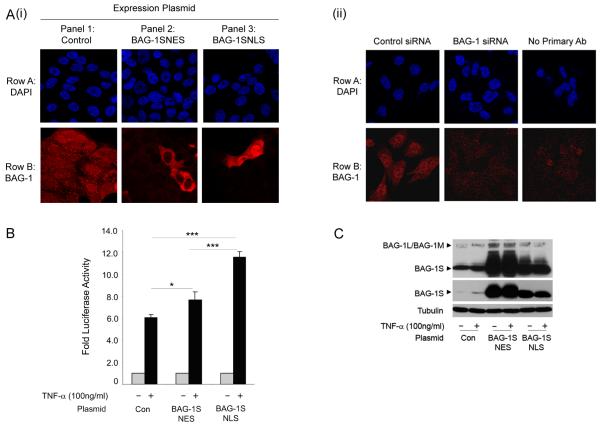
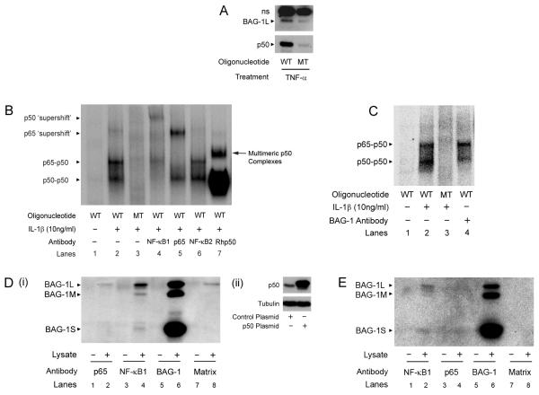
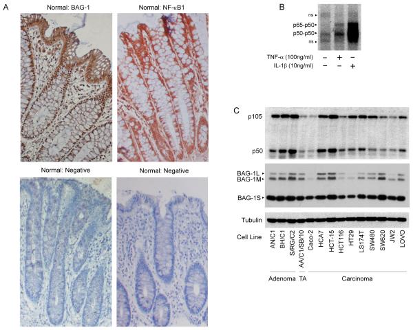
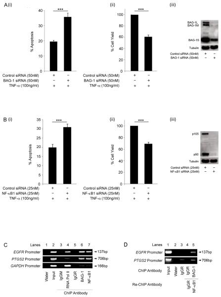
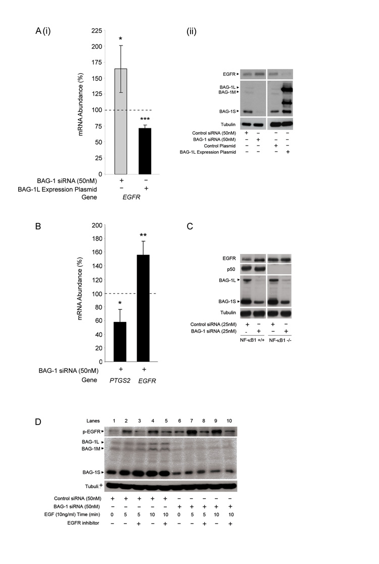
Understanding the mechanisms that promote aberrant tumour cell survival is critical for the determination of novel strategies to combat colorectal cancer (CRC). We have recently shown that the anti-apoptotic protein BAG-1, highly expressed in pre-malignant and CRC tissue, can potentiate cell survival through regulating NF-κB transcriptional activity. In this study, we identify a novel complex between BAG-1 and the p50-p50 NF-κB homodimers, implicating BAG-1 as a co-regulator of an atypical NF-κB pathway. Importantly, the BAG-1-p50 complex was detected at gene regulatory sequences including the epidermal growth factor receptor (EGFR) and COX-2 (PTGS2) genes. Suppression of BAG-1 expression using small interfering RNA was shown to increase EGFR and suppress COX-2 expression in CRC cells. Furthermore, mouse embryonic fibroblasts derived from the NF-κB1 (p105/p50) knock-out mouse were used to demonstrate that p50 expression was required for BAG-1 to suppress EGFR expression. This was shown to be functionally relevant as attenuation of BAG-1 expression increased ligand activated phosphorylation of EGFR in CRC cells. In summary, this paper identifies a novel role for BAG-1 in modulating gene expression through interaction with the p50-p50 NF-κB complexes. Data presented led us to propose that BAG-1 can act as a selective regulator of p50-p50 NF-κB responsive genes in colorectal tumour cells, potentially important for the promotion of cell survival in the context of the fluctuating tumour microenvironment. As BAG-1 expression is increased in the developing adenoma through to metastatic lesions, understanding the function of the BAG-1-p50 NF-κB complexes may aid in identifying strategies for both the prevention and treatment of CRC.


Figures

Figure 1
Figure 1. Enforced nuclear localisation of the BAG-1S isoform increases TNF-α induced NF-κB transcriptional activity
(A) (i) Confocal immunofluorescence microscopy was used to show the localisation of BAG-1 in HCA7 cells transfected with the control IRES plasmid, BAG-1SNES or BAG-1SNLS expression plasmids. Row A: DAPI staining showing the cell nuclei; Row B: Alexa Fluor-546 TRITC fluorescence showing BAG-1 staining. It is important to note that endogenous BAG-1 expression could not be detected in panel 2 or panel 3 as the intensity of the confocal microscope was decreased in order to visualise the localisation of the exogenous fusion proteins. (ii) The specificity of the antibody was confirmed in cells transfected with the control negative / BAG-1 siRNA , as previously reported (Clemo et al., 2008) Row A: DAPI staining showing the cell nuclei; Row B: Alexa Fluor-546 TRITC fluorescence showing BAG-1 staining. (B) Transcriptional activation of the NF-κB luciferase reporter in HCT116 cells transfected with control plasmid, BAG-1SNES or BAG-1SNLS expression plasmids and treated with TNF-α (100ng/ml) for 16 hours. The results shown are representative of three separate experiments done in triplicate (± standard deviation). Statistical analysis was carried out using a Student’s t-test * = P < 0.05; ** = P < 0.01; *** = P < 0.001. (C) Western analysis confirmed over-expression of the BAG-1SNES and BAG-1SNLS fusion proteins (middle panel shows lower exposure to demonstrate expression of the exogenous BAG-1S fusion proteins). With TNF-α treatment, an increase in endogenous BAG-1 expression was detected. Equal loading was confirmed by α-tubulin. Of note, expression of exogenous BAG-1SNES or BAG-1SNLS proteins increased expression of the endogenous BAG-1L/BAG-1M proteins (as previously reported, Hinnit et al., 2010).
Figure 2
Figure 2. BAG-1 interacts with the p50-p50 homodimeric NF-κB complex
(A) BAG-1 interacts with a consensus NF-κB DNA binding site as shown by an oligonucleotide pulldown assay. ns denotes a non-specific band. HCT116 cells were transfected with the BAG-1L expression plasmid and treated with TNF-α (100ng/ml) for 16 hours. After cellular fractionation, the nuclear lysate was incubated with either the wild-type (GGGGACTTTCCC, WT) or mutant (GGCGACTTTCCC, MT) oligonucleotide. (B) IL-1β (10ng/ml) treated HCT116 nuclear fractions were analysed by EMSA. Nuclear lysates were incubated with the mutant (MT) oligonucleotide to control for non-specific interactions (Lane 3). 1μg of NF-κB1, p65 or NF-κB2 antibody were incubated with nuclear lysate and wild-type (WT) oligonucleotide (Lanes 4, 5, 6). 0.5μg of recombinant human p50 (Rhp50) was incubated with the nuclear lysate and wild-type oligonucleotide to show the position of the p50-p50 homodimeric NF-κB complex (Lane 7), and other multimeric complexes (Duckett et al. 1993). (C) HCT116 nuclear fractions were treated with IL-1β (10ng/ml) for 16 hours. G3E2 BAG-1 antibody was added to the HCT116 nuclear lysate, followed by incubation with the wild-type (WT) radiolabelled oligonucleotide (Lane 4). Results are representative of three independent experiments. BAG-1 interacts with the p50 protein. (D) (i) HCT116 cells were seeded for 72 hours and transfected with the control or p50 expression plasmid (shown by Western analysis, ii). 48 hours following transfection, cells were lysed and proteins immunoprecipitated using antibodies against p65 (Lane 2), NF-κB1 (Lane 4) and BAG-1 (Lane 6); (Lane 8 is the no antibody control). Western analysis identified BAG-1 protein interactions within the whole cell lysate. Results are representative of three separate experiments. (E) BAG-1 interacts with the p50 protein in untransfected HCT116 cells. Endogenous protein was immunoprecipitated as before. Western analysis was carried out to determine endogenous BAG-1 protein interactions within the whole cell lysate. Proteins were immunoprecipitated using antibodies against NF-κB1 (Lane 2), p65 (Lane 4) and BAG-1 (Lane 6). Results are representative of three independent experiments.
Figure 3
Figure 3. Expression of BAG-1 and NF-κB1 in normal colorectal epithelium and cell lines
(A) BAG-1 and NF-κB1 expression in paraffin-embedded normal colorectal epithelial tissue. BAG-1 expression was detected using the BAG-1 (TB3; Brimmel et al., 1999) antibody, NF-κB1 expression was detected using the anti-NF-κB1 (E10) antibody. Both were visualised using DAB (brown staining) and counter-stained with haematoxylin (blue staining), objective x10. (The negative control for staining is normal colonic epithelium without primary BAG-1 or NF-κB1 antibody; objective x10). (B) DNA bound NF-κB complexes in cytokine stimulated HCT116 cells. 3μg of HCT116 nuclear fractions were treated with TNF-α (100ng/ml) or IL-1β (10ng/ml) for 16 hours and analysed by EMSA using the wild type [WT, GGGGACTTTCCG] labelled oligonucleotide to detect active NF-κB DNA binding complexes. Non-specific bands are denoted with ns. (C) Western analysis to determine NF-κB1 and BAG-1 protein expression in a panel of colorectal cell lines (TA denotes transformed adenoma). Equal loading was confirmed by α-tubulin. Results are representative of three independent experiments.
Figure 4
Figure 4. BAG-1 and NF-κB1 promote cell survival in HCT116 cells
HCT116 cells were transfected with (A) control negative / BAG-1 or (B) control negative / NF-κB1 siRNA; suppression of protein expression was confirmed by Western analysis (iii). Cells were treated with TNF-α (100ng/ml) for 16h and apoptosis (i) and attached cell yield (ii) assessed. The results shown are the mean of three independent experiments done in triplicate (± standard deviation). Statistical analysis was carried out using a Student’s t-test * = P < 0.05; ** = P < 0.01; *** = P < 0.001. BAG-1 and p50 are present at the same binding site on the EGFR and PTGS2 promoters. (C) ChIP analysis of the EGFR, PTGS2 and GAPDH promoters in HCT116 cells using antibodies to immunoprecipitate the indicated proteins. The IgGM and IgGR antbodies are used as negative controls for non-specific binding of the antibody to the chromatin fragments. Results are representative of four independent experiments. p50 and BAG-1 are part of the same DNA complex at gene promoters. (D) Re-ChIP analysis of the EGFR and PTGS2 promoters in the HCT116 cell line using the indicated combination of antibodies.
Figure 5
Figure 5. BAG-1 regulation of EGFR expression and function is dependent on NF-κB1 expression
(A) (i) HCT116 cells were transfected with control negative / BAG-1 siRNA or control plasmid / BAG-1L expression plasmid for 48 hours. Q-RT-PCR was carried out to show EGFR mRNA abundance; all mRNA values were normalised to the housekeeping gene, TBP. Data is presented as a percentage change of levels in control transfected cells, represented by the dotted line. Data points are of three independent experiments carried out in triplicate (± standard deviation). Statistical analysis was carried out using a Student’s t-test * = P < 0.05; ** = P < 0.01; *** = P < 0.001. (ii) Western analysis to show regulation of EGFR protein +/− BAG-1 expression. Equal loading was confirmed by α-tubulin. Data is representative of three independent experiments. (B) HT29 cells (positive for COX-2 expression) were transfected with control negative or BAG-1 siRNA for 48 hours. Q-RT-PCR was carried out to show PTGS2 and EGFR mRNA abundance; all mRNA values were normalised to the housekeeping gene, TBP. Data is presented as a percentage change of levels in control transfected cells, represented by the dotted line. Data points are of three independent experiments carried out in triplicate (± standard deviation). Statistical analysis was carried out using a Student’s t-test * = P < 0.05; ** = P < 0.01; *** = P < 0.001. (C) NF-κB1+/+ and NF-κB1−/− MEF cells were transfected with 25nM of control siRNA or mBAG-1 siRNA. Western analysis was carried out to show the regulation of EGFR protein expression following suppression of the BAG-1 protein in NF-κB1+/+ and −/− MEF cells. Equal loading was confirmed by α-tubulin. Results are representative of three independent experiments. (D) HCT116 cells treated with EGF (10ng/ml) for 72 hr after siRNA transfection either in the presence or absence of an EGFR inhibitor CL-387.785 (10μM) added 1h prior to EGF treatment. Activation of the EGFR upon EGF treatment was shown by the phosphorylation status of the receptor, specificity confirmed by the blockade with the EGFR inhibitor. Western blotting was used to confirm suppression of BAG-1 expression by siRNA. Equal loading was confirmed by α-tubulin.

References

    1. Arhel NJ, Packham G, Townsend PA, Collard TJ, H-Zadeh AM, Sharp A, et al. The retinoblastoma protein interacts with BAG-1 in human colonic adenoma and carcinoma derived cell lines. Int J Cancer. 2003;106:364–71. - PubMed
    1. Arlt A, Vorndamm J, Muerkoster S, Yu H, Schmidt WE, Folsch UR, et al. Autocrine production of interleukin 1beta confers constitutive nuclear factor kappaB activity and chemoresistance in pancreatic carcinoma cell lines. Cancer Res. 2002;62:910–916. - PubMed
    1. Baer M, Dillner A, Schwartz RC, Sedon C, Nedospasov S, Johnson PF. Tumor necrosis factor alpha transcription in macrophages is attenuated by an autocrine factor that preferentially induces NF-kappaB p50. Mol Cell Biol. 1998;18:5678–5689. - PMC - PubMed
    1. Baeuerle PA, Baltimore D. I kappa B: a specific inhibitor of the NF-kappa B transcription factor. Science. 1988;242:540–546. - PubMed
    1. Barnes JD, Arhel NJ, Lee SS, Sharp A, Al-Okail M, Packham G, et al. Nuclear BAG-1 expression inhibits apoptosis in colorectal adenoma-derived epithelial cells. Apoptosis. 2005;10:301–11. - PubMed

Publication types

MeSH terms