. 2011 Dec;32(4):472-83.
doi: 10.1016/j.reprotox.2011.09.006. Epub 2011 Sep 24.

Neonatal diethylstilbestrol exposure disrupts female reproductive tract structure/function via both direct and indirect mechanisms in the hamster

Affiliations

Neonatal diethylstilbestrol exposure disrupts female reproductive tract structure/function via both direct and indirect mechanisms in the hamster

Imala D Alwis et al. Reprod Toxicol. 2011 Dec.

Abstract

We assessed neonatal diethylstilbestrol (DES)-induced disruption at various endocrine levels in the hamster. In particular, we used organ transplantation into the hamster cheek pouch to determine whether abnormalities observed in the post-pubertal ovary are due to: (a) a direct (early) mechanism or (b) an indirect (late) mechanism that involves altered development and function of the hypothalamus and/or pituitary. Of the various disruption endpoints and attributes assessed: (1) some were consistent with the direct mechanism (altered uterine and cervical dimensions/organization, ovarian polyovular follicles, vaginal hypospadius, endometrial hyperplasia/dysplasia); (2) some were consistent with the indirect mechanism (ovarian/oviductal salpingitis, cystic ovarian follicles); (3) some were consistent with a combination of the direct and indirect mechanisms (altered endocrine status); and (4) the mechanism(s) for one (lack of corpora lutea) was uncertain. This study also generated some surprising observations regarding vaginal estrous assessments as a means to monitor periodicity of ovarian function in the hamster.


Figures

Figure 1
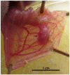
Figure 1. Appearance of a viable ovarian transplant in an everted cheek pouch
Shown is the everted and pinned cheek pouch with a viable and well-vascularized ovarian transplant mass (arrow). It is representative of those transplant sites judged successful during weekly post-surgical assessments. Scale bar at bottom represents 1 cm.
Figure 2
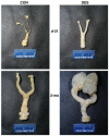
Figure 2. Effects of neonatal DES treatment on complete reproductive tracts from prepubertal and mature hamsters
The paraformaldehyde-fixed and trimmed tracts (vagina at the bottom to ovary/oviduct at the top of each panel) were removed from control (left panels) and neonatally DES-exposed (right panels) 21-day old prepubertal (upper panels) and 2-month old mature (lower panels) hamsters.
Figure 3
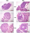
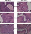
Figure 3. Effects of neonatal DES treatment on the histomorphology of oviducts and ovaries in prepubertal and mature hamsters
Shown are hematoxylin and eosin-stained sections of ovary (Ov) and oviduct (Od) regions from control (left panels) and neonatally DES-exposed (right panels) hamsters at day-21 (upper panels), 1-month (middle panels), and 2-months (lower panels) of age. Indicated are polyovular follicles (arrows), cystic follicles (Cy), oviductal luminal regions exhibiting salpingitis often with massive infiltration of inflammatory cells (**), and corpora lutea (CL). Scale bars in each panel represent 1 mm.
Figure 4
Figure 4. Effects of neonatal DES treatment on serum estradiol levels in prepubertal hamsters and in mature hamsters that were subjected or not to various prepubertal surgical procedures
Shown are the levels (mean ± S.E.) of estradiol-17β in serum collected from the indicated animal treatment groups (see Materials and Methods for detailed descriptions of each group). The number of observations in each treatment group were: [Table: see text]
Figure 5
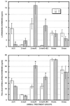
Figure 5. Effects of neonatal DES treatment on serum gonadotropin levels in prepubertal hamsters and in mature hamsters that were subjected or not to various prepubertal surgical procedures
Shown are the levels (mean ± S.E.) of luteinizing hormone (upper panel) and follicle-stimulating hormone (lower panel) in serum collected from the indicated animal treatment groups (see Materials and Methods for detailed descriptions of each group). Asterisks indicate means for the neonatally DES-exposed animals (D) that were significantly (p <0.05) different from the control animals (C) in a given group. The number of observations in each treatment group were: [Table: see text]
Figure 6
Figure 6. Effects of neonatal DES treatment on the cross-sectional area of uterine horns in prepubertal hamsters and in mature hamsters that were subjected or not to various prepubertal surgical procedures
Shown are the dimensions in mm2 (mean ± S.E.) of uterine cross sections from the indicated animal treatment groups (see Materials and Methods for detailed descriptions of each group). For all six groups, the differences in mean areas between control (C) and neonatally DES-exposed (D) animals were significant (p <0.05). The number of observations in each treatment group were: [Table: see text]
Figure 7
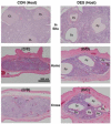
Figure 7. Effects of neonatal DES treatment on uterine histomorphology in mature, ovariectomized, and estrogen-replaced hamsters and in both groups of host hamsters with viable ovarian transplant tissue masses
Shown are hematoxylin and eosin-stained uterine cross sections (all taken at the same magnification) from mature (2-mo) animals that: 1) on the day of birth (d-0) were not (control or CON, left panels) or were treated with DES (right panels) and then were prepubertally (d-21) ovariectomized and either 2) received an estradiol-releasing implant (O+E2, upper panels) or received cheek pouch transplants of ovaries from 3) the same (Homo, middle panels) or 4) the other (Cross, lower panels) neonatal treatment group of donor animals (see Table 1 for further transplant group descriptions). Scale bar in upper right panel represents 100 μm.
Figure 8
Figure 8. Effects of neonatal DES treatment on ovarian histomorphology in mature hamsters as located in situ and after prepubertal transplantation into the cheek pouch
Shown are hematoxylin and eosin-stained ovarian tissue sections (all taken at the same magnification) from mature (2-mo) animals that: 1) on the day of birth (d-0) were not (control or CON, left panels) or were treated with DES (right panels) and then were 2) left intact (In Situ, upper panels) or were prepubertally (d-21) ovariectomized and received cheek pouch transplants of ovaries from 3) the same (Homo, middle panels) or 4) the other (Cross, lower panels) neonatal treatment group of donor animals (see Table 1 for further transplant group descriptions). Scale bar in middle left panel represents 500 μm.
Figure 9
Figure 9. Ovarian follicle histomorphology in mature hamsters after transplantation into the cheek pouch
Shown are hematoxylin and eosin-stained ovarian tissue sections (all taken at the same magnification, but higher than that used in Fig. 8) from mature (2-mo) animals that: 1) on the day of birth (d-0) were not (control or CON, left panels) or were treated with DES (right panels) and then were prepubertally (d-21) ovariectomized and received cheek pouch transplants of ovaries from 2) the same (Homo, upper panels) or 3) the other (Cross, lower panels) neonatal treatment group of donor animals (see Table 1 for further transplant group descriptions). Indicated are primary (1°), secondary (2°), tertiary (3°), atretic (At), late antral/preovulatory (LA/PO), and Cystic (Cy) follicles along with oogonial nests (*). Scale bar in lower right panel represents 100 μm.

Similar articles

Cited by

References

    1. Dodds EC, Goldberg L, Lawson W, Robinson R. Estrogenic activity of certain synthetic compounds. Nature. 1938;141:247–248.
    1. Swan SH. Intrauterine exposure to diethylstilbestrol: long-term effects in humans. Apmis. 2000;108:793–804. - PubMed
    1. Herbst AL, Ulfelder H, Poskanzer DC. Adenocarcinoma of the vagina. Association of maternal stilbestrol therapy with tumor appearance in young women. N Engl J Med. 1971;284:878–881. - PubMed
    1. Blunt E. Diethylstilbestrol exposure: it’s still an issue. Holist Nurs Pract. 2004;18:187–191. - PubMed
    1. Kaufman RH, Adam E. Findings in female offspring of women exposed in utero to diethylstilbestrol. Obstet Gynecol. 2002;99:197–200. - PubMed

Publication types

MeSH terms

LinkOut - more resources