. 2011 Nov 11;413(5):1016-27.
doi: 10.1016/j.jmb.2011.09.029. Epub 2011 Sep 21.

Molecular mechanism of transcription inhibition by phage T7 gp2 protein

Affiliations

Molecular mechanism of transcription inhibition by phage T7 gp2 protein

Vladimir Mekler et al. J Mol Biol. .

Abstract

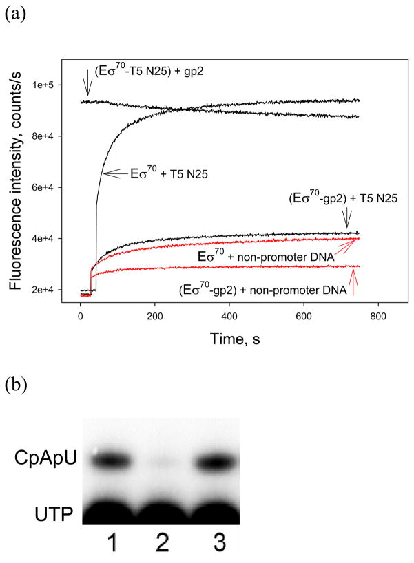
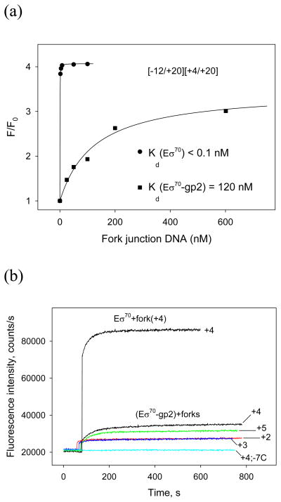
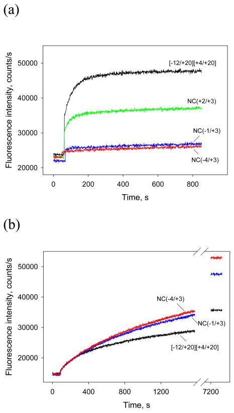
Escherichia coli T7 bacteriophage gp2 protein is a potent inhibitor of host RNA polymerase (RNAP). gp2 inhibits formation of open promoter complex by binding to the β' jaw, an RNAP domain that interacts with downstream promoter DNA. Here, we used an engineered promoter with an optimized sequence to obtain and characterize a specific promoter complex containing RNAP and gp2. In this complex, localized melting of promoter DNA is initiated but does not propagate to include the point of the transcription start. As a result, the complex is transcriptionally inactive. Using a highly sensitive RNAP beacon assay, we performed quantitative real-time measurements of specific binding of the RNAP-gp2 complex to promoter DNA and various promoter fragments. In this way, the effect of gp2 on RNAP interaction with promoters was dissected. As expected, gp2 greatly decreased RNAP affinity to downstream promoter duplex. However, gp2 also inhibited RNAP binding to promoter fragments that lacked downstream promoter DNA that interacts with the β' jaw. The inhibition was caused by gp2-mediated decrease of the RNAP binding affinity to template and non-template strand segments of the transcription bubble downstream of the -10 promoter element. The inhibition of RNAP interactions with single-stranded segments of the transcription bubble by gp2 is a novel effect, which may occur via allosteric mechanism that is set in motion by the gp2 binding to the β' jaw.


Figures

Figure 1
Figure 1. Structures of DNA probes used
The probe names used in the text are in red. In fork junction probe names, numbers in left and right parenthesis correspond to borders of upper and bottom strands of fork junctions, with respect to the transcription start position located at +1 (underlined), “SP” stands for the SP18 linker that joins non-template strand oligos with template strand oligos in short hairpin fork junctions (shown as a vertical line in corresponding probe structures). Asterisks above the [−50/+14] probe indicate positions where the fragments used in experiment shown in Fig. 8 were truncated.
Figure 2
Figure 2. T7 gp2 inhibits RNAP interaction with the T5 N25 promoter
(a) Time dependence of the change in fluorescence upon mixing 1 nM [211Cys-TMR] σ70 RNAP holoenzyme with 2 nM −65/+35 T5N25 (black) or a non-promoter DNA fragment (red) in the presence or in the absence of gp2. The upper curve (labeled as “(RNAP+T5N25)+gp2” was obtained in experiment where gp2 was added to the RNAP beacon preincubated with 2 nM T5N25 promoter fragment for 30 min. In other experiments, gp2 was added to preformed RNAP-T5N25 or RNAP-non-promoter DNA complexes. (b) Inhibition of abortive transcript synthesis from T5N25 promoter fragment by gp2. Reactions contained Eσ70 alone (lane 1) or Eσ70 with gp2 added before (lane 2) or after (lane 3) open complex formation. An autoradiograph of denaturing polyacrylamide gel is presented.
Figure 3
Figure 3. Eσ70 forms complexes on the N25cons promoter in the presence of gp2
Reactions containing indicated proteins and N25cons promoter DNA fragment terminally labeled at the template (“t”) or non-template (“nt”) strands were footprinted with DNase I or probed with KMnO4 as described in Materials and Methods. Gp2 was added prior to (lanes 3, 7, 12, 16) or after (lanes 4, 8, 13, 17) open complex formation. Reaction products were resolved by denaturing PAGE and revealed using Phosphorimager.
Figure 4
Figure 4. Binding of downstream fork junction probes to the Eσ70-gp2 complex
(a) Titration of Eσ70 and Eσ70-gp2 with [−12/+20][+4/+20] downstream fork junction. The Kd values were determined by fitting the dependence of relative fluorescence signal amplitude (F/Fo) on probe concentration to a chemical equilibrium equation. (b) The effect of fork junction point position on Eσ70-gp2 interaction with downstream fork junction probes. Time dependence of the increase in fluorescence upon mixing the (RNAP beacon-gp2) complex with 50 nM of [−12/+20][+2/+20], [−12/+20][+3/+20], [−12/+20][+4/+20], [−12/+20][+5/+20], and [−12/+20; −7C][+4/+20] probes abbreviated as +2, +3, +4, +5, and +4; −7C, correspondingly. The upper curve corresponds to RNAP beacon binding to 50 nM [−12/+20][+4/+20] probe in the absence of gp2.
Figure 5
Figure 5. Binding of downstream fork junction probes with template strand overhangs to the Eσ70-gp2 complex and Δβ(1149–1190)Eσ70
(a) Time dependence of the fluorescence intensity in samples containing 1 nM [211Cys-TMR] σ70 holoenzyme, 200 nM gp2 and 50 nM of indicated downstream fork junction probes. (b) Time dependence of the fluorescence intensity upon the addition of 3 nM of indicated downstream fork junction probes to 1 nM [211Cys-TMR] σ70 holoenzyme bearing the Δβ′(1149–1190) deletion.
Figure 6
Figure 6. Binding of non-template oligos containing the −10 promoter element consensus sequence to free Eσ70 and the Eσ70-gp2 complex
Titration of [211Cys-TMR] σ70 holoenzyme and the gp2-[211Cys-TMR] σ70 holoenzyme complex with oligonucleotide probes. Solid lines correspond to nonlinear regression fit of the data.
Figure 7
Figure 7. The effect of T7 gp2 and T4 AsiA on Eσ70 binding to [−59/−12] probe
2 nM of [−59/−12] probe was combined with RNAP beacon (1 nM) preincubated for 15 min with or without 0.2 μM T7 gp2 or T4 AsiA and increase in fluorescence was monitored.
Figure 8
Figure 8. The effect of gp2 on Eσ70 interaction with double-stranded promoter fragments truncated at various positions downstream of the transcription start point
(a) Time dependence of fluorescence intensity in samples containing 1 nM [211Cys-TMR] σ70 holoenzyme, 200 nM gp2 and 2 nM of either of [−50/+14], [−50/+12], [−50/+8], or [−50/+6] fragments. Upper curves correspond to experiments in which gp2 was added to the RNAP beacon preincubated with a DNA probe for 30 min; bottom curves show fluorescence intensity changes upon addition of the DNA probes to the RNAP beacon preincubated with gp2. The addition of gp2 or DNA probe is indicated by vertical arrows. (b) Inhibition of abortive transcript synthesis from the [−50/+6] probe by gp2. Reactions contained Eσ70 alone (lane 1) or RNAP with gp2 added before (lane 2) or after (lane 3) open complex formation.

Similar articles

Cited by

References

    1. Nechaev S, Severinov K. Bacteriophage-induced modifications of host RNA polymerase. Annu Rev Microbiol. 2003;57:301–322. - PubMed
    1. Haugen SP, Ross W, Gourse RL. Advances in bacterial promoter recognition and its control by factors that do not bind DNA. Nat Rev Microbiol. 2008;6:507–519. - PMC - PubMed
    1. Molineux IJ. The T7 group. In: Calendar RL, editor. The Bacteriophages. 2. Oxford University Press; New York: 2005. pp. 277–301.
    1. Hessellbach B, Nakada D. Inactive complex formation between E. coli RNA polymerase and inhibitor protein purified from T7 phage infected cells. Nature. 1975;258:354–357. - PubMed
    1. Nechaev S, Severinov K. Inhibition of Escherichia coli RNA polymerase by bacteriophage T7 gene 2 protein. J Mol Biol. 1999;289:815–826. - PubMed

Publication types

MeSH terms