. 2012 Jan;24(1):316-24.
doi: 10.1016/j.cellsig.2011.09.017. Epub 2011 Sep 22.

The p53-p21WAF1 checkpoint pathway plays a protective role in preventing DNA rereplication induced by abrogation of FOXF1 function

Affiliations

The p53-p21WAF1 checkpoint pathway plays a protective role in preventing DNA rereplication induced by abrogation of FOXF1 function

Pang-Kuo Lo et al. Cell Signal. 2012 Jan.

Abstract

We previously identified FOXF1 as a potential tumor suppressor gene with an essential role in preventing DNA rereplication to maintain genomic stability, which is frequently inactivated in breast cancer through the epigenetic mechanism. Here we further addressed the role of the p53-p21(WAF1) checkpoint pathway in DNA rereplication induced by silencing of FOXF1. Knockdown of FOXF1 by small interference RNA (siRNA) rendered colorectal p53-null and p21(WAF1)-null HCT116 cancer cells more susceptible to rereplication and apoptosis than the wild-type parental cells. In parental HCT116 cells with a functional p53 checkpoint, the p53-p21(WAF1) checkpoint pathway was activated upon FOXF1 knockdown, which was concurrent with suppression of the CDK2-Rb cascade and induction of G(1) arrest. In contrast, these events were not observed in FOXF1-depleted HCT116-p53-/- and HCT116-p21-/- cells, indicating that the p53-dependent checkpoint function is vital for inhibiting CDK2 to induce G(1) arrest and protect cells from rereplication. The pharmacologic inhibitor (caffeine) of ataxia telangiectasia mutated (ATM) and ataxia telangiectasia and Rad3 related (ATR) protein kinases abolished activation of the p53-p21(WAF1) pathway upon FOXF1 knockdown, suggesting that suppression of FOXF1 function triggered the ATM/ATR-mediated DNA damage response. Cosilencing of p53 by siRNA synergistically enhanced the effect of FOXF1 depletion on the stimulation of DNA rereplication and apoptosis in wild-type HCT116. Finally, we show that FOXF1 expression is predominantly silenced in breast and colorectal cancer cell lines with inactive p53. Our study demonstrated that the p53-p21(WAF1) checkpoint pathway is an intrinsically protective mechanism to prevent DNA rereplication induced by silencing of FOXF1.


Figures

Fig. 1
Fig. 1
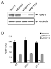
Analysis of the knockdown efficiency of FOXF1 siRNAs. (A) Analysis of the efficacy of FOXF1 siRNAs to knockdown endogenous FOXF1 protein expression. Western blot analysis was performed on nuclear protein lysates from HCT116 cells transfected with FOXF1 siRNAs (or the control siRNA) using antibodies specific to FOXF1 and nucleolin. Nucleolin was used as a loading control for nuclear proteins. (B) Analysis of the efficiency of siRNA-mediated knockdown of endogenous FOXF1 mRNA expression. The colon cancer cell line HCT116 (HCT116-WT) as well as its derived p53-null and p21WAF1-null cell lines (HCT116-p53−/− and HCT116-p21−/−) were transfected with FOXF1 siRNA or control siRNA. 48 h later, total RNA was isolated from siRNA-transfected cells and subjected to real-time qRT-PCR for quantitative analysis of FOXF1 mRNA expression. For relative quantitation, the expression level of FOXF1 mRNA in siControl-transfected cells was set as 100%.
Fig. 2
Fig. 2
Disruption of the p53-p21WAF1 checkpoint function promotes DNA rereplication induced by silencing of FOXF1. FACS analysis was performed on HCT116-WT and its derived p53−/− and p21−/− cell lines transfected with the negative control siRNA (siControl) or FOXF1 siRNA (either siFOXF1-1 or siFOXF1-2) for 48 h. Before harvesting, the cells were pulsed with BrdU for 30 min and then fixed immediately. BrdU incorporation was detected by immunostaining with the anti-BrdU antibody and nuclear DNA was detected by staining with propidium iodide. The FACS analyses of BrdU incorporation and DNA content of siRNA-transfected cells are shown here as: y axis, fluorescent intensity of staining with the anti-BrdU antibody; x axis, propidium iodide fluorescence (DNA content). Cells with DNA content >4N are boxed in FACS dot plots. The percentage indicated inside the box is a measure of the number of cells displaying DNA rereplication.
Fig. 3
Fig. 3
Knockdown of endogenous FOXF1 triggers ATM/ATR-mediated activation of the p53-p21WAF1 checkpoint pathway. (A) Analysis of the functional status of Chk1, Chk2, CDK2 and Rb as well as expression levels of p53 and p21WAF1 in FOXF1-depeleted cells. Western blot analysis was performed on protein extracts isolated from HCT116-WT, HCT116-p53−/− and HCT116-p21−/− cells transfected with control or FOXF1 siRNA using antibodies as indicated in the figure. On the right side of the Western blot gel data, p-Chk1 (S-345), p-Chk2 (T-68), p-CDK2 (T-160) and p-Rb (S-795) indicate their respective phosphorylated proteins. (B) Analysis of PIG3 mRNA expression in HCT116-WT and HCT116-p53−/− cells transfected with control or FOXF1 siRNA. Triplicate siRNA transfection experiments were performed for analysis. The star symbol (*) indicates the statistically significant difference (p<0.05) of PIG3 mRNA expression between siControl- and siFOXF1-1-transfected cells. (C) Analysis of p53 mRNA expression in HCT116-WT and HCT116-p21−/− cells transfected with control or FOXF1 siRNA. Means and standard deviations were calculated from triplicate transfection experiments. (D) Activation of the p53-p21WAF1 checkpoint pathway by siRNA-mediated depletion of FOXF1 is abolished by the inhibitor of the ATM/ATR kinases. HCT116-WT cells were transfected with siControl or siFOXF1-1. At 24 h after siRNA transfection, cells were treated with caffeine (5 mM) or UCN-01 (300 nM) for another 24 h, and cells were then harvested for Western blot analysis of p53, p21WAF1 and α-tubulin protein expression.
Fig. 4
Fig. 4
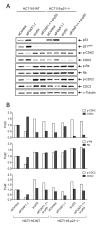
Cosilencing of p53 ablates FOXF1 knockdown-induced suppression of the CDK2-Rb cascade and concurrently induces inhibition of CDC2 kinase activity. (A) Western blot analysis of p53, p21WAF1, phospho-CDK2 (Thr-160), CDK2, phospho-Rb (Ser-795), Rb, phospho-CDC2 (Tyr-15) and CDC2 protein expression in HCT116-WT and HCT116-p21−/− cells transfected with the control siRNA (siControl), FOXF1 siRNA (siFOXF1-1), p53 siRNA (si-p53), or the combination of both FOXF1 and p53 siRNAs for 48 h. Protein extracts isolated from siRNA-transfected cells were subjected to Western blot analysis using antibodies as indicated. (B) Quantitative analysis of Western blot results in (A) by computer-assisted densitometry. After normalization by α-tubulin, expression levels of phospho-CDK2 (Thr-160), CDK2, phospho-Rb (Ser-795), Rb, phospho-CDC2 (Tyr-15) and CDC2 are presented as folds relative to siControl-transfected controls (set as default 1).
Fig. 5
Fig. 5
Concurrent knockdown of both FOXF1 and p53 by siRNAs leads to a synergistic increase in rereplicated and apoptotic cells in HCT116-WT. Flow cytometric analysis was performed on BrdU-labeled, propidium iodide-stained HCT116-WT and HCT116-p21−/− cells transfected with the same siRNA regimen as described in Fig. 4A. The percentage of cells showing rereplicated (A), apoptotic (B), G1 (C), S (D) or G2/M (E) nuclei is presented as mean ± SD of triplicate transfection experiments. Percentages of apoptosis and cell cycle phases were determined as described in “Materials and Methods”. DNA rereplication was scored by the same method as described in Fig. 2. A synergistic increase in apoptosis or DNA rereplication in HCT116-WT or HCT116-p21−/− cells cotransfected with siFOXF1-1 and si-p53 is statistically significant (*, p < 0.05) compared to single siRNA-transfected cells.
Fig. 6
Fig. 6
Expression analysis of FOXF1 gene in colorectal cancer cell lines. (A) In silico analysis of FOXF1 expression in 18 colorectal cancer cell lines. FOXF1 expression data were fetched from Wagner’s microarray dataset [27] through Oncomine. For comparison, the FOXF1 expression value of HCT116 cells was set as 100% to normalize expression values of all the other colorectal cancer cell lines. The p53 states of these lines were indicated according to studies of Liu et al. [28]. (B) Statistical analysis of correlation between FOXF1 expression and the p53 status. The FOXF1 expression data in (A) were replotted into two groups based on the p53 status in the scatter plot and statistical analysis was performed according to the method described in “Materials and Methods”.
Fig. 7
Fig. 7
A model to show collaboration between aberrant silencing of FOXF1 and genetic inactivation of p53 to promote DNA rereplication, which in turn leads to genomic instability and tumorigenesis.

Similar articles

Cited by

References

    1. Lai E, Clark KL, Burley SK, Darnell JE., Jr. Proc. Natl. Acad. Sci. U.S.A. 1993;90:10421–10423. - PMC - PubMed
    1. Katoh M, Katoh M. Int. J. Oncol. 2004;25:1495–1500. - PubMed
    1. Hannenhalli S, Kaestner KH. Nat. Rev. Genet. 2009;10:233–240. - PMC - PubMed
    1. Carlsson P, Mahlapuu M. Dev. Biol. 2002;250:1–23. - PubMed
    1. van der Horst A, Burgering BM. Nat. Rev. Mol. Cell Biol. 2007;8:440–450. - PubMed

Publication types

MeSH terms