Review
. 2012 Jan;52(1):48-61.
doi: 10.1016/j.yjmcc.2011.08.030. Epub 2011 Sep 22.

Measuring mitochondrial function in intact cardiac myocytes

Affiliations
Review

Measuring mitochondrial function in intact cardiac myocytes

Elena N Dedkova et al. J Mol Cell Cardiol. 2012 Jan.

Abstract

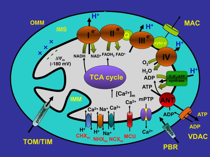
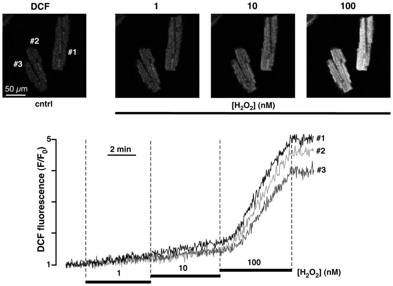
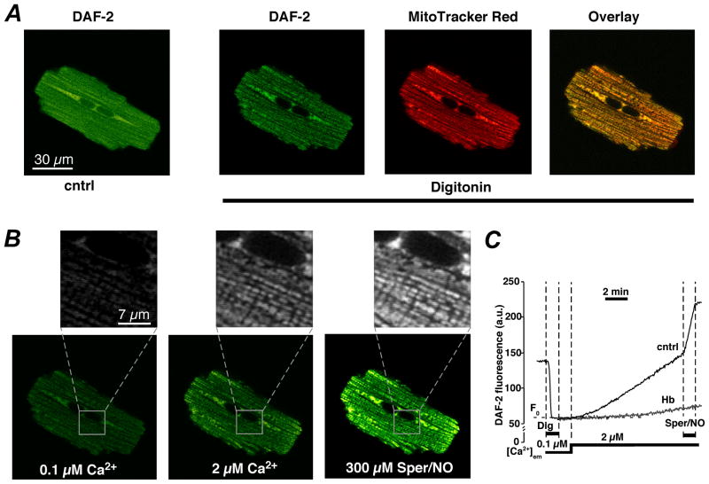
Mitochondria are involved in cellular functions that go beyond the traditional role of these organelles as the power plants of the cell. Mitochondria have been implicated in several human diseases, including cardiac dysfunction, and play a role in the aging process. Many aspects of our knowledge of mitochondria stem from studies performed on the isolated organelle. Their relative inaccessibility imposes experimental difficulties to study mitochondria in their natural environment-the cytosol of intact cells-and has hampered a comprehensive understanding of the plethora of mitochondrial functions. Here we review currently available methods to study mitochondrial function in intact cardiomyocytes. These methods primarily use different flavors of fluorescent dyes and genetically encoded fluorescent proteins in conjunction with high-resolution imaging techniques. We review methods to study mitochondrial morphology, mitochondrial membrane potential, Ca(2+) and Na(+) signaling, mitochondrial pH regulation, redox state and ROS production, NO signaling, oxygen consumption, ATP generation and the activity of the mitochondrial permeability transition pore. Where appropriate we complement this review on intact myocytes with seminal studies that were performed on isolated mitochondria, permeabilized cells, and in whole hearts.


Figures

Figure 1
Figure 1. Schematic overview of mitochondrial structure and transport mechanisms
For details and abbreviations see text.
Figure 2
Figure 2. Measurements ofΔΨm and mPTP activity in permeabilized ventricular myocytes and isolated mitochondria
A, ΔΨm monitored with the membrane potential sensitive dye TMRM in permeabilized rabbit ventricular myocytes upon inhibition of ETC complex I with rotenone and subsequent exposure to ETC complex II substrate succinate. The mitochondrial uncoupler FCCP completely depolarized Ψm. B, mPTP activity monitored with mitochondria entrapped calcein (620 Da) in permeabilized cat ventricular myocytes. Opening of mPTP was induced by enhanced ROS formation (prevented by superoxide and peroxynitrite scavenger MnTBAP, 50 μM) resulting from Ca2+-induced stimulation of mtNOS in the absence of L-arginine. Opening of mPTP resulted in the loss of calcein and a decrease of fluorescence. The pore forming antibiotic alamethicin induced maximum calcein release. Cyclosporine A (CsA, 5 μM) blocked mPTP opening. C, mPTP openings recorded from isolated mitochondria with TMRE. PTP opening can occur as brief incomplete (arrows) and prolonged maximal depolarizations. D, simultaneous monitoring of mPTP activity as changes in ΔΨm (TMRE) and loss of calcein in isolated mitochondria. Brief incomplete depolarizations (arrow) did not cause loss of calcein, however prolonged and complete depolarizations were accompanied by loss of calcein. The decline of calcein fluorescence was preceded by a transient increase (asterisk) caused by calcein dequenching. (Panel B: from Dedkova and Blatter, The Journal of Physiology, 2009, with permission; panels C and D: from Hüser, Rechenmacher and Blatter, Biophysical Journal, 1998, with permission).
Figure 3
Figure 3. Measurements of [Ca2+]m with compartmentalized fluo-3 in single permeabilized ventricular myocytes
A, top: changes in fluo-3 fluorescence following membrane permeabilization with digitonin and increase in extramitochondrial [Ca2+] ([Ca2+]em). Images a to c (bottom) were taken at times indicated by arrows. B, changes of [Ca2+]m in response to rapid cytosolic Ca2+ transients. a and b, technique used to generate rapid changes in [Ca2+]em to simulate cytosolic Ca2+ transients. Cells were placed in the laminar flow of zero-Ca2+ solution (1 mM EGTA) and computer controlled pressure ejections of 10 μM Ca2+-containing solution from a glass micropipette. c, changes of [Ca2+]m in a permeabilized myocyte in response to stimulation with Ca2+ pulses applied at 0.5 Hz (pulse duration = 0.5 s; [Na+]em = 40 mM; [Ca2+]pip = 10 μM). (From Sedova, Dedkova & Blatter, Am. J. Physiol. Cell Physiol., 2006; Am. Physiol. Soc., used with permission)
Figure 4
Figure 4. Monitoring mitochondrial redox potential in intact ventricular myocytes by measuring FAD autofluorescence
Top, from left to right: confocal images of ventricular myocyte FAD autofluorescence (excitation 488 nm, emission 510–525 nm) before (cntrl) and after exposure to ETC complex II substrate methyl succinate (Met-Succinate), ETC complex IV inhibitor sodium cyanide (NaCN), and mitochondrial uncoupler FCCP. Bottom, time course of changes in FAD/FADH2 fluorescence recorded from the regions of interest shown on top. Exposure to NaCN represents maximal reduction of FAD (i.e., 0% fluorescence from FAD with maximal formation of FADH2), while exposure to FCCP reflects maximal oxidation of FAD.
Figure 5
Figure 5. Measurements of the mitochondrial reactive oxygen species (ROS) generation with DCF in intact ventricular myocytes
Top, from left to right: confocal images of ventricular myocytes loaded with ROS-sensitive dye DCF (excitation 488 nm, emission 510–525 nm) before (cntrl) and after exposure to hydrogene peroxide (H2O2; 1 – 100 nM). Bottom, time course of DCF fluorescence in response to 1, 10 and 100 nM H2O2.
Figure 6
Figure 6. Measurements of mitochondrial NO production with DAF-2
A, from left to right: confocal images of a ventricular myocyte loaded simultaneously with DAF-2 (green, emission 510–525 nm) before (cntrl) and after cell permeabilization with digitonin, and MitoTracker Red (red, emission >590 nm). The right panel shows colocalization of DAF-2 and MitoTracker Red represented by shades of yellow. B, confocal images of a permeabilized DAF-2 loaded cell under unstimulated conditions ([Ca2+]em = 0.1 μM), after an increase in [Ca2+]em to 2 μM, and after subsequent addition of the exogenous NO donor Sper/NO. Insets revel NO signals from individual mitochondria. C, time course of DAF-2 fluorescence recorded from a subcellular region of interest (≤40 μm2) under conditions shown in A and B. Treatment with the NO scavenger hemoglobin (Hb, 10 μM) prevented the Ca2+- and Sper/NO-induced increase in DAF-2 fluorescence. (From: Dedkova and Blatter, The Journal of Physiology 2009; with permission).

References

    1. Stanley WC, Recchia FA, Lopaschuk GD. Myocardial substrate metabolism in the normal and failing heart. Physiol Rev. 2005 Jul;85(3):1093–129. - PubMed
    1. Saks V, Favier R, Guzun R, Schlattner U, Wallimann T. Molecular system bioenergetics: regulation of substrate supply in response to heart energy demands. J Physiol. 2006 Dec 15;577(Pt 3):769–77. - PMC - PubMed
    1. McBride HM, Neuspiel M, Wasiak S. Mitochondria: more than just a powerhouse. Curr Biol. 2006 Jul 25;16(14):R551–60. - PubMed
    1. Barth E, Stammler G, Speiser B, Schaper J. Ultrastructural quantitation of mitochondria and myofilaments in cardiac muscle from 10 different animal species including man. J Mol Cell Cardiol. 1992 Jul;24(7):669–81. - PubMed
    1. Bers DM. Excitation-Contraction Coupling and Cardiac Contractile Force. 2. Dordrecht, Netherlands: Kluwer; 2001.

Publication types

MeSH terms