. 2011 Nov 11;413(5):985-1000.
doi: 10.1016/j.jmb.2011.09.028. Epub 2011 Sep 22.

Crystal structure of the passenger domain of the Escherichia coli autotransporter EspP

Affiliations

Crystal structure of the passenger domain of the Escherichia coli autotransporter EspP

Shekeb Khan et al. J Mol Biol. .

Abstract

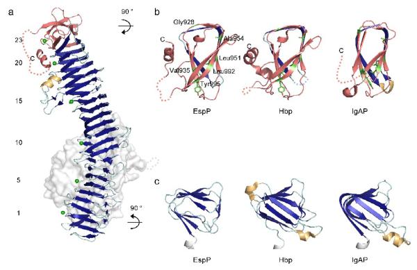
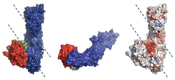
Autotransporters represent a large superfamily of known and putative virulence factors produced by Gram-negative bacteria. They consist of an N-terminal "passenger domain" responsible for the specific effector functions of the molecule and a C-terminal "β-domain" responsible for translocation of the passenger across the bacterial outer membrane. Here, we present the 2.5-Å crystal structure of the passenger domain of the extracellular serine protease EspP, produced by the pathogen Escherichia coli O157:H7 and a member of the serine protease autotransporters of Enterobacteriaceae (SPATEs). Like the previously structurally characterized SPATE passenger domains, the EspP passenger domain contains an extended right-handed parallel β-helix preceded by an N-terminal globular domain housing the catalytic function of the protease. Of note, however, is the absence of a second globular domain protruding from this β-helix. We describe the structure of the EspP passenger domain in the context of previous results and provide an alternative hypothesis for the function of the β-helix within SPATEs.


Figures

Figure 1
Figure 1. Crystal structure of the EspP passenger domain
a) Linear schematic representation of the EspP primary sequence, with the globular subdomain coloured red (subdomain 1 or SD1, residues 56-313) and green (subdomain 3 or SD3, residues 569-630), subdomain 4 (or SD4, residues 671-699) coloured yellow and the β-helical stalk subdomain coloured blue. The signal sequence and the translocator domain (β-domain) are coloured in gray. For ease of comparison with the other structurally known SPATE autotransporter passenger domains, the established nomenclature for subdomains is used although EspP does not contain the equivalent of subdomain 2. b) Wall-eyed stereo ribbon diagram of the EspP passenger coloured as in (a). Encircled in light blue and rendered as stick models are the catalytic triad residues (His127, Asp156 and Ser263Ala) of the passenger. c) Ribbon diagram of the EspP passenger rotated 90° clockwise about a vertical axis relative to (b).Represented as dotted loops are residues 581-593 (green) and 1000-1011 (blue); these residues are disordered in the crystal and not visible in the electron density map. d) Ribbon diagram of the passenger domain of the SPATE autotransporter Hbp (PDB ID: 1WXR) shown in the same orientation and to the same scale as the EspP passenger domain in (c). Subdomain 2 of the Hbp passenger is labeled and coloured purple. e) Wall-eyed stereo representation of the electrostatic surface of the EspP passenger oriented as in (b), with red indicating electronegative potential and blue indicating electropositive potential. Note the patch of negative charge at the bottom of the groove where the globular and β-helical domains meet.
Figure 2
Figure 2. Globular subdomain architecture of the EspP passenger
a) Subdomain 3 (green) forms a helix-turn-helix motif that rests on one face of subdomain 1 (red). Residues 581-593 within subdomain 3 are disordered in the crystal structure and are presented here as a green dotted loop. b) A tetramethionine cluster lies at the interface of subdomains 1 and 3, likely providing further structural stability to the globular subdomain. Interatomic distances between the sulfur atoms of the methionine cluster are indicated by yellow dashed lines and marked in Å units.
Figure 3
Figure 3. Subdomains 3 of(a) EspP with that of(b) Hbp and (c) IgAP for comparison
Subdomains 3 of EspP, Hbp and IgAP are presented as cartoons and coloured green with the remainder of each molecule rendered as a faded gray molecular surface.
Figure 4
Figure 4. Subdomains 1 of (a) EspP with that of (b) Hbp and (c) IgAP and (d) δ-chymotrypsin for comparison
Subdomains 1 of EspP, Hbp and IgAP and the structure of δ-chymotrypsin are presented as cartoons, with α-helices coloured red and β-strands coloured dark blue. The sidechains of the serine, histidine and aspartate residues forming the catalytic triad of each protease are presented as stick models and coloured with carbon atoms in silver, nitrogen atoms in blue and oxygen atoms in red. The bound inhibitor present in the crystal structure of δ-chymotrypsin is presented as a stick model and coloured purple. All images are rendered from the same view. To orient the reader, subdomain 1 of the EspP passenger is presented in the context of subdomains 3 (green cartoon) and 4 (yellow surface) and the β-helical spine subdomain (light blue surface) of EspP.
Figure 5
Figure 5. Comparison of the putative S1 binding pocket subsite of EspP with the S1 subsites of bovine chymotrypsin, Hbp, and IgAP
a) Wall-eyed stereo view of the S1 binding pocket subsite of bovine trypsin in complex with bovine pancreatic trypsin inhibitor (BPTI; PDB ID 1P2N). b) Bovine chymotrypsin was matched onto EspP by least-squares fitting residues 56-58, 101-103, 137-139, 183-196 and 212- 229 in bovine chymotrypsin with residues126-128, 155-157, 202-204, 247-264 and 282-299 in EspP. This match produces a root mean square deviation of 1.5 Å over all backbone atoms. The position of BPTI from its complex with bovine chymotrypsin was then transposed onto the structure of EspP to highlight the putative S1 binding pocket of EspP. This panel is a wall-eyed stereo view of BPTI as bound in bovine trypsin superimposed onto the putative S1 binding pocket of EspP. (a and b) BPTI (green) is depicted from positions P4-P4’ (Gly12-Ile19) with the residue at position P1 (Leu15) together with the backbone atoms spanning positions P2-P2’ (Cys14-Arg17) rendered as stick models. The catalytic triad of bovine chymotrypsin and EspP are also rendered as stick models and labeled. Protein residues surrounding the inhibitor position P1 (Leu15) are rendered as line models and labeled. c-f) Electrostatic surface representation of the substrate binding pocket of (c) bovine chymotrypsin in complex with BPTI (BPTI rendered as in (a and b)), (d) EspP, (e) Hbp and (f) IgAP, with red indicating electronegative potential and blue indicating electropositive potential. The S1 binding pocket subsites in (c-f) are highlighted by yellow circles.
Figure 6
Figure 6. The β-helical stalk subdomain of EspP
a) The β-helical stalk subdomain of EspP is presented as a ribbon diagram with α-helices coloured yellow and β-strands coloured blue. Subdomains 1, 3 and 4 are presented as faded gray molecular surfaces. Dotted loops represent residues disordered in the crystal structure: In EspP these are residues 581-593 (faded gray) and 1000-1011 (orange). The whole autochaperone (AC) region of EspP (residues 925-1023) is coloured orange. Every fifth turn of the β-helix along with the first (1) and last (23) turns are marked by green dots and numbered at the far left. b) Close-up view of the EspP AC region and comparison to the same region of Hbp (PDB ID: 1WXR) and IgAP (PDB ID: 3H09). One turn of the β-helix immediately preceding the AC region in each autotransporter is shown for reference. Residues 938-940 (light blue dotted loop) and 1025-1030 (orange dotted loop) in Hbp and residues 990-1014 (orange dotted loop) in IgAP are not present in the respective models. Shown in green with side chains rendered as sticks, are those residues that are absolutely conserved between the AC regions of EspP, Hbp and IgAP. c) Close-up view of the EspP β-helical stalk N-terminal cap and comparison to that of Hbp and IgAP. Shown are the first three turns of the β-helix in each protein, with α-helices coloured yellow and β-strands coloured blue. In white are the last 4-5 residues of subdomain 1 of each protein leading into the β-helix.
Figure 7
Figure 7. Proposed binding cleft within the EspP passenger domain
The EspP passenger molecular surface is presented with the same colour scheme as in Figure 1a and as an electrostatic surface. The proposed binding cleft within the EspP passenger is marked with dashed lines.

References

    1. Dautin N, Bernstein HD. Protein secretion in gram-negative bacteria via the autotransporter pathway. Annu Rev Microbiol. 2007;61:89–112. - PubMed
    1. Henderson IR, Navarro-Garcia F, Desvaux M, Fernandez RC, Ala’Aldeen D. Type V protein secretion pathway: the autotransporter story. Microbiol Mol Biol Rev. 2004;68:692–744. - PMC - PubMed
    1. Jose J, Jähnig F, Meyer TF. Common structural features of IgA1 protease-like outer membrane protein autotransporters. Mol Microbiol. 1995;18:378–380. - PubMed
    1. Pohlner J, Halter R, Beyreuther K, Meyer T. Gene structure and extracellular secretion of Neisseria gonorrhoeae IgA protease. Nature. 1987;325:458–462. - PubMed
    1. Henderson IR, Navarro-Garcia F, Nataro JP. The great escape: structure and function of the autotransporter proteins. Trends Microbiol. 1998;6:370–378. - PubMed

Publication types

MeSH terms

Associated data

LinkOut - more resources