. 2011 Oct 2;478(7370):529-33.
doi: 10.1038/nature10509.

Inhibition of BET recruitment to chromatin as an effective treatment for MLL-fusion leukaemia

Affiliations

Inhibition of BET recruitment to chromatin as an effective treatment for MLL-fusion leukaemia

Mark A Dawson et al. Nature. .

Abstract

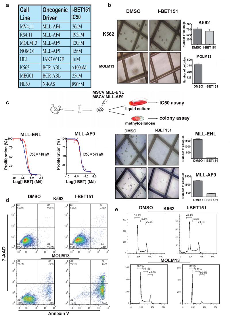
Recurrent chromosomal translocations involving the mixed lineage leukaemia (MLL) gene initiate aggressive forms of leukaemia, which are often refractory to conventional therapies. Many MLL-fusion partners are members of the super elongation complex (SEC), a critical regulator of transcriptional elongation, suggesting that aberrant control of this process has an important role in leukaemia induction. Here we use a global proteomic strategy to demonstrate that MLL fusions, as part of SEC and the polymerase-associated factor complex (PAFc), are associated with the BET family of acetyl-lysine recognizing, chromatin 'adaptor' proteins. These data provided the basis for therapeutic intervention in MLL-fusion leukaemia, via the displacement of the BET family of proteins from chromatin. We show that a novel small molecule inhibitor of the BET family, GSK1210151A (I-BET151), has profound efficacy against human and murine MLL-fusion leukaemic cell lines, through the induction of early cell cycle arrest and apoptosis. I-BET151 treatment in two human leukaemia cell lines with different MLL fusions alters the expression of a common set of genes whose function may account for these phenotypic changes. The mode of action of I-BET151 is, at least in part, due to the inhibition of transcription at key genes (BCL2, C-MYC and CDK6) through the displacement of BRD3/4, PAFc and SEC components from chromatin. In vivo studies indicate that I-BET151 has significant therapeutic value, providing survival benefit in two distinct mouse models of murine MLL-AF9 and human MLL-AF4 leukaemia. Finally, the efficacy of I-BET151 against human leukaemia stem cells is demonstrated, providing further evidence of its potent therapeutic potential. These findings establish the displacement of BET proteins from chromatin as a promising epigenetic therapy for these aggressive leukaemias.


Figures

Figure 1
Figure 1. A global proteomic survey identifies BET proteins as part of the PAFc and SEC
(A). Proteomic strategy (B) Left: Cytoscape representation of the BET protein complex network (discussed in detail in supplementary figure 3). Bold circles indicate associations confirmed by the three orthogonal methods. Right: Heat map representing quantitative-MS data following co-IP of BETs, PAF and SEC complex members. (C) Differential proteomic analysis of the proteins interacting with I-BET and triple acetylated histone H4 tail. Left: Affinity matrices with immobilized I-BET762 or Histone H4(K5acK8acK12ac) peptide bind to the same set of BET complexes. Right: competitive inhibition of the binding of BET isoforms, and SEC and PAF complex components, to the I-BET762 matrix showing matching concentration dependence. (D) Brd4 and MLLT1 interact in HL60, MV4;11 and RS4;11 cells and binding to the I-BET762 matrix is blocked by excess I-BET151. (E) GSK1210151A (I-BET151). (F) I-BET151 binding to the acetyl-binding pocket of BRD4-BD1 (cyan) overlaid with H3K14-Acetyl peptide (green) (3jvk.pdb). A surface representation of the BRD4-BD1 is shown with key recognition and the specificity WPF shelf identified. (G) Ribbon representation of the BRD4-BD1 (cyan) crystal structure complexed with I-BET151 (shown in magenta stick format) overlaid with H3(12-19)K14ac peptide (green) taken from its complex with BRD4-BD1(3jvk.pdb). Secondary elements of the BRD4-BD1 structure have been highlighted. (H) Selectivity profile of IBET-151 showing average temperature shifts (Tm) using a fluorescent thermal shift assay. Numbering inside the spheres, e.g. 12 signifies both bromodomains 1 and 2 have been assessed. Overlaid is the selectivity profile generated using a proteomic approach (shown as boxes around proteins, discussed in Supplementary Fig. 5). Where the bromodomains have been profiled by both thermal shift and proteomic approaches the agreement is excellent. Proteins not assessed by either technique are shown in grey. (I) Comparison of I-BET762 and I-BET151 potency in ligand displacement assays, direct BIAcore binding and LPS stimulated IL6 cytokine production from human PBMCs or whole blood (WB).
Figure 2
Figure 2. I-BET151 selectively and potently inhibits MLL-fusion leukaemic cell lines in vitro
(A) Human leukaemia cell lines tested using I-BET151. (B) Clonogenic assays performed in the presence of DMSO or I-BET151. (C) Haematopoietic progenitors were isolated from mouse bone marrow and retrovirally transformed with MLL-ENL or MLL-AF9. These cells were used in both proliferation and clonogenic assays. (D) Apoptosis was assessed by FACS analysis following 72 hours incubation with DMSO or I-BET151. (E) Cell cycle progression was assessed by FACS analysis 24 hours after incubation with DMSO or I-BET151. Bar graphs are represented as the mean and error bars reflect standard deviation of results derived from triplicate experiments.
Figure 3
Figure 3. Transcriptome and ChIP analyses provide mechanistic insights for the efficacy of I-BET151
(A) Volcano plots for DMSO vs I-BET151 treated samples, showing the adjusted significance p-value (log10) vs. fold change (log2). (B) Correlation of log2 fold change between MV411/MOLM131 across all genes. Notably no genes show opposing expression changes. Lines represent the identity line (black solid), the line of best fit (black dotted), or log2 fold-change threshold values (green dotted). (C) Heatmap of top 100 genes down-regulated following treatment with IBET151. (D) BCL2 gene-expression (normalized to B2M expression) is shown. Expression level of BCL2 in DMSO was assigned a value of 1. (E) Immunoblotting demonstrating a decrease in BCL2 and an increase in cleaved PARP (*) after IBET151 treatment. (F) ChIP analysis at the TSS 3′-end of BCL2 is illustrated. Bar graphs are represented as the mean enrichment relative to input and error bars reflect standard deviation of results derived from biological triplicate experiments.
Figure 4
Figure 4. I-BET151 is efficacious in in vivo murine models and primary patients samples of MLL-fusion leukaemia
(A) Murine pharmacokinetic studies (mean ± SD (n = 4 per compound)) comparing the blood concentration of I-BET151 with I-BET762 and JQ1. (B) Kaplan-Meier curve of control and treated NOD-SCID mice transplanted with 1 × 10⁷ MV4;11 cells. Green arrowhead = treatment commencement D21. (C) Haematoxylin and eosin (H&E) stained histological sections of the renal parenchyma of control and treated mice. Black arrows highlight leukaemic infiltration. (D) Representative FACS analysis from the peripheral blood of control or I-BET151 treated mice. (E) Kaplan-Meier curve of control and treated C57BL/6 mice transplanted with 2.5 × 10⁶ syngeneic MLL-AF9 leukaemic cells. Green arrowhead = treatment commencement D9. (F) Photomicrograph of the spleen size from 5/8 control and 1/12 I-BET151 treated mice that died on day 12. (G) H&E stained histological sections of the liver parenchyma from control and IBET151 treated mice demonstrating reduced disease burden in the treated animal. (H) Peripheral blood white cell count, (I) liver weight and (J) spleen weights from all the control and treated mice at the time of necropsy. (K) Representative FACS analysis assessing apoptosis from a patient with MLL-AF6 leukaemia. (L) Clonogenic assays with human MLL-fusion LSC isolated by FACS sorting (CD34+/CD38-) and plated in the presence of DMSO or I-BET151. (M) Gene-expression changes in human MLL-fusion leukaemia cells following treatment with I-BET151 or DMSO. The log2 fold change in the expression level for all genes (expression level with IBET151 treatment/expression level with DMSO) is represented. (N) Schematic model proposing the mode of action for I-BET151 in MLL-fusion leukaemia.

References

    1. Krivtsov AV, Armstrong SA. MLL translocations, histone modifications and leukaemia stem-cell development. Nat Rev Cancer. 2007;7:823–833. - PubMed
    1. Lin C, et al. AFF4, a component of the ELL/P-TEFb elongation complex and a shared subunit of MLL chimeras, can link transcription elongation to leukemia. Mol Cell. 2010;37:429–437. - PMC - PubMed
    1. Yokoyama A, Lin M, Naresh A, Kitabayashi I, Cleary ML. A higher-order complex containing AF4 and ENL family proteins with P-TEFb facilitates oncogenic and physiologic MLL-dependent transcription. Cancer Cell. 2010;17:198–212. - PMC - PubMed
    1. Milne TA, et al. Multiple interactions recruit MLL1 and MLL1 fusion proteins to the HOXA9 locus in leukemogenesis. Mol Cell. 2010;38:853–863. - PMC - PubMed
    1. Muntean AG, et al. The PAF complex synergizes with MLL fusion proteins at HOX loci to promote leukemogenesis. Cancer Cell. 2010;17:609–621. - PMC - PubMed

Publication types

MeSH terms