. 2011 Oct 2;14(11):1421-9.
doi: 10.1038/nn.2931.

A dual shaping mechanism for postsynaptic ephrin-B3 as a receptor that sculpts dendrites and synapses

Affiliations

A dual shaping mechanism for postsynaptic ephrin-B3 as a receptor that sculpts dendrites and synapses

Nan-Jie Xu et al. Nat Neurosci. .

Abstract

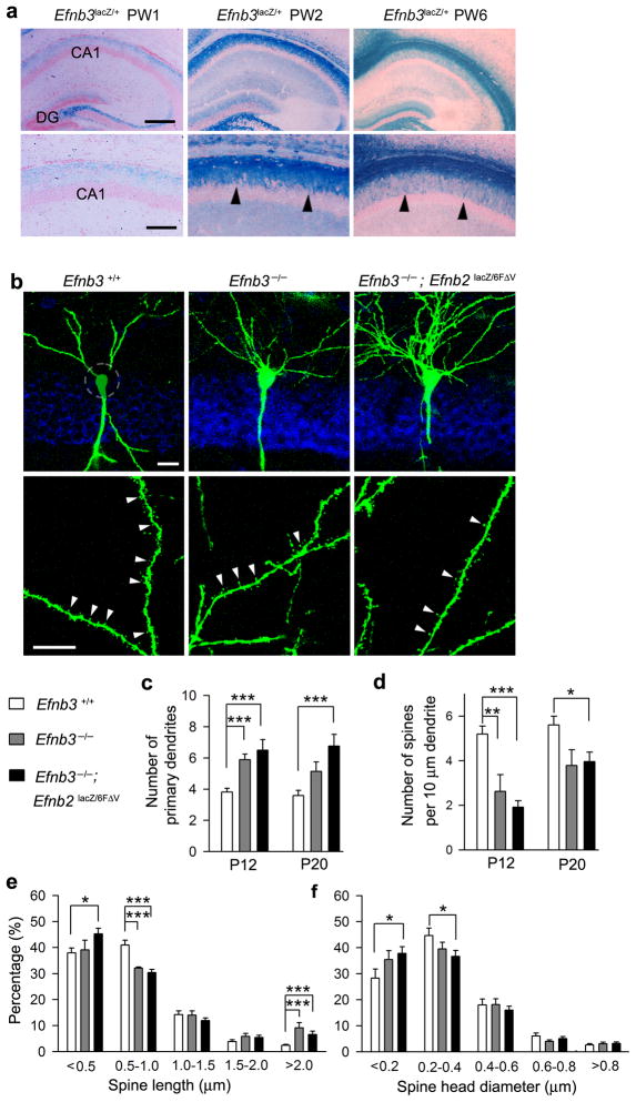
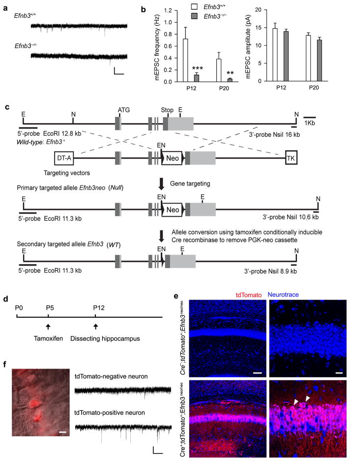
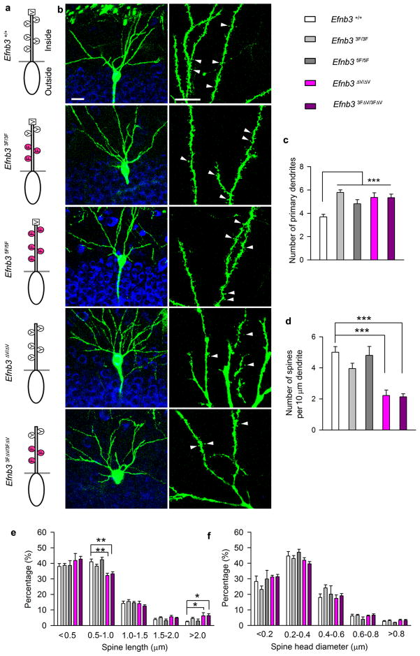
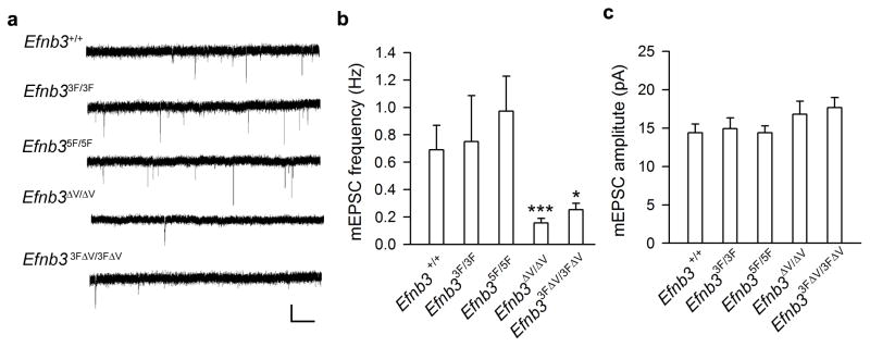
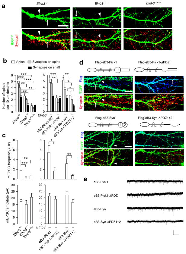
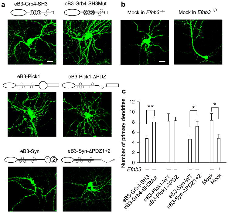
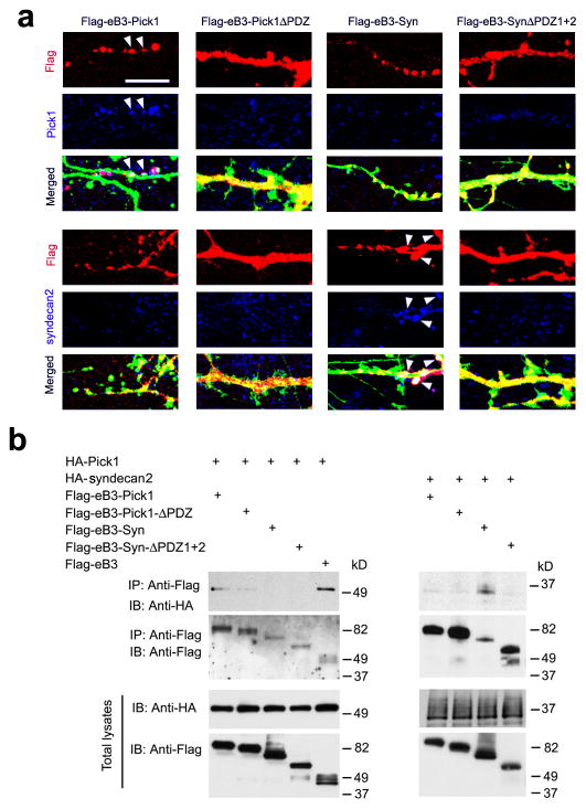
As the neural network becomes wired, postsynaptic signaling molecules are thought to control the growth of dendrites and synapses. However, how these molecules are coordinated to sculpt postsynaptic structures is less well understood. We find that ephrin-B3, a transmembrane ligand for Eph receptors, functions postsynaptically as a receptor to transduce reverse signals into developing dendrites of mouse hippocampal neurons. Both tyrosine phosphorylation-dependent GRB4 SH2/SH3 adaptor-mediated signals and PSD-95-discs large-zona occludens-1 (PDZ) domain-dependent signals are required for inhibition of dendrite branching, whereas only PDZ interactions are necessary for spine formation and excitatory synaptic function. PICK1 and syntenin, two PDZ domain proteins, participate with ephrin-B3 in these postsynaptic activities. PICK1 has a specific role in spine and synapse formation, and syntenin promotes both dendrite pruning and synapse formation to build postsynaptic structures that are essential for neural circuits. The study thus dissects ephrin-B reverse signaling into three distinct intracellular pathways and protein-protein interactions that mediate the maturation of postsynaptic neurons.


Conflict of interest statement

Competing interests statement: The authors declare no competing financial interests.

Figures

Figure 1
Figure 1. Ephrin-B3 is required for dendrite pruning and spine formation in hippocampal CA1 neurons
(a) Developmental expression of eB3 in the CA1 pyramidal cell layer visualized by X-gal staining of coronal sections for the eB3-β-gal fusion protein (blue) in Efb3lacZ mice from one week (PW1) to six weeks age (PW6). Structure of the hippocampus is visualized by eosin counterstaining (red). Arrowheads indicate the localization of eB3 protein in the CA1 dendritic field. There is also strong expression of eB3 in granule cells of the dentate gyrus (DG) where the eB3-β-gal fusion localizes to mossy fiber axons and dendrites. Scale bars, 300 μm in the upper panel and 150 in the lower panel. (b) Efnb3−/− null and Efnb3−/−;Efnb2lacZ/6FΔV mutants at postnatal day 12 (P12) showed excessive dendrites and reduced spine density in CA1 pyramidal neurons. White circle in the upper left panel indicates the crossed primary dendrites. Green: Thy1-GFP-M fluorescence; Blue: neurotrace to stain CA1 pyramidal cell layer. Scale bars, 20 μm in upper panel and 10 μm in lower panel. (c, d, e, f) Quantification of primary dendrites (c), spine density (d), spine length (e) and spine head diameter (f) in wild-type (WT), Efnb3−/− null, and Efnb3−/−;Efnb2lacZ/6FΔV mutants at P12 and P20 (n = 12 per group). Mean ± s.e.m. * P < 0.05; ** P< 0.01; *** P < 0.001.
Figure 2
Figure 2. Ephrin-B3 is required and sufficient for excitatory synaptic function in hippocampal CA1 neurons
(a,b) Efnb3−/− null mutant showed reduced mEPSC frequency in CA1 pyramidal neurons at P12 (a) and P20. Scale bar: 20 pA (vertical) × 1 s (horizontal). Mean ± s.e.m. ** P< 0.01; *** P < 0.001. (c) Generation of conditional floxed knock-in Efnb3neo mutant. Exons (coding/ non-coding segments are dark/ gray filled boxes) and introns (lines) are flanked by a diphtheria toxin (DT-A) and thymidine kinase (TK) expression cassette for negative selection. E (EcoRI) and N (NsiI) indicate restriction sites. The primary targeted allele, Efnb3neo homozygous was crossed with CAGG-CreERT2M driver and a tdTomato reporter to identify cells exposed to active Cre. eB3 expression in the initial Efnb3neo/neo null is restored upon tamoxifen administration to induces Cre-mediated excision of the loxP-flanked PGK-neo cassette. (d) Schedule of tamoxifen treatment and hippocampus dissection during postnatal development. (e) Cre-mediated recombination in CA1 area was detected in P12 hippocampal sections by td-Tomato fluorescence (arrowheads) in Cre+; tdTomato+; Efnb3neo/neo mice but not in Cre; tdTomato+; Efnb3neo/neo mice following tamoxifen administration at P5. Scale bar: 100 μm in the left panel and 20 μm in the right panel. (f) mEPSC recording were performed in tdTomato positive (indicated by a glass electrodes) and negative neurons in hippocampal CA1 area of Cre+; tdTomato+; Efnb3neo/neo mice after tamoxifen treatment. Scale bar: 10 μm for the left panel and 20 pA (vertical) × 1 s (horizontal) for the right panel.
Figure 3
Figure 3. Ephrin-B3 tyrosine phosphorylation and PDZ binding are differentially required for dendrite morphogenesis and synaptic function
(a) Point mutations in the Efnb3 gene that eliminate tyrosine phosphorylation and SH2 binding (3F and 5F), PDZ binding (ΔV), or both SH2 and PDZ binding (3FΔV). (b) Thy1-GFP-M fluorescence (green) was used to visualize the morphology of CA1 neurons at P12. Scale bar: 20 μm for left panels and 10 μm for right panels. (c, d, e, f) Quantification of primary dendrites (c), spine density (d), spine length (e) and spine head diameter (f) in WT, Efnb33F/3F, Efnb35F/5F, Efnb3ΔV/ΔV, and Efnb33FΔV/3FΔV mutants at P12 (n = 25 per group). Mean ± s.e.m, **, P < 0.01; *** P < 0.001.
Figure 4
Figure 4. PDZ binding of ephrin-B3 are required for synaptic function
(a) mEPSCs were recorded in CA1 pyramidal neurons from WT, Efnb33F/3F, Efnb35F/5F, Efnb3ΔV/ΔV, and Efnb33FΔV/3FΔV mutants at P12. Scale bar: 20 pA (vertical) × 1s (horizontal). (b, c) Quantification of mEPSC frequency (b) and amplitude (c) for CA1 pyramidal neurons from the different Efnb3 mutants (n = 15). Mean ± s.e.m. * P < 0.05; *** P< 0.001.
Figure 5
Figure 5. Pick1 and syntenin mediate ephrin-B3 reverse signaling through PDZ binding to control spine/synapse formation and synaptic function
(a) Spine and synapse formation were indicated with a transfected f-EGFP reporter and presynaptic marker synapsin in 12d cultured hippocampal neurons from WT, Efnb3−/−, and Efnb3ΔV/ΔV mice. Arrowhead and arrow indicate synapse on spine and synapse on shaft, respectively. Scale bar: 10 μm. (b) The density of spines and synapses on spines (arrowhead)/shafts (arrow) were quantified. n = 10. (c) mEPSCs were recorded in 12–14 day cultured neurons from Efnb3 mutants and WT (left), and in Efnb3−/− hippocampal neurons that were infected with lentivirus packaged eB3-Pick1 or eB3-syntenin (Syn) expression vectors. Quantification of mEPSC frequency (upper) and amplitude (lower) is shown. n =15–20. Mean ± s.e.m. * P < 0.05; ** P < 0.01; *** P < 0.001 in b and c. (d) Expression of Flag-tagged WT eB3-Pick1 or eB3-syntenin (Syn) chimeric fusion proteins (left panels) in transfected Efnb3−/− neurons rescues spine and synapse formation as visualized with f-EGFP and synapsin in chimeric protein expressing neurons labeled with anti-Flag antibodies (upper panels). Expression of the eB3-Pick1-ΔPDZ or eB3-Syn-ΔPDZ1+2 fusion proteins deleted for the respective PDZ domains (right panels) had little if any effect on spine and synapse formation in Efnb3−/− neurons. Scale bar: 10 μm. (e) Example of mEPSC recorded in Efnb3−/− neurons expressing eB3-Pick1 or eB3-syntenin (Syn) fusion proteins and their PDZ deleted mutant forms. Scale bar: 40 pA (vertical) × 2 s (horizontal).
Figure 6
Figure 6. Grb4, Pick1 and syntenin mediated distinct reverse signaling to prune primary dendrites in cultured hippocampal neurons
(a) Expression of eB3-Grb4-SH3 or eB3-syntenin (Syn) chimeric fusion proteins in transfected Efnb3−/− hippocampal neurons reduces primary dendrites as visualized with f-EGFP. Expression of eB3-Pick1 or its mutant eB3-Pick1-ΔPDZ or mutants of eB3-Grb4 or eB3-Syn fusion proteins had little if any effect on the number of primary dendrites in Efnb3−/− neurons. Scale bar: 10 μm. (b) Cultured hippocampal neurons at P12 from Efnb3−/− mice show more primary dendritic branches comparable to that from WT littermates as visualized with f-EGFP. Scale bar: 10 μm. (c) Quantitative analysis for number of the primary dendrites in neurons expressing different chimeric fusion proteins. Mean ± s.e.m. n =10–12. * P < 0.05; ** P< 0.01.
Figure 7
Figure 7. Association of eB3-Pick1 or eB3-Syn with downstream signaling molecules
(a) Following EphB2-Fc treatment for 16 h in cultured hippocampal neurons to cluster the eB3 proteins to spots on the plasma membrane, WT Flag-eB3-Pick1 co-localized with Pick1 while WT Flag-eB3-syntenin (Syn) colocalized with syndecan-2 (SDC2), which is indicated by arrowheads. The PDZ domain deleted proteins showed highly diminished or no ability to form protein-protein interactions with Pick1 or syndecan-2. Scale bar: 5 μm. (b) In transfected Cos-1 cells, HA-Pick1 or HA-syndecan2 was co-immunoprecipitated with WT Flag-eB3-Pick1 or Flag-eB3-syntenin (Syn), respectively, but little if any was precipitated with the eB3-Pick1-ΔPDZ or eB3-Syn-ΔPDZ1+2 PDZ deleted counterparts. IB, immunoblot; IP, immunoprecipitation. The presented bolts were cropped and the full-length blots are presented in Supplementary Fig. 15.

Comment in

  • EphrinBs send mixed messages.
    Soskis M, Salogiannis J, Greenberg M. Soskis M, et al. Nat Neurosci. 2011 Oct 26;14(11):1356-8. doi: 10.1038/nn.2968. Nat Neurosci. 2011. PMID: 22030542 No abstract available.

References

    1. Ethell IM, Pasquale EB. Molecular mechanisms of dendritic spine development and remodeling. Prog Neurobiol. 2005;75:161–205. - PubMed
    1. Parrish JZ, Emoto K, Kim MD, Jan YN. Mechanisms that regulate establishment, maintenance, and remodeling of dendritic fields. Annu Rev Neurosci. 2007;30:399–423. - PubMed
    1. Klein R. Bidirectional modulation of synaptic functions by Eph/ephrin signaling. Nat Neurosci. 2009;12:15–20. - PubMed
    1. Lai KO, Ip NY. Synapse development and plasticity: roles of ephrin/Eph receptor signaling. Curr Opin Neurobiol. 2009;19:275–283. - PubMed
    1. Cowan CA, Henkemeyer M. Ephrins in reverse, park and drive. Trends Cell Biol. 2002;12:339–346. - PubMed

Publication types

MeSH terms