. 2012 Mar;12(1):115-39.
doi: 10.3758/s13415-011-0062-x.

Beyond arousal and valence: the importance of the biological versus social relevance of emotional stimuli

Affiliations

Beyond arousal and valence: the importance of the biological versus social relevance of emotional stimuli

Michiko Sakaki et al. Cogn Affect Behav Neurosci. 2012 Mar.

Abstract

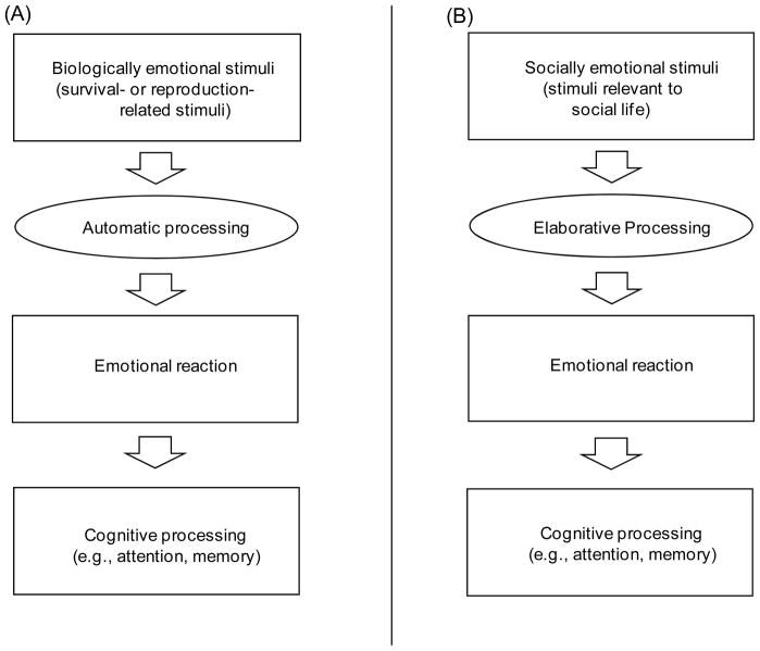
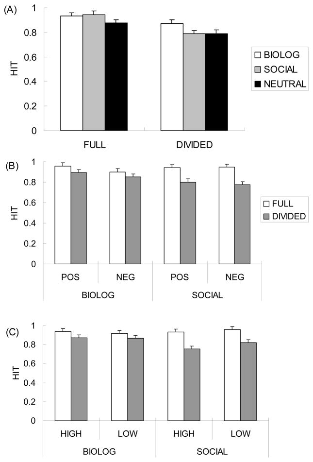
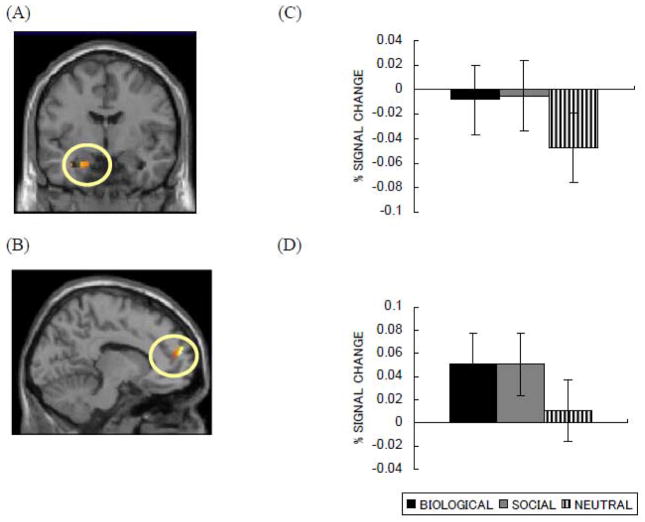
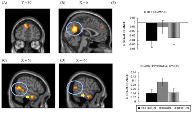
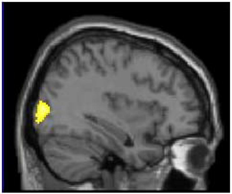
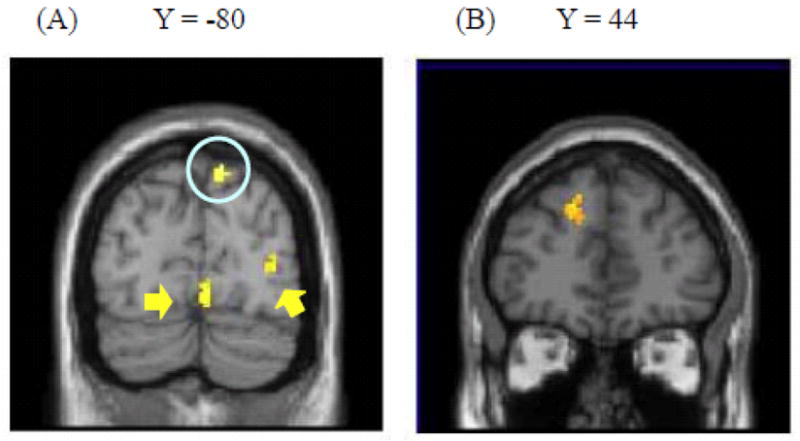
The present study addressed the hypothesis that emotional stimuli relevant to survival or reproduction (biologically emotional stimuli) automatically affect cognitive processing (e.g., attention, memory), while those relevant to social life (socially emotional stimuli) require elaborative processing to modulate attention and memory. Results of our behavioral studies showed that (1) biologically emotional images hold attention more strongly than do socially emotional images, (2) memory for biologically emotional images was enhanced even with limited cognitive resources, but (3) memory for socially emotional images was enhanced only when people had sufficient cognitive resources at encoding. Neither images' subjective arousal nor their valence modulated these patterns. A subsequent functional magnetic resonance imaging study revealed that biologically emotional images induced stronger activity in the visual cortex and greater functional connectivity between the amygdala and visual cortex than did socially emotional images. These results suggest that the interconnection between the amygdala and visual cortex supports enhanced attention allocation to biological stimuli. In contrast, socially emotional images evoked greater activity in the medial prefrontal cortex (MPFC) and yielded stronger functional connectivity between the amygdala and MPFC than did biological images. Thus, it appears that emotional processing of social stimuli involves elaborative processing requiring frontal lobe activity.


Figures

Figure 1
Figure 1
Proposed mechanisms by which biologically and socially emotional stimuli modulate cognitive processing. (A) Biologically emotional stimuli imply clear/direct physical outcomes. Therefore, their emotional nature can be detected even with just automatic processing, and they can modulate cognitive processing without elaborative processing. (B) In contrast, socially emotional stimuli have ambiguous meanings and outcomes. Thus, each social stimulus has to be interpreted by each individual in each context in order to elicit an emotion and to modulate cognitive processing.
Figure 2
Figure 2
(A) A schematic representation of procedures in Study 1. On each trial, participants viewed a riddle and then saw a picture (in the biological, social, and neutral conditions) or asterisks (in the control condition). After 150 ms of the picture or the asterisks (150 ms), the red dot appeared at one of eight possible locations. Participants were asked to indicate the location of the dot as quickly and as accurately as possible. Immediately after they answered the correct location of the dot, the dot was replaced by the solution to the riddle. (B) Eight possible locations for the dot probe and the correct key response for each of them.
Figure 3
Figure 3
Effects of stimulus type (biologically vs. socially emotional stimuli) on attention in Study 1. Error bars represent standard errors. (A) Reaction times to detect the dot-probe were slower after biologically emotional pictures than other conditions, while the reaction times did not differ across social, neutral and control conditions. (B) The valence category and subjective arousal did not modulate the results in reaction times.
Figure 4
Figure 4
Results of the hit rates from the picture memory test in Study 2. Error bars represent standard errors. (A)When people have enough cognitive resources at encoding, they remembered both biologically and socially emotional stimuli better than neutral stimuli. In contrast, when participants’ attention was focused elsewhere, it impaired their memory for social stimuli, but not for biological stimuli. (B) Valence and (C) subjective arousal did not modulate the patterns.
Figure 5
Figure 5
Results of the Remember rates from the picture memory test in Study 2. Error bars represent standard errors. (A) Attentional resources influenced encoding detailed memories for socially emotional stimuli more than for biologically emotional stimuli; although social stimuli produced higher remember rates than biological stimuli in the full attention condition, dividing attention impaired memory for socially emotional stimuli, but not for biologically emotional stimuli. Similar results were obtained regardless of (B) valence and (C) subjective arousal.
Figure 6
Figure 6
Brain areas in which activity was associated with both biological and social pictures in Study 3: (A) left amygdala (Y = −5) and (B) left MPFC (x = −11). ROI analyses also revealed that both biologically and socially emotional pictures produced similar activity in (C) left and (D) right amygdala.
Figure 7
Figure 7
Brain regions showing greater activity for socially emotional pictures than biological pictures (Study 3). Socially emotional pictures induced activity in dorsal MPFC (A), posterior cingulate (circled in (B)), bilateral temporo-parietal junction (circled in (C) and (D)), and bilateral anterior temporal gyri (pointed to with arrows in (C) and (D)). (E) Right hippocampus and right parahippocampal gyrus showed greater activity in response to social pictures than to biological pictures.
Figure 8
Figure 8
Right occipital gyrus showed greater activity for biologically emotional pictures than for social ones (x = 34) in Study 3.
Figure 9
Figure 9
Brain regions showing differential functional connectivity with the amygdala across biologically and socially emotional pictures (Study 3). (A) Inferior parietal lobe (circled), and occipital cortex (pointed to with arrows) showed stronger connectivity with left amygdala for biological pictures than for social pictures. (B) In contrast, dorsal MPFC showed stronger connectivity for social pictures than for biological pictures.
Figure 10
Figure 10
Results from the conjunction analysis between socially emotional words and socially emotional pictures in Study 3. Posterior cingulate (circled in (A)), MPFC (pointed to by an arrow in (A) and (B)), temporo- parietal junction (circled in (C)), and anterior temporal gyrus (pointed to by an arrow in (C)) showed greater activity for both socially emotional words and socially emotional pictures.

References

    1. Adolphs R. Cognitive neuroscience of human social behavior. Nature Reviews: Neuroscience. 2003;4:165–178. doi: 10.1038/nrn1056. - DOI - PubMed
    1. Amano S, Kondo T. Lexical properties of japanese. Tokyo: Sanshodo; 2000.
    1. Amaral DG, Behniea H, Kelly JL. Topographic organization of projections from the amygdala to the visual cortex in the macaque monkey. Neuroscience. 2003;118:1099–1120. doi: 10.1016/S0306-4522(02)01001-1. - DOI - PubMed
    1. Amodio DM, Frith CD. Meeting of minds: The medial frontal cortex and social cognition. Nature Reviews: Neuroscience. 2006;7:268–277. doi: 10.1038/nrn1884. - DOI - PubMed
    1. Anderson AK. Affective influences on the attentional dynamics supporting awareness. Journal of Experimental Psychology: General. 2005;134:258–281. doi: 10.1037/0096-3445.134.2.258. - DOI - PubMed

Publication types

MeSH terms