. 2011 Oct 2;43(11):1074-81.
doi: 10.1038/ng.944.

Inverted genomic segments and complex triplication rearrangements are mediated by inverted repeats in the human genome

Affiliations

Inverted genomic segments and complex triplication rearrangements are mediated by inverted repeats in the human genome

Claudia M B Carvalho et al. Nat Genet. .

Abstract

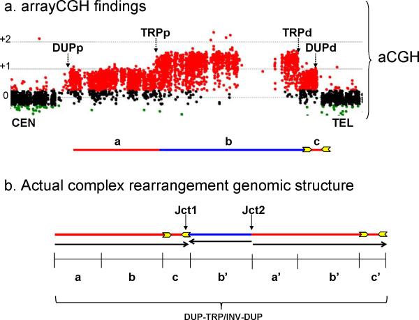
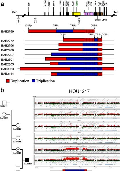
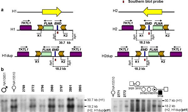
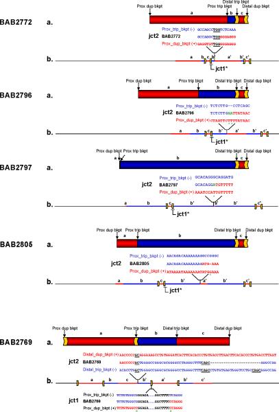
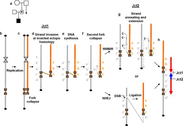
We identified complex genomic rearrangements consisting of intermixed duplications and triplications of genomic segments at the MECP2 and PLP1 loci. These complex rearrangements were characterized by a triplicated segment embedded within a duplication in 11 unrelated subjects. Notably, only two breakpoint junctions were generated during each rearrangement formation. All the complex rearrangement products share a common genomic organization, duplication-inverted triplication-duplication (DUP-TRP/INV-DUP), in which the triplicated segment is inverted and located between directly oriented duplicated genomic segments. We provide evidence that the DUP-TRP/INV-DUP structures are mediated by inverted repeats that can be separated by >300 kb, a genomic architecture that apparently leads to susceptibility to such complex rearrangements. A similar inverted repeat-mediated mechanism may underlie structural variation in many other regions of the human genome. We propose a mechanism that involves both homology-driven events, via inverted repeats, and microhomologous or nonhomologous events.


Figures

Figure 1
Figure 1. General genomic structure of the complex rearrangements triplications embedded in duplications
(a) Copy number dosage alteration inferred from aCGH. A typical aCGH experiment is shown for a complex DUP-TRP-DUP rearrangement. Transitions of copy number dosage alterations are demonstrated by black vertical dotted arrows; the size of genomic segments defined by those boundaries vary in each individual complex rearrangement and are denoted a, b, c. The horizontal line below depicts the array data. Duplications are represented in red and triplications in blue; yellow arrows represent inverted repeats. (b) Representative figure of the genomic structure as determined by further analysis of copy number breakpoint junctions (jct1 and jct2) in five independent rearrangements using multiple molecular experimental approaches. The genomic segments involved are denoted a, b, c whereas the respective copy number gains are denoted as a’, b’, c’. These findings were corroborated by three additional independent rearrangements for which we obtained information for either jct1 or jct2 from two genomic loci. Change in orientation of the genomic segment (represented by black arrows) occurred at each of the junctions formed. DUPp: proximal transition/breakpoint of duplications; TRPp: proximal transition/breakpoint of triplication; DUPd: distal transition/breakpoint of duplications; TRPd: distal transition/breakpoint of triplications.
Figure 2
Figure 2. Individuals carrying complex triplications of Xq28
(a) Genomic region harboring the alterations involving MECP2. Duplications are represented in red and triplications in blue. Arrows on top of the BAB2769 and BAB2772 rearrangements indicate the position of the transitions to gain according to array-based Comparative Genomic Hybridization (aCGH); in 6 out of 8 cases (BAB2796 and BAB2980 are brothers), the distal transition/breakpoint of both duplications (DUPd) and triplications (TRPd) (indicated by arrows) cluster within a pair of LCRs termed K1 and K2 contrasting with the scattered nature of the proximal breakpoints of both duplications (DUPp) and triplications (TRPp) in the same group of patients. Vertical lines embedded within the rearrangement bars represent low copy repeat regions (LCRs) for which the copy numbers were not inferred due to poor probe coverage. (b) aCGH (Agilent Technologies, Santa Clara, CA) result for family HOU1217. Carrier mother BAB3115 with a de novo complex triplication that was transmitted to her son (BAB3114).
Figure 3
Figure 3. Southern blot analysis of the region flanked by LCRs K1 and K2 at the Xq28 chromosome
LCRs K1 and K2 are approximately 11.3 kb in length, and are located ~38 kb apart in inverted orientation. Their 99% nucleotide sequence identity is likely maintained by frequent gene conversion. These LCRs flank two genes FLNA and EMD. (a) Yellow arrows indicate an inversion: cent-FLNA/EMD-tel (H1) and the alternative genomic orientation cent-EMD/FLNA-tel (H2). An 18.2 kb band is expected to be produced in either inversion haplotype background (H1dup or H2dup) upon duplication and inversion; we hypothesize that the 18.2 kb band includes the breakpoint junction 1 (jct1). To test the haplotype of our cohort and to map the duplication breakpoints, we performed a Southern blot assay as shown here. Genomic DNA was digested with BglII; EMD was targeted using a PCR-based probe. The reference genome H1 produces a 30.7 kb band whereas the inversion haplotype (H2) yields an 18.2 kb band. (b) Southern blot results for patients carrying triplications embedded within duplications: BAB2769, BAB2772, BAB2796/BAB2980, BAB2797, BAB2801, BAB2805 (left) and family HOU1217 (right). NA10851: male carrying the reference haplotype (H1); NA15510 heterozygous female carrying both reference and inversion haplotypes (H1 and H2). BAB2771: patient carrying MECP2 duplication not involving LCR K1 and LCR K2 (H1).
Figure 4
Figure 4. Rearrangement structure for patients BAB2769, BAB2772, BAB2796/BAB2980, BAB2797 and BAB2805 based on aCGH, Southern blotting and breakpoint sequencing
(a) Genomic region harboring duplications and triplications spanning chromosome Xq28 according to aCGH: duplications are represented in red, triplications in blue. Arrows on top of the rearrangements indicate the position of the breakpoints; inverted repeats involved in the rearrangement are represented as yellow arrows. Letters a, b and c represent the segments with copy-number gain. (b) Individual genomic structure of the region involved in the rearrangement as inferred by analysis of breakpoint junction 1 (jct1) and breakpoint junction 2 (jct2) for each patient. Jct1* represents those junctions analyzed by Southern blotting (please refer to Fig. 3); all others were sequenced. Genomic positions of each of the junctions are shown. Breakpoint junction sequences are color-coded to highlight their segment of origin in the reference genome (duplications in red and triplications in blue). The triplicated segment (b’) is inserted amid a normal (a, b, c) and a duplicated copy (a’, c’) in inverted orientation as supported by jct1 and jct2 analysis. This structure was further confirmed by FISH for patient BAB2805 (Supplementary Fig. 6). Microhomologies observed at the junctions are represented by underlined black letters; insertions or mismatches at the junctions are represented in green; deletions are represented by dashes; mismatches between the reference sequences and patient sequences are marked with a green asterisk underneath.
Figure 5
Figure 5. Proposed model for generation of the common DUP-TRP/INV-DUP rearrangement product
(a) The rearrangement may have occurred during spermatogenesis in the ancestral male on his X chromosome, likely during S or G2 phase. (b) During replication of the sister chromatid, the replication fork may collapse and induce break-induced replication (BIR) (c) BIR uses homologous recombination to re-establish a new fork using ectopic homology provided by inverted repeats forming jct1. (d) This event initiates replication that forms a length of chromatid back in the opposite direction from that in which the fork had been traveling before the collapse, forming a large inverted duplication (e). If the reversed replication again collapses, or if there is a double-strand break in the chromatid carrying the inverted duplication, then there is a new DNA end (f). In our patient cohort, this new end rejoining (jct2 formation) occurred by either non-homologous end joining (NHEJ) requiring a double-strand break (DSB) on the original strand followed by ligation of that segment to the end of the rearranged newly replicated strand, or by a replication mechanism such as microhomology-mediated break-induced replication (MMBIR) (g) that requires a new strand annealing and extension to the end of the replicon or the chromosome. (h) Representative structure obtained after two steps of homologous and nonhomologous mechanisms. Duplicated and triplicated segments are represented in red and blue colors, respectively.

References

    1. Morrow DM, Connelly C, Hieter P. “Break copy” duplication: a model for chromosome fragment formation in Saccharomyces cerevisiae. Genetics. 1997;147:371–82. - PMC - PubMed
    1. Smith CE, Llorente B, Symington LS. Template switching during break-induced replication. Nature. 2007;447:102–5. - PubMed
    1. McEachern MJ, Haber JE. Break-induced replication and recombinational telomere elongation in yeast. Annu Rev Biochem. 2006;75:111–35. - PubMed
    1. Hastings PJ, Ira G, Lupski JR. A microhomology-mediated break-induced replication model for the origin of human copy number variation. PLoS Genet. 2009;5:e1000327. - PMC - PubMed
    1. Hastings PJ, Lupski JR, Rosenberg SM, Ira G. Mechanisms of change in gene copy number. Nat Rev Genet. 2009;10:551–64. - PMC - PubMed

Publication types

Substances