. 2011 Oct 2;12(11):1086-95.
doi: 10.1038/ni.2106.

Mucosal memory CD8⁺ T cells are selected in the periphery by an MHC class I molecule

Affiliations

Mucosal memory CD8⁺ T cells are selected in the periphery by an MHC class I molecule

Yujun Huang et al. Nat Immunol. .

Abstract

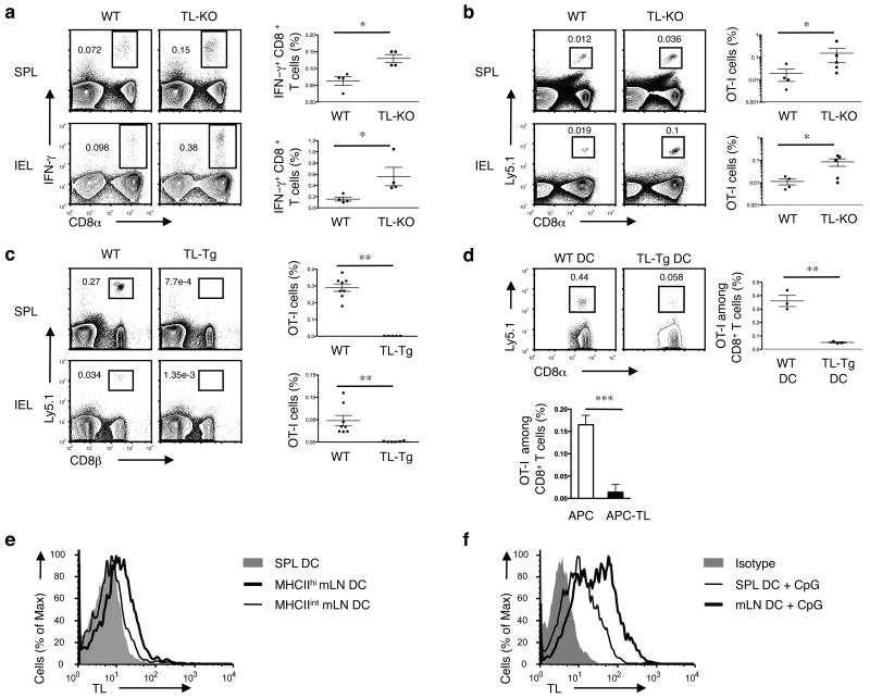
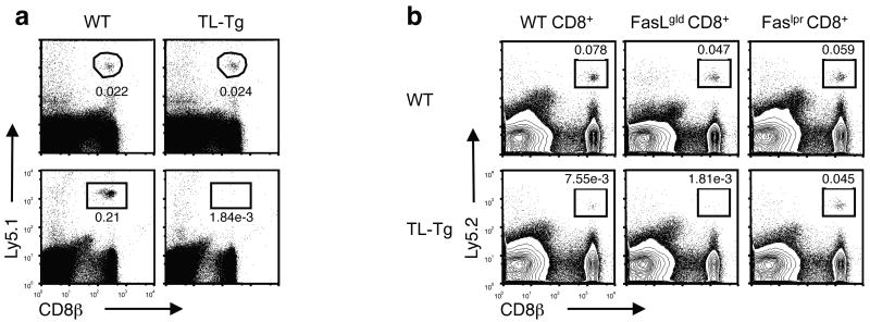
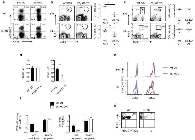
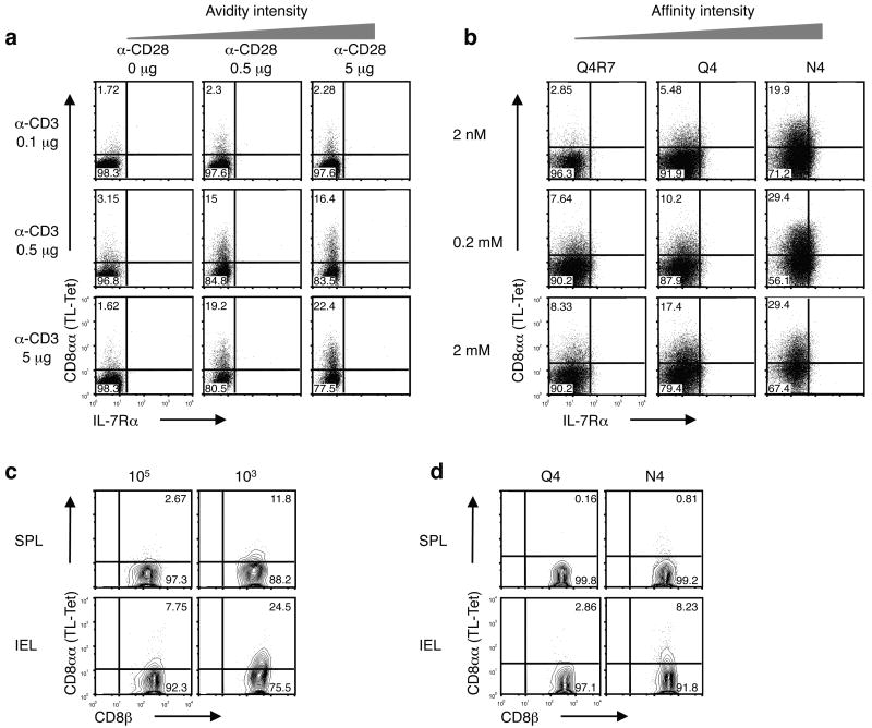
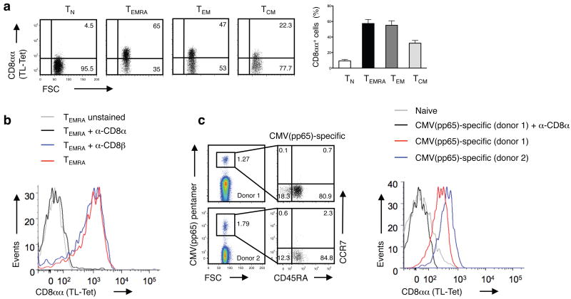
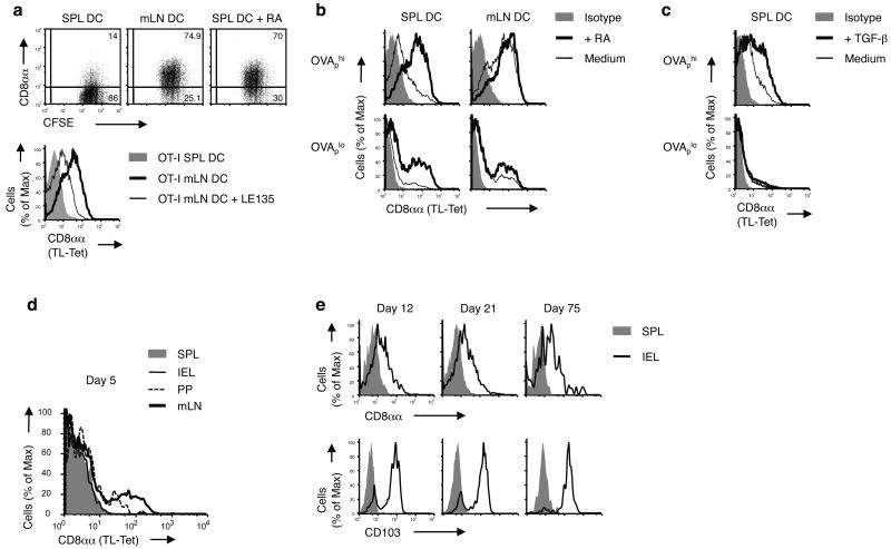
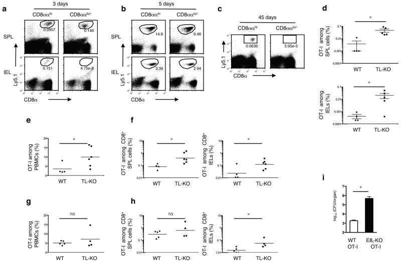
The presence of immune memory at pathogen-entry sites is a prerequisite for protection. Nevertheless, the mechanisms that warrant immunity at peripheral interfaces are not understood. Here we show that the nonclassical major histocompatibility complex (MHC) class I molecule thymus leukemia antigen (TL), induced on dendritic cells interacting with CD8αα on activated CD8αβ(+) T cells, mediated affinity-based selection of memory precursor cells. Furthermore, constitutive expression of TL on epithelial cells led to continued selection of mature CD8αβ(+) memory T cells. The memory process driven by TL and CD8αα was essential for the generation of CD8αβ(+) memory T cells in the intestine and the accumulation of highly antigen-sensitive CD8αβ(+) memory T cells that form the first line of defense at the largest entry port for pathogens.


Figures

Figure 1
Figure 1. TL negatively affects memory generation of CD8αβ+ T cells
(a) WT or TL- mice were orally infected with 1 × 10⁹ ActA- Lm-OVA. 30 days p.i., splenocytes (SPL) and IEL were isolated for IFN-γ intracellular staining and CD8α cell surface staining after ex vivo re-stimulation with OVA257-264 peptide. Graph depicts pooled data ± s.e.m.. (b) 5 × 10⁴ naïve Ly5.1+ CD8+ OT-I cells were adoptively transferred into Ly5.2+ WT or TL- recipients. One day after transfer, mice were orally infected with 1 × 10⁹ ActA- Lm-OVA. Donor OT-I cells were tracked in the spleen and IEL 2 months p.i. Graph depicts pooled data ± s.e.m.. (c) 1 × 10⁶ naïve Ly5.1+ OT-I cells were transferred into WT or TL-Tg recipients. One day after transfer, mice were orally infected with 1 × 10⁹ ActA- Lm-OVA. The donor OT-I cells in the spleens and IEL were tracked two months p.i. Graph depicts pooled data ± s.e.m.. (d) 5 × 10⁴ Ly5.1+ naïve OT-I cells were transferred into C57BL/6 mice which were subsequently immunized i.v. with 5 × 10⁵ OVAp-loaded DCs generated from bone marrow cells (BMDC) of WT or TL-Tg mice (top). Alternatively, OT-I cells were primed in vitro with TL negative APC or APC transfected with TL and then transferred to B6 recipients (bottom). Memory OT-I cells in the spleen were analyzed 2 months after DC immunization or after transfer of in vitro activated OT-I cells. Graph depicts pooled data ± s.e.m.. (e) Spleen or mLN DC were sorted based on the CD11c and MHC II expression and assessed for TL expression by flow cytometry. (f) Freshly isolated SPL or mLN DC were activated with CpG for 1 d and then analyzed for TL expression. * P < 0.05 and ** P < 0.001 (unpaired t-test). All data are representative of three independent experiments.
Figure 2
Figure 2. TL mediates death of activated CD8αβ+ T cells
(a) 1 × 10⁶ naïve Ly5.1+ OT-I cells were transferred into WT or TL-Tg recipients and the donor cells in the spleens were tracked one month after transfer (top, n = 5 per group). 1 × 10⁶ in vitro activated Ly5.1+ OT-I cells were transferred into WT or TL-Tg recipients and the donor cells in the spleen were tracked one month after transfer (bottom, n = 8 per group). (b) Naïve CD8 T cells sorted from WT, FasLgld or Faslpr mice were stimulated in vitro by anti-CD3/-CD28 beads for 3 d. 0.5 × 10⁶ activated CD8 T cells were transferred into Ly5.1+ WT or TL-Tg recipients and the donor cells in the spleens were tracked 1 month after transfer. Representative data are shown of four mice analyzed in each group. Data are representative of two independent experiments (a, b).
Figure 3
Figure 3. Activation-induced CD8αα rescues CD8αβ primary effector T cells from TICD
(a) 2 × 10⁵ CFSE-labeled naïve ΔE8I OT-I cells were cultured with 4 × 10⁴ OVAp-pulsed DCs from SPL or mLN of WT or TL- mice that were previously orally infected with ActA- Lm-OVA. After 2 d culture, OT-I cells were harvested and analyzed for cell death by Annexin V staining. (b) 5 × 10⁴ naïve Ly5.2+ WT or Ly5.2+ ΔE8I OT-I cells were adoptively transferred into Ly5.1 recipient mice. 1 d after transfer, mice were orally infected with 1 × 10⁹ ActA- Lm-OVA. Donor OT-I cells were tracked in the spleen and IEL, 2 months p.i.. Graph depicts pooled data ± s.e.m.. (c) 5 × 10⁴ naïve Ly5.2+ WT or Ly5.2+ ΔE8I OT-I cells were adoptively transferred into Ly5.1 recipient mice. 1 d after transfer, mice were intravenously infected with 2.5 × 10⁵ ActA- Lm-OVA. Donor OT-I cells were tracked in the spleen and IEL, 2 months p.i.. Graph depicts pooled data ± s.e.m.. (d) As described in (b) and (c), CD8β expression (MFI) was measured on effector WT OT-I or ΔE8I OT-I cells 7 days post oral infection (left) or i.v. infection (right). (e) As described in (c), CD8β expression was measured on memory WT OT-I or ΔE8I OT-I cells 2 m post i.v. infection in the spleen and IEL (f, g) 5 × 10⁴ naïve Ly5.1+Ly5.2+ WT OT-I and 5 × 10⁴ naïve Ly5.2+ ΔE8I OT-I cells were co-transferred into Ly5.1+ WT or Ly5.1+ TL- mice. One day after transfer, the mice were orally infected with 1 × 10⁹ ActA- Lm-OVA. Two months p.i., memory OT-I cells were tracked in the spleen and IEL. (f) Graph depicts pooled data ± s.e.m. (n = 5 per group). (g) Staining for CD8αα expression, using TL-tetramers, on gated memory WT OT-I cells in IEL of WT or TL- recipient mice 2m p.i. * P < 0.001 and ** P < 0.01 (unpaired t-test). Data are representative of three (a, b, c, e, f, g) and two (d) independent experiments.
Figure 4
Figure 4. CD8αα expression correlates with the intensity of TCR activation
(a) Total splenocytes were cultured in the presence of graded concentration of soluble anti-CD3 and anti-CD28. CD8αα expression, as measured by TL tetramer staining, was analyzed 3 d after in vitro culture. Representative data on gated CD8+ T cells are shown. IL-7Rα expression is also depicted. Three independent experiments were performed. (b) Naïve OT-I cells were cultured with artificial APC (MEC.B7) in the presence of graded concentration of OVA257-264 SIINFEKL (N4) or altered peptide ligands (Q4R7 and Q4). CD8αα expression was detected 2 d after in vitro culture. Three independent experiments were performed. (c) 1 x10⁵ or 1 × 10³ sorted naïve Ly5.1+ CD8+ OT-I cells were transferred into B6 recipient mice. 1 d after transfer, mice were orally infected with 1 × 10⁹ ActA- Lm-OVA. 7 d p.i., CD8αα expression was analyzed on Ly5.1+ CD8+ OT-I cells from the spleen and IEL (representative data from a single mouse is shown, n = 4 mice per group). (d) 5 × 10⁴ naïve Ly5.1+ CD8+ OT-I cells were transferred into WT recipient mice. 1 d after transfer, mice were orally infected with 2 × 10⁸ WT Lm-Q4OVA or Lm-N4OVA. 7 d p.i., CD8αα expression was analyzed on donor OT-I cells from the spleen and IEL (representative data from a single mouse is shown, n = 5 mice per group). Data are representative of three (a, b) and two (c, d) independent experiments.
Figure 5
Figure 5. CD8αα expression marks effector memory CD8αβ T cells in humans
(a) Expression of CD8αα on polyclonal human naive (TN; CCR7+CD45RA+), recently activated effector-memory (TEMRA; CCR7CD45RA+), effector-memory (TEM; CCR7CD45RA) and central-memory (TCM; CCR7+CD45RA) CD8+ T cells was measured by TL-tetramer staining. The numbers indicate the percentage of TL-tetramerhi cells. Graph depicts pooled data ± s.e.m. on percentage of CD8αα expression on human peripheral blood CD8+ T cells (n = 9). The differences between TN and TEMRA, TEM or TCM were significant (P < 0.001, unpaired t-test). (b) TL-tetramer staining of human TEMRA CD8+ T cells is blocked by anti-CD8α but not anti-CD8β antibody. Data are representative of two independent experiments. (c) CMVpp65-specific CD8+ T cells display a TEM/TEMRA phenotype and persist at high frequency in humans. Data from two representative donors from a total of six persons are shown. The TL-tetramer staining was absent on naive CD8+ T cells and was blocked by an anti-CD8α.
Figure 6
Figure 6. Retinoic acid promotes the affinity-based accumulation of CD8αα+ CD8αβ T cells in the intestine
(a) OT-I cells were stimulated by OVAp-loaded DCs from SPL or mLN of WT mice with or without 100 nM RA (dot plots) or with LE135 (histogram) in vitro for 3 d and CD8αα expression was analyzed. Data are representative of five independent experiments. (b,c) OT-I cells were stimulated by SPL or mLN DCs pulsed with OVAp (high, 1 nM; low, 0.01 nM) in the presence or absence of 100 nM RA (b) or 5 ng/ml TGF-β (c) in vitro for 3 d and CD8αα expression was analyzed. Data are representative of more than five independent experiments. (d,e) 0.5 × 10⁶ CD8+ OT-I cells isolated from naïve Ly5.1+ OT-I+ Rag-/- mice were adoptively transferred into B6 recipient mice. 1 d after transfer, mice were orally infected by 0.5 × 10⁹ ActA- Lm-OVA. CD8αα expression was measured on gated donor OT-I cells from the SPL, mLN, PP and IEL, 5 d p.i. (d). CD8αα and CD103 expression is shown on gated donor OT-I cells from the spleen and IEL on days 12, 21 and 75 p.i. (e). Representative data from two to three mice per group are shown. At least three independent experiments were performed.
Figure 7
Figure 7. Constitutive expression of TL on intestinal epithelial cells mediates selection of mature memory CD8αβ T cells
(a,b) Naïve Ly5.1+ CD8+OT-I cells were cultured in the presence of APC (MEC.B7.SigOVA). After 2 days' culture, CD8ααhi and CD8ααlo/- OT-I cells were sorted and cultured for 3 more days in vitro. Then 0.5 × 10⁶ CD8ααhi or CD8ααlo/- cells were adoptively transferred into B6 recipients. One month after transfer, mice were orally infected with 5 × 10⁸ ActA- Lm-OVA. Donor Ly5.1+ OT-I cells were tracked in the spleen and IEL 3 d (a) and 5 d (b) p.i.. Representative data from 3-4 mice in each group are shown. At least five independent experiments were performed. (c) As shown in (a), secondary OT-I memory cells were assessed in the IEL 45 d p.i.. Representative data from three to four mice in each group are shown. At least three independent experiments were performed. (d) Sorted in vitro activated Ly5.1+ CD8ααlo/- OT-I cells were cultured for 3 d and 0.5 × 10⁶ primary effector cells were transferred into WT or TL-recipients. One month after transfer, mice were orally infected with 5 × 10⁸ ActA- Lm-OVA. 4 months p.i., memory OT-I cells were tracked in the spleens and IEL. Pooled data ± s.e.m. are shown. At least two independent experiments were performed. (e, f) 5 × 10⁴ naïve CD8+ OT-I cells were transferred into Ly5.1+ WT or Ly5.1+ TL- recipient mice. 1 d after transfer, mice were orally infected with Lm-Q4OVA. Effector OT-I cells in the peripheral blood (7 d p.i.) and memory OT-I cells (2 m p.i.) in the spleen and IEL were analyzed. Pooled data ± s.e.m. are shown. (g, h) 5 × 10⁴ naïve CD8+ OT-I cells were transferred into Ly5.1+ WT or Ly5.1+ TL- recipient mice. 1 d after transfer, mice were intravenously infected with Lm-Q4OVA. Effector OT-I cells in the peripheral blood (7 d p.i.) and memory OT-I cells (2 m p.i.) in the spleen and IEL were analyzed. Pooled data ± s.e.m. are shown. Data are representative of three independent experiments(e, f, g, h). (i) Ly5.1 mice adoptively transferred with 5 × 10⁴ naïve WT or ΔE8I OT-I cells were orally immunized with 1 × 10⁹ ActA- Lm-OVA. Two months after immunization, mice were re-challenged orally with 1 × 10¹⁰ WT Lm-OVA. Bacterial loads in the livers were assessed day 3 p.i.. Pooled data ± s.e.m. are shown (n = 6). Representative data are shown of three independent experiments. * P < 0.05; NS: not significant (unpaired t-test).

Comment in

  • TL we meet again.
    Nagler C, Wroblewska J. Nagler C, et al. Nat Immunol. 2011 Oct 19;12(11):1027-8. doi: 10.1038/ni.2138. Nat Immunol. 2011. PMID: 22012434 No abstract available.

References

    1. Sallusto F, Lanzavecchia A, Araki K, Ahmed R. From vaccines to memory and back. Immunity. 2010;33:451–463. - PMC - PubMed
    1. Cheroutre H, Madakamutil L. Mucosal effector memory T cells: the other side of the coin. Cell Mol Life Sci. 2005;62:2853–2866. - PMC - PubMed
    1. Masopust D, Vezys V, Marzo AL, Lefrancois L. Preferential localization of effector memory cells in nonlymphoid tissue. Science. 2001;291:2413–2417. - PubMed
    1. Masopust D, Vezys V, Wherry EJ, Barber DL, Ahmed R. Cutting edge: gut microenvironment promotes differentiation of a unique memory CD8 T cell population. J Immunol. 2006;176:2079–2083. - PubMed
    1. Sallusto F, Lenig D, Forster R, Lipp M, Lanzavecchia A. Two subsets of memory T lymphocytes with distinct homing potentials and effector functions. Nature. 1999;401:708–712. - PubMed

Publication types

MeSH terms