. 2011 Dec;32(12):1881-9.
doi: 10.1093/carcin/bgr218. Epub 2011 Sep 28.

Green tea polyphenol EGCG suppresses lung cancer cell growth through upregulating miR-210 expression caused by stabilizing HIF-1α

Affiliations

Green tea polyphenol EGCG suppresses lung cancer cell growth through upregulating miR-210 expression caused by stabilizing HIF-1α

Hong Wang et al. Carcinogenesis. 2011 Dec.

Abstract

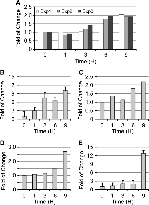
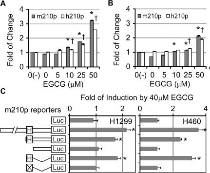
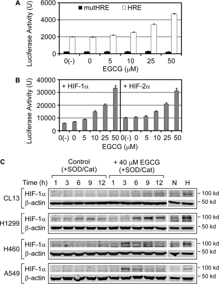
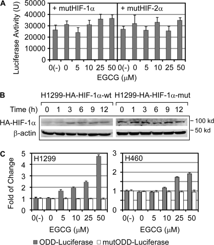
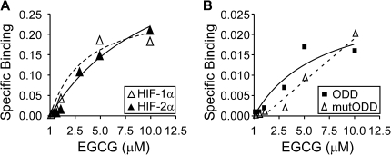
(-)-Epigallocatechin-3-gallate (EGCG) has been reported to affect many cellular regulatory pathways. This study aims to determine whether EGCG could target microRNA (miRNA), one of the mechanisms for cells to achieve subtle change in multiple targets. We found that, in both human and mouse lung cancer cells in culture, EGCG specifically upregulated the expression of miR-210, a major miRNA regulated by HIF-1α. Furthermore, we found that overexpression of miR-210 led to reduced cell proliferation rate and anchorage-independent growth as well as reduced sensitivity to EGCG. On the mechanisms of miR-210 regulation by EGCG, we demonstrated that the regulation was mediated through the hypoxia-response element in miR-210 promoter. Consistently, the upregulation of miR-210 was found to be correlated with the stabilized HIF-1α in lung cancer cell lines after EGCG treatment. This EGCG-induced stabilization of HIF-1α was further shown by the stabilization of HA-tagged HIF-1α but not the P402A/P564A-mutated HIF-1α by EGCG, suggesting that EGCG targets the oxygen-dependent degradation (ODD) domain. Direct evidence was obtained by affinity binding assay showing that EGCG specifically binds HIF-1α with a K(d) = 3.47 μM. This result suggests that EGCG binding interferes with the hydroxylation of key Pro residues in the ODD domain, preventing HIF-1α from the Pro hydroxylation-dependent ubiquitination and subsequent proteosome-mediated degradation. In summary, our results demonstrated, for the first time, the elevation of miR-210 by EGCG in lung cancer cell lines and this is mediated by the stabilization of HIF-1α. This event contributes to the anticancer activity of EGCG.


Figures

Fig. 1.
Fig. 1.
MiR-210 was upregulated in lung cancer cells upon EGCG treatement. The expression levels of miR-210 in lung cancer cells after EGCG (40 μM) treatment as compared with the control (0 h). (A). Results of three individual experiments using CL13 cells as determined by miRNA microarray (Exp. 1–3). (B). In CL13 cells as determined by real-time PCR (n = 3). (C) In CL13 cells treated with EGCG in the absence of SOD/catalase as determined by miRNA microarray. (D) In H1299 cells as determined by miRNA microarray. (E) In H1299 cells as determined by real-time PCR (n = 3).
Fig. 2.
Fig. 2.
Ectopic expression of miR-210 in lung cancer cells reduced proliferation, sensitivity to EGCG and anchorage-independent growth. H1299 and H460 cells infected with retrovirus expressing human miR-210 were compared with the cells infected with the control virus for their proliferation in 5% fetal bovine serum medium (A and B; n = 8), responses to different concentrations of EGCG at 48 h (C and D; n = 8) and growth abilities on the semi-solid medium (E and F; n = 3). *indicates statistical difference from the controls at each time point (A and B) or concentration of EGCG (C and D) (Student’s t-test; P < 0.05).
Fig. 3.
Fig. 3.
HRE in miR-210 promoter was responsive to the EGCG stimulation. H1299 (A) and H460 (B) cells transfected with human and mouse miR-210 gene promoter-luciferase reporter (h210p and m210p) were assayed for reporter activities at 12 h after EGCG treatment in the presence of SOD/catalase (n = 3). * and † indicate statistical difference between levels at different concentrations of EGCG and the control (EGCG = 0) (by one-way analysis of variance; P < 0.05); * is for m210p and † is for h210p.The responses of mouse miR-210 gene promoter fragment to EGCG treatment were analyzed by the luciferase activities in H1299 and H460 cells transfected with different reporter constructs and treated with EGCG for 12 h (n = 3) (C). 0(−) represents for the treatment without SOD/catalase. H and X stand for HRE and mutHRE, respectively. *indicates statistical difference from the control empty vector (by one-way analysis of variance; P < 0.05).
Fig. 4.
Fig. 4.
EGCG enhanced the activities of HIF-1α and HIF-2α. The upregulation of the HRE-luciferase reporter by EGCG was determined in H1299 cells: (A) transfected with phosphoglycerate kinase HRE or mut-HRE-luciferase reporter plus Rellina-luciferase as internal control (n = 3) and (B) transfected with the HIF-1α or HIF-2α expression vector. (C) HIF-1α levels in lung cancer cell lines after treatment with EGCG (40 μM) were determined by western blots. The cells without treatment were also collected at the match time points as the controls. Sample loading was monitored by β-actin level. As negative and positive controls for determining the hypoxia-induced accumulation of HIF-1α, protein samples collected from cells grown under normoxia (N) and hypoxia (H) were used.
Fig. 5.
Fig. 5.
The ODD domain was responsive to EGCG. (A) H1299 cells transfected with the mutHIF1α or mutHIF2α expression vector with HRE-luciferase reporter were incubated with EGCG for 12 h and luciferase activity were measured. (B) H1299 cells stably expressing HA-tagged HIF-1α or mutHIF1α (H1299-HA-HIF-1α-wt and -mut) were treated with EGCG (40 μM) and analyzed for HA-HIF-1α and -mutHIF1α levels by western blots using anti-HA antibody. Sample loading was monitored by β-actin level. (C) The ODD-luciferase fusion protein or mutODD-luciferase expression vector was transfected into H1299 and H460 cells with Renilla-luciferase as the internal control. Luciferase activity of ODD- or mutODD-luciferase was normalized by Renilla-luciferase activity (n = 3).
Fig. 6.
Fig. 6.
EGCG directly bound HIFα proteins. (A) HA-tagged HIF-1α and 2α proteins bound to the anti-HA agarose beads were used to study the binding of EGCG. The beads without HIF protein were used to determine the non-specific binding. Specific binding after correction for non-specific binding was plotted in the figure. The empty and solid triangles represent the specific binding of EGCG on HIF-1α and 2α, respectively. The dashed and solid line lines represent the saturation curves by non-linear regression analysis. (B) The specific binding of EGCG to Flag-ODD or Flag-mutODD bound to the anti-Flag agarose beads was measured. The solid square and solid line represent the specific binding of EGCG on ODD. The empty triangle and dashed line represent the binding of EGCG on mutODD.

Similar articles

Cited by

References

    1. Khan N, et al. Targeting multiple signaling pathways by green tea polyphenol (-)-epigallocatechin-3-gallate. Cancer Res. 2006;66:2500–2505. - PubMed
    1. Yang CS, et al. Cancer prevention by tea: animal studies, molecular mechanisms and human relevance. Nat. Rev. Cancer. 2009;9:429–439. - PMC - PubMed
    1. Liang G, et al. Green tea catechins augment the antitumor activity of doxorubicin in an in vivo mouse model for chemoresistant liver cancer. Int. J. Oncol. 2010;37:111–123. - PubMed
    1. Shimizu M, et al. (-)-Epigallocatechin gallate inhibits growth and activation of the VEGF/VEGFR axis in human colorectal cancer cells. Chem. Biol. Interact. 2010;185:247–252. - PubMed
    1. Zhang Q, et al. Green tea extract and (-)-epigallocatechin-3-gallate inhibit hypoxia- and serum-induced HIF-1alpha protein accumulation and VEGF expression in human cervical carcinoma and hepatoma cells. Mol. Cancer Ther. 2006;5:1227–1238. - PubMed

Publication types