. 2011 Nov;22(22):4424-34.
doi: 10.1091/mbc.E10-12-0971. Epub 2011 Sep 30.

NOX4 mediates activation of FoxO3a and matrix metalloproteinase-2 expression by urotensin-II

Affiliations

NOX4 mediates activation of FoxO3a and matrix metalloproteinase-2 expression by urotensin-II

Isabel Diebold et al. Mol Biol Cell. 2011 Nov.

Abstract

The vasoactive peptide urotensin-II (U-II) has been associated with vascular remodeling in different cardiovascular disorders. Although U-II can induce reactive oxygen species (ROS) by the NADPH oxidase NOX4 and stimulate smooth muscle cell (SMC) proliferation, the precise mechanisms linking U-II to vascular remodeling processes remain unclear. Forkhead Box O (FoxO) transcription factors have been associated with redox signaling and control of proliferation and apoptosis. We thus hypothesized that FoxOs are involved in the SMC response toward U-II and NOX4. We found that U-II and NOX4 stimulated FoxO activity and identified matrix metalloproteinase-2 (MMP2) as target gene of FoxO3a. FoxO3a activation by U-II was preceded by NOX4-dependent phosphorylation of c-Jun NH(2)-terminal kinase and 14-3-3 and decreased interaction of FoxO3a with its inhibitor 14-3-3, allowing MMP2 transcription. Functional studies in FoxO3a-depleted SMCs and in FoxO3a(-/-) mice showed that FoxO3a was important for basal and U-II-stimulated proliferation and vascular outgrowth, whereas treatment with an MMP2 inhibitor blocked these responses. Our study identified U-II and NOX4 as new activators of FoxO3a, and MMP2 as a novel target gene of FoxO3a, and showed that activation of FoxO3a by this pathway promotes vascular growth. FoxO3a may thus contribute to progression of cardiovascular diseases associated with vascular remodeling.


Figures

FIGURE 1:
FIGURE 1:
U-II stimulates MMP2 expression and activity. (A) Pulmonary artery SMC were stimulated with U-II (100 nM) or 10% fetal calf serum (FCS) for different time periods. Supernatants were subjected to Western blot analysis using antibodies against MMP2 or MMP9 or to zymography to determine MMP2 or MMP9 activity. (B) SMC were stimulated with U-II for the indicated time periods. MMP2 mRNA and protein were determined by RT-PCR using primers for MMP2 or GAPDH or by Western blot analyses using an antibody against MMP2, respectively. PonceauS staining served as loading control. Data are presented as relative change to unstimulated cells (100%; n = 3, *p < 0.05 vs. unstimulated cells [0]). (C and D) SMC were pretreated with actinomycin D (Act, 5 μM) or dimethyl sulfoxide (DMSO) (Ctr) for 1 h and stimulated with U-II for 2 h. (C) Western blot analyses were performed with an antibody against MMP2. PonceauS staining served as loading control. (D) RT-PCR was performed using primers for MMP2 or GAPDH. Data are presented as relative change to control (100%) (n = 3, *p < 0.05 vs. unstimulated Ctr, #p < 0.05 vs. U-II–stimulated Ctr).
FIGURE 2:
FIGURE 2:
NOX4 and ROS promote U-II–induced MMP2 expression. (A) Pulmonary artery SMC were transfected with a human MMP2 promoter luciferase construct (MMP2-1709) and cotransfected with a NOX4 expression vector or control vector (Ctr). Cells were stimulated with U-II (100 nM) for 8 h or left untreated. Luciferase activity under control conditions was set to 100% (n = 5; *p < 0.05 vs. Ctr). (B) SMC were preincubated with NAC (10 mM) for 30 min or left untreated and stimulated with U-II for 4 h. Western blot analysis was performed with cell culture supernatants (sup) using an MMP2 antibody. PonceauS staining served as loading control. (C) SMC were transfected with vectors coding for NOX4 or for shRNA against NOX4 (siN4) or the respective control vectors (Ctr), and stimulated with U-II for 4 h. Western blot analyses were performed with antibodies against MMP2 and NOX4. PonceauS staining served as loading control. Blots are representative of three independent experiments.
FIGURE 3:
FIGURE 3:
FoxO is activated by U-II and mediates MMP2 expression. (A) Pulmonary artery SMC were transfected with luciferase constructs containing either six DBE (6xDBE) or three Forkhead responsive elements from the FasL gene (3xFHRE) in front of the SV40 promoter. Cells were stimulated with U-II (100 nM) or PDGF (10 nM) for 8 h or left untreated. In some cases SMC were cotransfected with plasmids coding for NOX4 or for shRNA against NOX4 (siNOX4), FoxO1 (siF1), FoxO3a (siF3), or FoxO4 (siF4) or with respective control vectors, or were pretreated with NAC (10 mM) for 30 min. Luciferase activity under control conditions (Ctr) was set equal to 100% (n = 4; *p < 0.05 vs. Ctr or #p < 0.05 vs. U-II-stimulated Ctr). (B) SMC were transfected with vectors coding for shRNA against FoxO1 (siF1), FoxO3a (siF3), or FoxO4 (siF4) or for control shRNA (Ctr) and exposed to U-II for 4 h before Western blot analysis using antibodies against MMP2, FoxO1, FoxO3a, or FoxO4. Actin levels served as loading control. Blots are representative of three independent experiments.
FIGURE 4:
FIGURE 4:
FoxO3a regulates MMP2 transcription and promoter activity. (A) Pulmonary artery SMC were transfected with a vector coding for FoxO3a or with control vector (–). RT-PCR was performed using primers for MMP2, FoxO3a, or GAPDH. Data are presented as relative change to control vector (100%; n = 3, *p < 0.05 vs. control vector [–]). (B) SMC were cotransfected with luciferase constructs containing either the WT MMP2 promoter (MMP2-1709) or the MMP2 promoter mutated at the FoxO consensus sequence (MMP2-1709mut) and with plasmids coding for WT (FoxO3a WT) or inactive (FoxO3a ΔCT) FoxO3a or for NOX4. Cells were stimulated with U-II (100 nM) for 8 h or left untreated. Luciferase activity under control conditions (Ctr) was set equal to 100% (n = 3; *p < 0.05 vs. Ctr; #p < 0.05 vs. U-II–stimulated Ctr). (C) SMC were stimulated with U-II for 2 h and subjected to chromatin immunoprecipitation with an antibody against FoxO3a. PCR amplifying a 310–base pair fragment of the proximal MMP2 promoter region containing a putative FoxO binding site was performed. Data are presented as relative change to control (Ctr, 100%; n = 3, *p < 0.05 vs. Ctr).
FIGURE 5:
FIGURE 5:
U-II decreases interaction of FoxO3a and 14-3-3. (A) Pulmonary artery SMC were stimulated with U-II (100 nM) for 15 min in the presence or absence of the JNK inhibitor SP600125 (SP, 25 μM). Immunoprecipitation was performed with an antibody against FoxO3a and revealed with an antibody against 14-3-3. Blots are representative of three independent experiments. (B) SMC were stimulated with U-II for increasing time periods. Western blot analysis was performed with antibodies against phosphorylated 14-3-3 (p14-3-3) and actin. Blots are representative of three independent experiments. (C) SMC were preincubated with SP600125 (SP, 25 μM), the PI3-kinase inhibitor LY294002 (LY, 10 μM), or DMSO (Ctr) for 30 min or (D) were transfected with shNOX4 (siN4) or control vector (Ctr) and stimulated with U-II for 5 min. Western blot analyses were performed using antibodies against phosphorylated 14-3-3 (p14-3-3), JNK (pJNK), NOX4, and actin. Blots are representative of three independent experiments. (E) SMC were transfected with luciferase constructs containing six DBE in front of the SV40 promoter (6xDBE) or the MMP2 promoter (MMP2-1709), preincubated with SP or DMSO (Ctr) for 30 min, and stimulated with U-II for 8 h. Luciferase activity under control conditions (Ctr) was set equal to 100% (n = 3; *p < 0.05 vs. Ctr or #p < 0.05 vs. U-II–stimulated Ctr). (F) SMC were preincubated for 30 min with the MEK1 inhibitor PD98059 (PD, 20 μM), the p38 MAP kinase inhibitor SB202190 (SB, 20 μM), LY294002 (LY, 10 μM), or SP600125 (25 μM) or with solvent (Ctr) and stimulated with U-II for 4 h. Western blot analysis was performed using antibodies against MMP2 and actin. Blots are representative of three independent experiments.
FIGURE 6:
FIGURE 6:
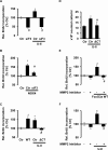
FoxO3a promotes U-II–induced SMC proliferation. (A and B) Pulmonary artery SMC were transfected with vectors coding for shRNA against FoxO3a (siF3) or control shRNA (Ctr), and stimulated with U-II (100 nM) for 2 h (A), or were cotransfected with a vector for NOX4 (B). DNA synthesis was evaluated by BrdU incorporation. Data are shown as relative change to control (Ctr, 100%; n = 3, *p < 0.05 vs. Ctr, #p < 0.05 vs. U-II–stimulated Ctr or NOX4 overexpression). (C and D) SMC were transfected with vectors for WT or inactive (ΔCT) FoxO3a and stimulated with U-II for 2 h. DNA synthesis was evaluated by BrdU incorporation, and control was set to 100% (C), or cell numbers were determined using a hemocytometer (D) (n = 3, *p < 0.05 vs. Ctr, #p < 0.05 vs. U-II–stimulated Ctr). (E and F) SMC were transfected with a vector encoding FoxO3a (E) or were stimulated with U-II for 2 h (F), after treatment with an MMP2 inhibitor (20 μM) (+) or DMSO (–) for 30 min. BrdU incorporation was evaluated. Data are shown as relative change to DMSO control (Ctr, 100%; n = 3, *p < 0.05 vs. Ctr, #p < 0.05 vs. overexpression of FoxO3a or U-II–stimulated Ctr).
FIGURE 7:
FIGURE 7:
FoxO3a is required for vascular outgrowth. (A) SMC were isolated from FoxO3a WT or FoxO3a knockout (KO) mice and stimulated with U-II (100 nM) for 72 h. Cell number was counted in a hemocytometer. To confirm the presence of SMC, cells were fixed and stained with an antibody against SMC α-actin (red). Nuclei were counterstained with DAPI (blue). Size bar = 20 μm for all panels. (B) Aortae or pulmonary arteries (PA) from FoxO3a WT or FoxO3a knockout (KO) mice were stimulated with U-II for 4 h. RT-PCR was performed from mRNA isolated from these vessels using primers for MMP2, FoxO3a, and 18S. (C) Vessel rings from aortae or pulmonary arteries (PA) from FoxO3a WT or KO mice were cultured in Matrigel with or without U-II to allow vessel sprouting. Tubule length was quantified using ImageJ software. Data are presented as relative change to control (WT, 100%; n = 3, *p < 0.05 vs. WT, #p < 0.05 vs. U-II–stimulated WT).
FIGURE 8:
FIGURE 8:
FoxO3a activation by U-II leads to vascular proliferation. The scheme summarizes the pathways described: U-II activates FoxO3a by NOX4-mediated ROS production and phosphorylation of JNK and 14-3-3. This pathway leads to up-regulation of MMP2 and enhanced proliferation of SMC and may thus play an important role in vascular remodeling.

Similar articles

Cited by

References

    1. Abid MR, Yano K, Guo S, Patel VI, Shrikhande G, Spokes KC, Ferran C, Aird WC. Forkhead transcription factors inhibit vascular smooth muscle cell proliferation and neointimal hyperplasia. J Biol Chem. 2005;280:29864–29873. - PubMed
    1. Accili D, Arden KC. FoxOs at the crossroads of cellular metabolism, differentiation, and transformation. Cell. 2004;117:421–426. - PubMed
    1. Back M, Ketelhuth DF, Agewall S. Matrix metalloproteinases in atherothrombosis. Prog Cardiovasc Dis. 2010;52:410–428. - PubMed
    1. Bonello S, Zahringer C, BelAiba RS, Djordjevic T, Hess J, Michiels C, Kietzmann T, Gorlach A. Reactive oxygen species activate the HIF-1alpha promoter via a functional NFkappaB site. Arterioscler Thromb Vasc Biol. 2007;27:755–761. - PubMed
    1. Brunet A, et al. Stress-dependent regulation of FOXO transcription factors by the SIRT1 deacetylase. Science. 2004;303:2011–2015. - PubMed

Publication types

MeSH terms

LinkOut - more resources