. 2012 Jan 15;21(2):237-50.
doi: 10.1093/hmg/ddr449. Epub 2011 Sep 30.

NMNAT suppresses tau-induced neurodegeneration by promoting clearance of hyperphosphorylated tau oligomers in a Drosophila model of tauopathy

Affiliations

NMNAT suppresses tau-induced neurodegeneration by promoting clearance of hyperphosphorylated tau oligomers in a Drosophila model of tauopathy

Yousuf O Ali et al. Hum Mol Genet. .

Abstract

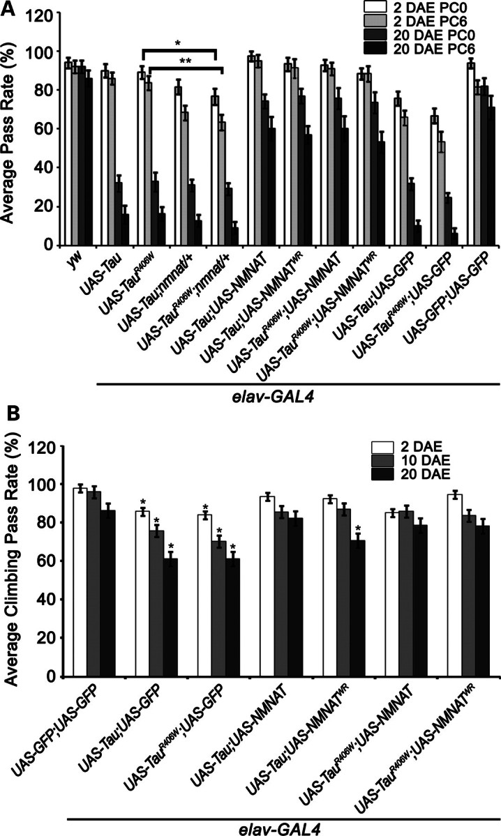
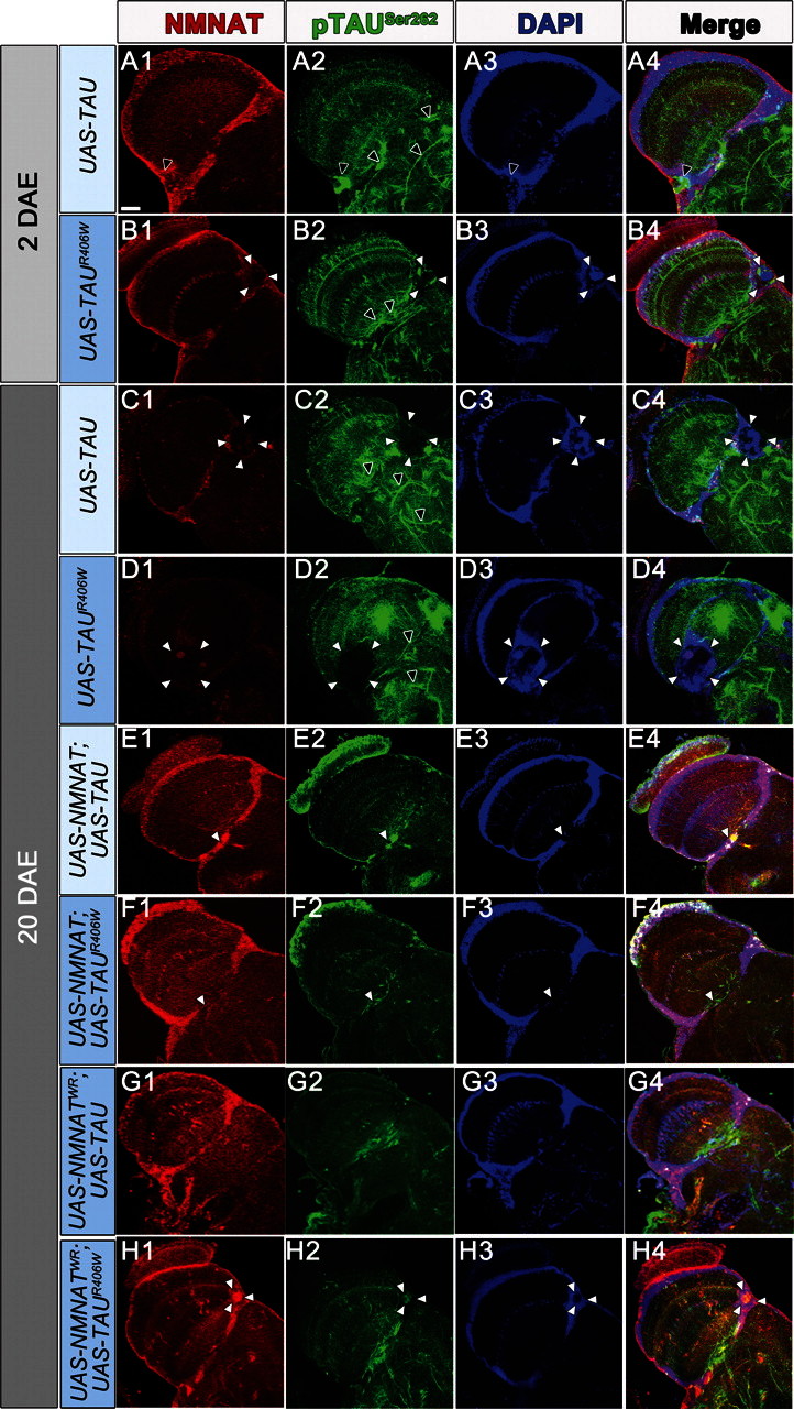
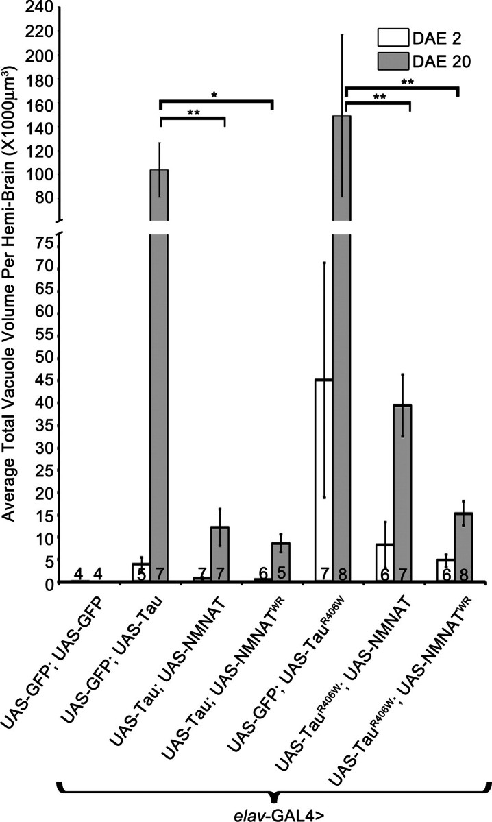
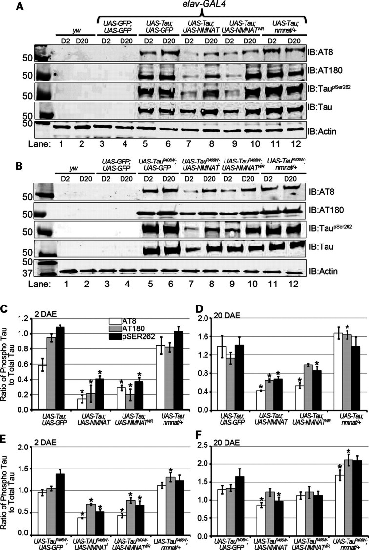
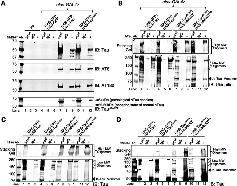
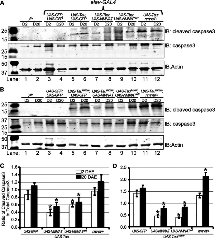
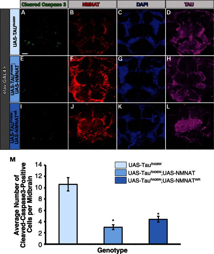
Tauopathies, including Alzheimer's disease, are a group of neurodegenerative diseases characterized by abnormal tau hyperphosphorylation that leads to formation of neurofibrillary tangles. Drosophila models of tauopathy display prominent features of the human disease including compromised lifespan, impairments of learning, memory and locomotor functions and age-dependent neurodegeneration visible as vacuolization. Here, we use a Drosophila model of frontotemporal dementia with parkinsonism linked to chromosome 17 (FTDP-17), in order to study the neuroprotective capacity of a recently identified neuronal maintenance factor, nicotinamide mononucleotide (NAD) adenylyl transferase (NMNAT), a protein that has both NAD synthase and chaperone function. NMNAT is essential for maintaining neuronal integrity under normal conditions and has been shown to protect against several neurodegenerative conditions. However, its protective role in tauopathy has not been examined. Here, we show that overexpression of NMNAT significantly suppresses both behavioral and morphological deficits associated with tauopathy by means of reducing the levels of hyperphosphorylated tau oligomers. Importantly, the protective activity of NMNAT protein is independent of its NAD synthesis activity, indicating a role for direct protein-protein interaction. Next, we show that NMNAT interacts with phosphorylated tau in vivo and promotes the ubiquitination and clearance of toxic tau species. Consequently, apoptosis activation was significantly reduced in brains overexpressing NMNAT, and neurodegeneration was suppressed. Our report on the molecular basis of NMNAT-mediated neuroprotection in tauopathies opens future investigation of this factor in other protein foldopathies.


Figures

Figure 1.
Figure 1.
NMNAT rescues learning and memory deficits and locomotor impairments induced in tauopathy. (A) Learning and memory functions were studied in flies of specified genotypes using APS assay, where flies were conditioned to associate light with the bitter taste of quinine. PC0, 0 h post-conditioning (learning index); PC6, 6 h post-conditioning (memory index). Overexpression of tau or tauR406W causes age-dependent decrements in learning and memory functions. NMNAT or NMNATWR overexpression rescues these deficits. Error bars represent results in five consecutive trials for 20 individual flies of each age and genotype (statistical significance noted in Supplementary Material, Tables 1–4). (B) Locomotor performance was investigated using negative geotaxis assay. Overexpression of tau or tauR406W causes significant deficit in locomotor activities which are rescued by NMNAT or WR. Each bar graph is an average of 10 trials of 100 flies of each genotype and age. *P< 0.05, **P< 0.005.
Figure 2.
Figure 2.
NMNAT suppresses tau-induced morphological vacuolization. Tau or tauR406W overexpression causes age-dependent vacuolization (A–D) demarcated by white arrowheads. Brains were stained for pTauSer262 to detect hyperphosphorylated tau and also NMNAT levels (red) and DAPI to mark cell bodies. Overexpression of wild-type NMNAT (E and F) or enzyme-inactive NMNATWR (G and H) significantly reduced the size and occurrence of vacuoles in 20-day-old flies. Scale bar = 20 μm. Black arrows: filamentous tau; white arrows demarcate vacuole boundary.
Figure 3.
Figure 3.
NMNAT reduces the total vacuole volume per brain induced by hTau overexpression. Overexpression of both wild-type and enzyme-deficient NMNAT reduces total vacuole volume per brain. Vacuole volume was quantified by using the FluoView10-ASW (Olympus) software, assuming each vacuole to be spherical in form. Numbers at the bottom of the bars indicate the number of brains quantified per age per genotype. *P< 0.001, **P< 0.05.
Figure 4.
Figure 4.
NMNAT reduces the levels of hyperphosphorylated, disease-associated tau oligomers. (A and B) Brain lysates of 2 or 20 DAE wild-type flies (lanes 1 and 2) or flies overexpressing GFP (lanes 3 and 4), hTau [WT (A) or R406W (B)] with GFP (lanes 5 and 6) or NMNAT (lanes 7 and 8) or NMNATWR (lanes 9 and 10), as well as in the nmnat/+ background (lanes 11 and 12) were probed with antibodies specific for disease-associated phospho-tau epitopes AT8, AT180 and TauSer262 as well as total hTau and actin for loading control. (C and D) Quantification of phosphorylated-tau species normalized to actin and total hTau from samples in (A) in 2 DAE (C) and 20 DAE (D) flies or in (B) in 2 DAE (E) or 20 DAE (F) flies. *P< 0.05.
Figure 5.
Figure 5.
Endogenous NMNAT interacts with hTau oligomers to promote proteasome-mediated clearance of ubiquitinated tau. (A) Immunoprecipitated whole-brain lysates with NMNAT antibody were probed with total hTau or disease-associated phospho-tau-specific antibodies AT8 and AT180. Lack of tau bands in the negative wild-type and GFP-overexpressing brains reveal that the NMNAT–tau interaction is specific to overexpressed hTau. (B) Total hTau was immunoprecipitated from brains of 2 DAE flies overexpressing wild-type hTau, hTauR406W, hTauR406W +NMNAT, hTauR406W + NMNATWR and probed for ubiquitin. Stacking gel shows HMW tau oligomers and lower gel shows LMW oligomers (note the arrowheads) that can be mono- or polyubiquitinated. Note in NMNAT-overexpressing flies, the band immunoprecipitated at 75 kDa probably corresponds to ubiquitinated hyperphosphorylated tau that can be directly cleared by the proteasome. (C) Blot in (B) probed with a human Tau antibody; stacking gel shows HMW tau oligomers, and lower gel shows LMW oligomers at similar size to those detected by ubiquitin antibody in (B). (D) Total NMNAT was immunoprecipitated from brains of 2 DAE flies overexpressing wild-type hTau, hTauR406W, hTauR406W +NMNAT or hTauR406W + NMNATWR and probed for Tau.
Figure 6.
Figure 6.
NMNAT-mediated protection of morphological defects in tauopathy is partly through reduction in apoptosis. (A and B) Levels of apoptosis were measured in 2 and 20 DAE wild-type flies (yw, lanes 1 and 2) and flies overexpressing GFP (lanes 3 and 4); wild-type hTau (A) or hTauR406W (B) with GFP (lanes 5 and 6) or NMNAT (lanes 7 and 8) or NMNATWR (lanes 9 and 10) or in the nmnat/+ background (lanes 11 and 12) by probing for cleaved caspase 3, total caspase 3 and actin as a loading control. (C and D) Quantification of cleaved caspase 3 levels in panel A (C) or B (D). The level of cleaved or total caspase 3 were normalized to that of actin. The ratio of cleaved caspase 3 to total caspase 3 was displayed. *P< 0.005.
Figure 7.
Figure 7.
Overexpression of NMNAT or NMNATWR reduces the total number of cleaved caspase 3-positive cells in TauR406W brains. Whole-mount adult brains overexpressing either TauR406W with GFP, or NMNAT or NMNATWR were stained for cleaved caspase 3 (green, A, E and I), NMNAT (red, B, F and J), DAPI (blue, C, G and K) and hTau (magenta, D, H and L). (M) The total number of cleaved caspase 3-positive cells per mid-brain were averaged per genotype and plotted. N= 8, *P< 0.005.

References

    1. Iqbal K., Alonso Adel C., Chen S., Chohan M.O., El-Akkad E., Gong C.X., Khatoon S., Li B., Liu F., Rahman A., et al. Tau pathology in Alzheimer disease and other tauopathies. Biochim. Biophys. Acta. 2005;1739:198–210. - PubMed
    1. Sergeant N., Delacourte A., Buee L. Tau protein as a differential biomarker of tauopathies. Biochim. Biophys. Acta. 2005;1739:179–197. - PubMed
    1. Alonso A.C., Grundke-Iqbal I., Iqbal K. Alzheimer's disease hyperphosphorylated tau sequesters normal tau into tangles of filaments and disassembles microtubules. Nat. Med. 1996;2:783–787. - PubMed
    1. Chatterjee S., Sang T.K., Lawless G.M., Jackson G.R. Dissociation of tau toxicity and phosphorylation: role of GSK-3beta, MARK and Cdk5 in a Drosophila model. Hum. Mol. Genet. 2009;18:164–177. - PMC - PubMed
    1. Khatoon S., Grundke-Iqbal I., Iqbal K. Brain levels of microtubule-associated protein tau are elevated in Alzheimer's disease: a radioimmuno-slot-blot assay for nanograms of the protein. J. Neurochem. 1992;59:750–753. - PubMed

Publication types

Substances