. 2011 Dec;211(3):285-95.
doi: 10.1530/JOE-11-0247. Epub 2011 Sep 29.

Cortisol, estradiol-17β, and progesterone secretion within the first hour after awakening in women with regular menstrual cycles

Affiliations

Cortisol, estradiol-17β, and progesterone secretion within the first hour after awakening in women with regular menstrual cycles

Ryun S Ahn et al. J Endocrinol. 2011 Dec.

Abstract

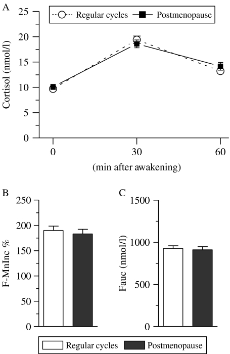
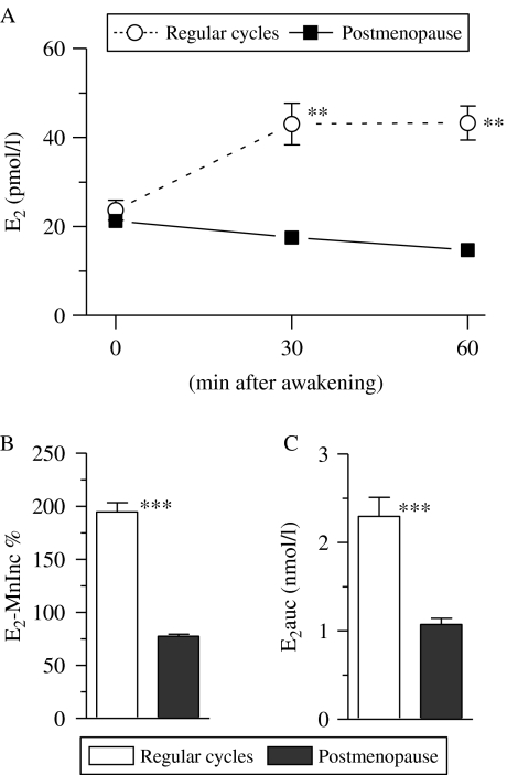
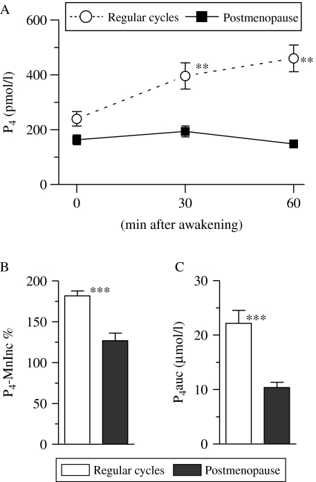
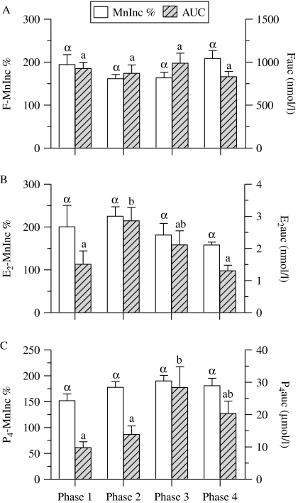
Cortisol concentration in both serum and saliva sharply increases and reaches a peak within the first hour after waking in the morning. This phenomenon is known as the cortisol awakening response (CAR) and is used as an index of hypothalamus-pituitary-adrenal (HPA) axis function. We examined whether ovarian steroid concentrations increased after awakening as with the CAR in the HPA axis. To do this, cortisol, estradiol-17β (E(2)), and progesterone (P(4)) concentrations were determined in saliva samples collected immediately upon awakening and 30 and 60 min after awakening in women with regular menstrual cycles and postmenopausal women. We found that both E(2) and P(4) concentrations increased during the post-awakening period in women with regular menstrual cycles, but these phenomena were not seen in any postmenopausal women. The area under the E(2) and P(4) curve from the time interval immediately after awakening to 60 min after awakening (i.e. E(2)auc and P(4)auc) in women with regular menstrual cycles were greater than those in the postmenopausal women. E(2) and P(4) secretory activity during the post-awakening period was influenced by the phase of the menstrual cycle. E(2)auc in the peri-ovulatory phase and P(4)auc in the early to mid-luteal phase were greater than in the menstrual phase. Meanwhile, cortisol secretory activity during the post-awakening period was not influenced by menstrual status or the phase of menstrual cycle. These findings indicate that, as with the CAR in the HPA axis function, ovarian steroidogenic activity increased after awakening and is closely associated with menstrual status and phase of menstrual cycle.


Figures

Figure 1
Figure 1
Changes in cortisol concentration within the first hour after awakening in women with regular menstrual cycles and postmenopausal women. Cortisol concentration at each examined time point is depicted in (A). The relative mean increase in cortisol concentration 30 and 60 min after awakening over the individual baseline (F-MnInc %) and the integrated cortisol concentration ranging from the time immediately upon awakening to 60 min after awakening (i.e. area under the cortisol curve with respect to baseline, Fauc) in each examined group are depicted in (B and C) respectively. Each data point in (A) and each bar in (B and C) represents mean±s.e.m.
Figure 2
Figure 2
Difference in estradiol-17β (E2) concentrations within the first hour after awakening in women with regular menstrual cycles and postmenopausal women. E2 concentration at each examined time point is depicted in (A). The relative mean increase in E2 concentration 30 and 60 min after awakening over the individual baseline (E2-MnInc %) and integrated E2 concentration ranging from the time immediately upon awakening to 60 min after awakening (i.e. area under the E2 curve with respect to baseline, E2auc) are depicted in (B and C) respectively. Each data point in (A) and each bar in (B and C) represents mean±s.e.m. Asterisks denote the level of significance between both groups: **P<0·01; ***P<0·001 (by the Mann–Whitney test).
Figure 3
Figure 3
Difference in progesterone (P4) concentration within the first hour after awakening in women with regular menstrual cycles and postmenopausal women. P4 concentration at each examined time point is depicted in (A). The relative mean increase in P4 concentration 30 and 60 min after awakening over the individual baseline (P4-MnInc %) and integrated P4 concentration ranging from the time immediately upon awakening to 60 min after awakening (i.e. area under the P4 curve with respect to baseline, P4auc) are depicted in (A and B) respectively. Each data point in (A) and each bar in (B and C) represents mean±s.e.m. Asterisks denote the level of significance between both groups: **P<0·01; ***P<0·001 (by the Mann–Whitney test).
Figure 4
Figure 4
Changes in cortisol, estradiol-17β, and progesterone (P4) secretory activity throughout phases of the menstrual cycles in women with regular menstrual cycles. Cortisol (F), estradiol-17β (E2), and P4 secretory activity in each phase is presented in panels A–C respectively. The relative mean increases in steroid concentrations 30 and 60 min after awakening over the individual baseline (MnInc %) and the integrated steroid concentrations ranging from the time immediately upon awakening to 60 min after awakening (i.e. area under the curve (AUC) with respect to baseline) are depicted as mean±s.e.m. Bars with different Greek or Latin letters in each panel are significantly different from each other (P<0·05) by the Kruskal–Wallis test with Dunn's post-hoc test.

Similar articles

Cited by

References

    1. Ahn RS, Lee YJ, Choi JY, Kwon HB, Chun SI. Salivary cortisol and DHEA levels in the Korean population: age-related differences, diurnal rhythm, and correlations with serum levels. Yonsei Medical Journal. 2007;48:379–388. doi: 10.3349/ymj.2007.48.3.379. - DOI - PMC - PubMed
    1. Atkinson HC, Waddell BJ. Circadian variation in basal plasma corticosterone and adrenocorticotropin in the rat: sexual dimorphism and changes across the estrous cycle. Endocrinology. 1997;138:3842–3848. doi: 10.1210/en.138.9.3842. - DOI - PubMed
    1. Bao AM, Liu RY, van Someren EJ, Hofman MA, Cao YX, Zhou JN. Diurnal rhythm of free estradiol during the menstrual cycle. European Journal of Endocrinology. 2003;148:227–232. doi: 10.1530/eje.0.1480227. - DOI - PubMed
    1. Bao AM, Ji YF, Van Someren EJ, Hofman MA, Liu RY, Zhou JN. Diurnal rhythms of free estradiol and cortisol during the normal menstrual cycle in women with major depression. Hormones and Behavior. 2004;45:93–102. doi: 10.1016/j.yhbeh.2003.09.004. - DOI - PubMed
    1. van der Beek EM, Wiegant VM, van Oudheusden HJ, van der Donk HA, van den Hurk R, Buijs RM. Synaptic contacts between gonadotropin-releasing hormone-containing fibers and neurons in the suprachiasmatic nucleus and perichiasmatic area: an anatomical substrate for feedback regulation? Brain Research. 1997;755:101–111. doi: 10.1016/S0006-8993(97)00086-3. - DOI - PubMed

Publication types

LinkOut - more resources