. 2011 Dec;193(23):6597-604.
doi: 10.1128/JB.05987-11. Epub 2011 Sep 30.

Phenol sensing by Escherichia coli chemoreceptors: a nonclassical mechanism

Affiliations

Phenol sensing by Escherichia coli chemoreceptors: a nonclassical mechanism

Hai The Pham et al. J Bacteriol. 2011 Dec.

Abstract

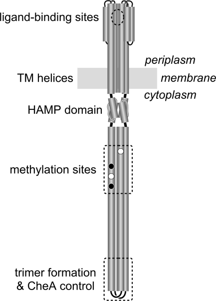
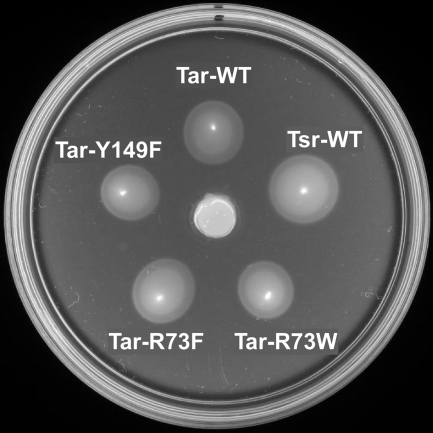
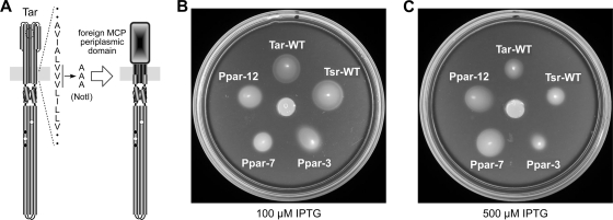
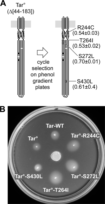
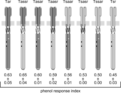
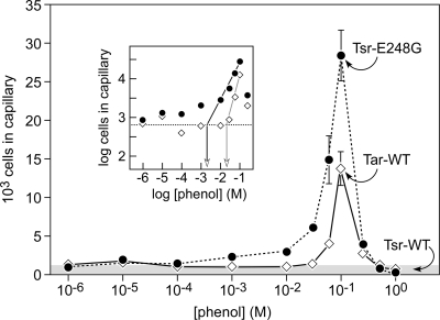
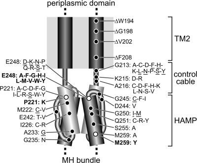
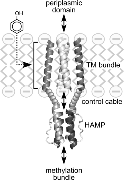
The four transmembrane chemoreceptors of Escherichia coli sense phenol as either an attractant (Tar) or a repellent (Tap, Trg, and Tsr). In this study, we investigated the Tar determinants that mediate its attractant response to phenol and the Tsr determinants that mediate its repellent response to phenol. Tar molecules with lesions in the aspartate-binding pocket of the periplasmic domain, with a foreign periplasmic domain (from Tsr or from several Pseudomonas chemoreceptors), or lacking nearly the entire periplasmic domain still mediated attractant responses to phenol. Similarly, Tar molecules with the cytoplasmic methylation and kinase control domains of Tsr still sensed phenol as an attractant. Additional hybrid receptors with signaling elements from both Tar and Tsr indicated that the transmembrane (TM) helices and HAMP domain determined the sign of the phenol-sensing response. Several amino acid replacements in the HAMP domain of Tsr, particularly attractant-mimic signaling lesions at residue E248, converted Tsr to an attractant sensor of phenol. These findings suggest that phenol may elicit chemotactic responses by diffusing into the cytoplasmic membrane and perturbing the structural stability or position of the TM bundle helices, in conjunction with structural input from the HAMP domain. We conclude that behavioral responses to phenol, and perhaps to temperature, cytoplasmic pH, and glycerol, as well, occur through a general sensing mechanism in chemoreceptors that detects changes in the structural stability or dynamic behavior of a receptor signaling element. The structurally sensitive target for phenol is probably the TM bundle, but other behaviors could target other receptor elements.


Figures

Fig. 1.
Fig. 1.
Functional architecture of Tar and Tsr receptors. These molecules function as homodimers of ∼550-residue subunits. The cylinders represent segments with α-helical secondary structure, drawn approximately to scale. The circles represent the four principal methylation sites in each subunit. The black circles denote glutamine residues that must first be deamidated by CheB before methylation; the white circles denote glutamic acid residues that undergo reversible CheR-mediated methylation. The methylation sites and kinase (CheA) control region operate as dynamic 4-helix bundles comprised of two interacting, antiparallel coiled coils.
Fig. 2.
Fig. 2.
Tar- and Tsr-mediated motility responses on soft-agar phenol gradient plates. UU1250 cells containing pHP1 (Tar-WT), pHP1 derivatives (Tar variants Y149F, R73F, and R73W), and pRR53 (Tsr-WT) were inoculated by toothpick stab from fresh transformants to a minimal soft agar plate containing a central agar plug of 100 mM phenol. The plate contained 100 μM IPTG to induce expression of the receptors and 50 μg/ml ampicillin to select for plasmid maintenance and was incubated for 20 h at 30°C. The response index values for these colonies are (reading clockwise from the top): 0.59, 0.47, 0.58, 0.62, and 0.63.
Fig. 3.
Fig. 3.
Construction and properties of Tar hybrids with periplasmic domains from P. putida MCPs. (A) Hybrid construction scheme. The tar gene in plasmid pHP1 encodes a stretch of three valine residues near the cytoplasmic end of TM2. Those codons were converted to alanine codons to create a unique NotI restriction site. Coding regions for foreign periplasmic domains were spliced to tar at this new site. (B and C) Colony morphologies of UU1250 cells expressing different Tar hybrids on semisolid-agar phenol gradient plates containing 100 μM (B) and 500 μM (C) IPTG. The plates also contained 50 μg/ml ampicillin and were incubated for 16 h at 30°C.
Fig. 4.
Fig. 4.
Selection and properties of Tar° receptor mutants on soft-agar phenol gradient plates. (A) Summary of Tar° amino acid changes obtained by three cycles of selection for phenol attractant responses. The phenol response index values for each Tar° mutant derivative are shown in parentheses. (B) Colony morphologies of UU1250 cells expressing various Tar° derivatives. See the legend to Fig. 2 for additional details.
Fig. 5.
Fig. 5.
Phenol responses mediated by Tar-Tsr hybrid receptors. See Materials and Methods for construction details. The three middle letters of the hybrid designation indicate the origin (Tar or Tsr) of the periplasmic domain and TM bundle, the HAMP domain, and the methylation bundle and kinase control tip. The RI values (bottom) represent the averages and standard deviations for three independent experiments.
Fig. 6.
Fig. 6.
Capillary assay of phenol attractant responses mediated by wild-type (WT) Tar and Tsr-E248G receptors. Plasmids expressing Tar-WT (pHP1), Tsr-WT (pRR53), and Tsr-E248G (pRR53 derivative) were transferred to strain UU1250 and tested for phenol chemotaxis as detailed in Materials and Methods. The data points represent averages of at least two independent experiments; the error bars indicate the standard deviations. Where error bars are not apparent, their size is less than that of the plot symbols. The shaded area represents the control accumulations due to background motility alone. The Tsr-WT response did not exceed this background range, and the individual data points are not shown. The inset contains a log-log plot for determining response threshold concentrations by back-extrapolating the rising portion of the curves to intercept the background motility level.
Fig. 7.
Fig. 7.
Summary of mutant Tsr receptors screened for attractant responses to phenol in soft-agar gradient plates. The HAMP structural elements are as follows: AS1 helices (light-gray cylinders), AS2 helices (dark-gray cylinders), and connector segments (thin gray tubes). Amino acid replacements in the TM2-control cable-HAMP segments of Tsr, expressed from pRR53 or pPA114 derivatives, were tested in strain UU1250. The underlined replacements retain a repellent response to phenol; the replacements in boldface produced an attractant response to phenol; all other mutant receptors produced neutral responses to phenol. Replacements at three HAMP residues (white circles) gave rise to attractant responses. With the exception of some E248 lesions, all of these mutant receptors still mediate an attractant response to serine (24, 60).
Fig. 8.
Fig. 8.
Model for phenol sensing by Tar and Tsr. Bidirectional conformational interactions (double-headed arrows) between contiguous signaling elements transmit stimulus information to the kinase control tip of the receptor molecule. The shading convention for HAMP structural elements follows that of Fig. 7. Phenol might diffuse into the cytoplasmic membrane to influence the equilibrium position or structural stability of the transmembrane helices, thereby modulating the packing stability of the HAMP bundle.

Similar articles

Cited by

References

    1. Adler J. 1973. A method for measuring chemotaxis and use of the method to determine optimum conditions for chemotaxis by Escherichia coli. J. Gen. Microbiol. 74:77–91 - PubMed
    1. Airola M., Watts K. J., Crane B. R. 2010. Structure of concatenated HAMP domains provides a mechanism for signal transduction. Structure 18:436–448 - PMC - PubMed
    1. Alexander R. P., Zhulin I. B. 2007. Evolutionary genomics reveals conserved structural determinants of signaling and adaptation in microbial chemoreceptors. Proc. Natl. Acad. Sci. U. S. A. 104:2885–2890 - PMC - PubMed
    1. Ames P., Parkinson J. S. 1994. Constitutively signaling fragments of Tsr, the Escherichia coli serine chemoreceptor. J. Bacteriol. 176:6340–6348 - PMC - PubMed
    1. Ames P., Studdert C. A., Reiser R. H., Parkinson J. S. 2002. Collaborative signaling by mixed chemoreceptor teams in Escherichia coli. Proc. Natl. Acad. Sci. U. S. A. 99:7060–7065 - PMC - PubMed

Publication types

MeSH terms