. 2011 Dec;193(23):6724-32.
doi: 10.1128/JB.06042-11. Epub 2011 Sep 30.

Probing the Borrelia burgdorferi surface lipoprotein secretion pathway using a conditionally folding protein domain

Affiliations

Probing the Borrelia burgdorferi surface lipoprotein secretion pathway using a conditionally folding protein domain

Shiyong Chen et al. J Bacteriol. 2011 Dec.

Abstract

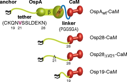
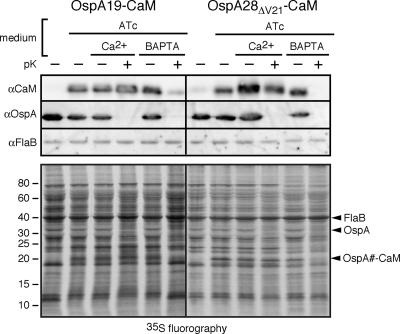
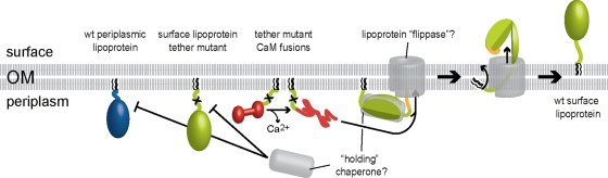
Surface lipoproteins of Borrelia spirochetes are important virulence determinants in the transmission and pathogenesis of Lyme disease and relapsing fever. To further define the conformational secretion requirements and to identify potential lipoprotein translocation intermediates associated with the bacterial outer membrane (OM), we generated constructs in which Borrelia burgdorferi outer surface lipoprotein A (OspA) was fused to calmodulin (CaM), a conserved eukaryotic protein undergoing calcium-dependent folding. Protein localization assays showed that constructs in which CaM was fused to full-length wild-type (wt) OspA or to an intact OspA N-terminal "tether" peptide retained their competence for OM translocation even in the presence of calcium. In contrast, constructs in which CaM was fused to truncated or mutant OspA N-terminal tether peptides were targeted to the periplasmic leaflet of the OM in the presence of calcium but could be flipped to the bacterial surface upon calcium chelation. This indicated that in the absence of an intact tether peptide, unfolding of the CaM moiety was required in order to facilitate OM traversal. Together, these data further support a periplasmic tether peptide-mediated mechanism to prevent premature folding of B. burgdorferi surface lipoproteins. The specific shift in the OM topology of sequence-identical lipopeptides due to a single-variable change in environmental conditions also indicates that surface-bound Borrelia lipoproteins can localize transiently to the periplasmic leaflet of the OM.


Figures

Fig. 1.
Fig. 1.
OspA-CaM fusion constructs. Cartoons of OspA-CaM fusion constructs generated and used in this study show triacyl anchor lipids in black, OspA peptides in green (N-terminal tether and β-strands) and orange (C-terminal α-helix), linker peptides in blue, and CaM peptides in red. Tether and linker peptide sequences are given in one-letter amino acid code. Mutant nomenclature follows that of earlier publications (50, 51). Briefly, modified residues are numbered according to their prolipoprotein positions; numbers in lipoprotein tether-CaM fusion constructs indicate the C-terminal tether residue present in the fusion.
Fig. 2.
Fig. 2.
Growth of B. burgdorferi in serum- and calcium-free minimal medium. B. burgdorferi was cultured in complete BSK-II medium and in various modifications of serum-free (SF) medium. Serum-free incomplete or basal BSK medium (BSK-SF) (60) was supplemented with lipids (+ lip), treated with Chelex-100 (CH), or supplemented with essential cations (+Mg/Mn/Zn). Cells were counted under phase-contrast microscopy using a Petroff-Hausser counting chamber. Representative growth curves are shown. Note that the 1-day lag phase for cells grown in BSK-SFcc persisted even when bacteria had been passed in BSK-SFcc previously (data not shown).
Fig. 3.
Fig. 3.
Chelation-dependent surface exposure of OspA-CaM fusion proteins at steady state. (A) Accessibility of full-length OspA-CaM or OspA tether-CaM fusion proteins to proteinase K. Cells expressing the fusion constructs were grown in BSK-SFcc containing either Ca2+ or BAPTA. Lipoprotein surface exposure was assessed by incubating intact cells with proteinase K (pK+) or a control buffer (pK−), followed by Western immunoblotting with antibodies against CaM, OspC (used as an OspA-independent surface control, due to the coexpression of endogenous OspAwt and OspAwt-CaM), and FlaB (periplasmic control) (αCaM, αOspC, and αFlaB, respectively). (B) Total-protein fractionation by Triton X-114 of B. burgdorferi cells expressing the OspA28ΔV21-CaM or OspA19-CaM fusion protein in the presence of Ca2+. A Coomassie-stained SDS-PAGE gel (Coom) and an immunoblot with αCaM are shown. D, detergent phase; Aq, aqueous phase. The positions of OspA and OspB are indicated on the right. Asterisks indicate the weakly Coomassie stained OspA-CaM fusion bands present in the respective detergent fractions. (C) Membrane fractionation immunoblots of the subsurface OspAΔV21-CaM and OspA19-CaM fusion proteins expressed by B. burgdorferi in the presence of Ca2+. OppAIV served as an inner membrane control, and OspA served as an OM control. PC, protoplasmic cylinder fraction; OMV, outer membrane vesicle fraction enriched for OM proteins. Note that the PC fraction also contains OM proteins, because OMVs were only partially separated from protoplasmic cylinders by treatment of Borrelia cells with a hypotonic citrate buffer; therefore, the PC fraction is similar to a whole-cell protein preparation (54). (D) Accessibility to proteinase K of partial or mutant OspA tether-CaM fusion proteins expressed by B. burgdorferi. The experimental procedures and labels described for panel A were used, except that OspA was used as the standard surface control. (E) Accessibility to proteinase K of previously described periplasmic OspA-mRFP fusion proteins (50, 51) under calcium-containing (Ca2+) or chelating (BAPTA) conditions. The experimental procedures and labels were the same as those for panel D.
Fig. 4.
Fig. 4.
Assessment of the folding states of the OspA-CaM fusion proteins by hydrophobic interaction chromatography (HIC) and immunoprecipitation (IP). Western blot analysis was performed on OspA-CaM fusion proteins purified by HIC or IP from whole-cell lysates of recombinant B. burgdorferi cultured in the presence of Ca2+ or BAPTA. Phenyl Sepharose was used to enrich for the folded CaM fusion proteins by HIC. For IP experiments, CaM fusion proteins were immunoprecipitated with a Ca2+-CaM complex-specific monoclonal antibody. For both the HIC and IP approaches, equal ratios of whole-cell lysates (input) were loaded for comparison. OspA-CaM fusion proteins in both the input and eluted pulldown samples were detected by immunoblotting with a non-conformation-specific antibody against CaM.
Fig. 5.
Fig. 5.
Chelation-dependent surface exposure of radiolabeled, conditionally expressed OspA-CaM fusion proteins. (Top) Western immunoblot analysis; (bottom) fluorography analysis. Prior to analysis, B. burgdorferi cells were cultured to early-log phase in BSK-SFcc containing Ca2+. Radiolabeling with [35S]Cys-Met amino acids and anhydrotetracycline (ATc)-mediated induction of Post promoter-driven expression were initiated simultaneously in RPMI 1640 medium. Subsequently, cells were incubated in BSK-SFcc containing either Ca2+ or BAPTA, followed by in situ surface proteolysis with proteinase K (+pK). Total fusion proteins expressed upon ATc induction were detected by Western immunoblotting with antibodies against CaM, OspA (surface control), and FlaB (subsurface control), while fluorography detected proteins expressed during the pulse. The proteins bands detected by Western immunoblotting are indicated on the right.
Fig. 6.
Fig. 6.
Proposed molecular events at the B. burgdorferi OM during surface lipoprotein secretion. Where possible, the color scheme follows that of Fig. 1. Hypothetical components are drawn in gray. Tether mutants of surface lipoproteins such as OspA (light green, with an X marking the tether mutation) fold prematurely in the periplasm, and their secretion through the OM is thereby prevented (17, 18). Periplasmic folding or interactions with periplasmic envelope components may be responsible for subsurface retention of wt periplasmic lipoproteins such as Lp6.6 (blue). Translocation of periplasmic mutant OspA tether-CaM fusion proteins (red) is dependent on chelation-mediated structural changes (this study). Intact surface lipoprotein tether peptides prevent premature folding of proteins—and thereby allow for OM translocation—via interaction with a hypothetical “holding” chaperone (gray). As shown for wt OspA (but also valid for the wt OspA-CaM fusion protein), translocation can initiate at the lipoprotein's C terminus if an unfolded peptide is provided (; also this study). The peptide portion travels via the hydrophilic lumen of a hypothetical OM lipoprotein “flippase” pore (gray), while the lipid anchor (black) flips from the periplasmic leaflet to the surface leaflet of the OM bilayer; this ultimate anchor topology was recently shown for OspA and B. turicatae Vsp1 (17). In the absence of ATP, the directionality of the process is likely driven by the folding and assembly of the protein on the bacterial surface. This figure updates the Borrelia envelope biogenesis model shown in the work of Bergström and Zückert (7).

Similar articles

Cited by

References

    1. Adams T. M., Wentzel A., Kolmar H. 2005. Intimin-mediated export of passenger proteins requires maintenance of a translocation-competent conformation. J. Bacteriol. 187:522–533 - PMC - PubMed
    1. Babb K., McAlister J. D., Miller J. C., Stevenson B. 2004. Molecular characterization of Borrelia burgdorferi erp promoter/operator elements. J. Bacteriol. 186:2745–2756 - PMC - PubMed
    1. Barbour A. G. 1984. Isolation and cultivation of Lyme disease spirochetes. Yale J. Biol. Med. 57:521–525 - PMC - PubMed
    1. Barbour A. G., Guo B. P. 2010. Pathogenesis of relapsing fever, p. 333–357 In Samuels D. S., Radolf J. D. (ed.), Borrelia: molecular biology, host interaction, and pathogenesis. Caister Academic Press, Norfolk, United Kingdom
    1. Barbour A. G., Hayes S. F., Heiland R. A., Schrumpf M. E., Tessier S. L. 1986. A Borrelia-specific monoclonal antibody binds to a flagellar epitope. Infect. Immun. 52:549–554 - PMC - PubMed

Publication types

MeSH terms

LinkOut - more resources