. 2011 Dec;193(23):6683-700.
doi: 10.1128/JB.00210-11. Epub 2011 Sep 30.

Impact of the N-terminal secretor domain on YopD translocator function in Yersinia pseudotuberculosis type III secretion

Affiliations

Impact of the N-terminal secretor domain on YopD translocator function in Yersinia pseudotuberculosis type III secretion

Ayad A A Amer et al. J Bacteriol. 2011 Dec.

Abstract

Type III secretion systems (T3SSs) secrete needle components, pore-forming translocators, and the translocated effectors. In part, effector recognition by a T3SS involves their N-terminal amino acids and their 5' mRNA. To investigate whether similar molecular constraints influence translocator secretion, we scrutinized this region within YopD from Yersinia pseudotuberculosis. Mutations in the 5' end of yopD that resulted in specific disruption of the mRNA sequence did not affect YopD secretion. On the other hand, a few mutations affecting the protein sequence reduced secretion. Translational reporter fusions identified the first five codons as a minimal N-terminal secretion signal and also indicated that the YopD N terminus might be important for yopD translation control. Hybrid proteins in which the N terminus of YopD was exchanged with the equivalent region of the YopE effector or the YopB translocator were also constructed. While the in vitro secretion profile was unaltered, these modified bacteria were all compromised with respect to T3SS activity in the presence of immune cells. Thus, the YopD N terminus does harbor a secretion signal that may also incorporate mechanisms of yopD translation control. This signal tolerates a high degree of variation while still maintaining secretion competence suggestive of inherent structural peculiarities that make it distinct from secretion signals of other T3SS substrates.


Figures

Fig. 1.
Fig. 1.
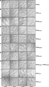
Intrabacterial stability of preformed pools of various YopD mutant proteins. Bacteria were first cultured for 1 h in noninducing (i.e., including 2.5 mM CaCl2) BHI broth at 37°C. The protein synthesis inhibitor chloramphenicol (50 μg/ml) was added at time point 0 min. Samples were then collected at subsequent time points. Protein levels associated with pelleted bacteria were detected by Western blotting using polyclonal anti-YopD antiserum. Panels: Parental (YopDwt), YPIII/pIB102; YopDFrame+1, YPIII/pIB62502; YopDFrame−1, YPIII/pIB62503; YopDScramble, YPIII/pIB62504; YopDI3,5N, YPIII/pIB62559; YopDFrame−1, I3,4N, YPIII/pIB62564; YopDhigh(x2), YPIII/pIB62537; YopDlow(x2), YPIII/pIB62538; YopDΔ4-20, YPIII/pIB625; YopDΔ5-19, YPIII/pIB62524; YopDΔ6-19, YPIII/pIB62525; YopDΔ7-19, YPIII/pIB62526; YopDΔ8-19, YPIII/pIB62527; YopDΔ9-19, YPIII/pIB62528; YopDΔ10-19, YPIII/pIB62529; YopDΔ11-19, YPIII/pIB62530. The asterisk (*) highlights YopDlow(x2) as the only visibly unstable variant.
Fig. 2.
Fig. 2.
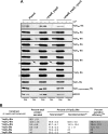
A YopD frameshift mutant altering the amino acid sequence of the N-terminal secretion signal specifically affects secretion. (A) Overnight cultures were subcultured into BHI broth either containing (+) or lacking (−) calcium and grown at 26°C for 1 h and then at 37°C for 3 h. Protein samples were fractionated by 12% SDS-PAGE and then transferred onto a membrane support for immune detection. Expression fractions (upper panels) represent total protein associated with bacteria and also released into the culture supernatant. Secretion fractions (lower panels) signify protein freely released into the culture supernatant. Lanes: Parental (YopDwt), YPIII/pIB102; ΔyscU, ΔlcrQ (YopDwt), YPIII/pIB75-26; YopDFrame+1, YPIII/pIB62502; YopDFrame−1, YPIII/pIB62503; YopDScramble, YPIII/pIB62504. Molecular mass values shown in parentheses were deduced from primary amino acid sequences. (B) At least three independent experiments were used to quantify relative YopD and YopE synthesis and secretion values ± standard errors of the means using Quantity One software, version 4.52 (Bio-Rad). Percent total secretion (lighter gray) values reflect the ratio (expressed as a percentage) of secreted protein to the amount synthesized in each respective strain. Percent secretion efficiency values reflect the extent of Yop secretion occurring in mutant bacteria relative to what occurs in parental bacteria (darker gray). It is calculated from the ratio of secreted protein seen with the parent (y) to the total protein seen with the parent (x). The median secretion efficiency of YopDFrame+1 was significantly lower (**, P = 0.079, two-tailed nonparametric Mann-Whitney U test; P < 0.05) than that of native YopD; it was also lower than the secretion efficiencies of both YopDFrame−1 and YopDScramble. In contrast, YopE secretion efficiency in the same strains was not statistically different from that of the parent bacteria (ns, not significant; P = 0.1143).
Fig. 3.
Fig. 3.
N-terminal isoleucine residues contribute to secretion of native YopD. (A) Overnight cultures were subcultured into BHI broth either containing (+) or lacking (−) calcium and grown at 26°C for 1 h and then at 37°C for 3 h. Protein samples were fractionated by 12% SDS-PAGE and then transferred onto a membrane support for immune detection. Expression fractions (upper panels) represent total protein associated with bacteria and also released into the culture supernatant. Secretion fractions (lower panels) signify protein freely released into the culture supernatant. Lanes: Parental (YopDwt), YPIII/pIB102; ΔyscU, ΔlcrQ (YopDwt), YPIII/pIB75-26; YopDFrame−1, YPIII/pIB62503; YopDI3,5N, YPIII/pIB62559; YopDFrame−1, I3,4N, YPIII/pIB62564. Molecular mass values shown in parentheses were deduced from primary amino acid sequences. (B) The quantification of YopD and YopE secretion efficiency ± standard error of the mean was calculated from a minimum of three independent experiments using Quantity One software, version 4.52 (Bio-Rad). See the legend to Fig. 2 for explanations of “Percent total secretion” (lighter gray) and “Percent secretion efficiency” (darker gray). Compared to native YopD, the median secretion efficiency of YopDI3,5N was significantly lower than that of YopDFrame−1, I3,4N (*, P = 0.0317, two-tailed nonparametric Mann-Whitney U test; P < 0.05) or YopDFrame−1 (**, P = 0.079). Conversely, the observed secretion deficiency of YopDFrame−1, I3,4N was not considered to be statistically different from that of YopDFrame−1 or native YopD (ns, not significant; P = 0.1508).
Fig. 4.
Fig. 4.
Amphipathicity of the N terminus is not an obvious mediator of YopD secretion. Overnight cultures were subcultured into BHI broth either containing (+) or lacking (−) calcium and grown at 26°C for 1 h and then at 37°C for 3 h. Protein samples were fractionated by 12% SDS-PAGE and then transferred onto a membrane support for immune detection. Expression fractions (upper panels) represent total protein associated with bacteria and also released into the culture supernatant. Secretion fractions (lower panels) signify protein freely released into the culture supernatant. Lanes: Parental (YopDwt), YPIII/pIB102; ΔyscU, ΔlcrQ (YopDwt), YPIII/pIB75-26; YopDhigh, YPIII/pIB62514; YopDlow YPIII/pIB62515; YopDhigh(x2), YPIII/pIB62537; YopDlow(x2), YPIII/pIB62538. Molecular mass values shown in parentheses were deduced from primary amino acid sequences.
Fig. 5.
Fig. 5.
RT-PCR of mRNA isolated from Y. pseudotuberculosis. RNA was isolated from log-phase bacterial cultures grown at 37°C in BHI medium with (+) and without (−) Ca2+. Samples were subjected to RT-PCR using primers specific for rpoA (used as a loading control) and the T3SS genes yopD and yopE. Lanes: Parental (YopDwt), YPIII/pIB102; YopDΔ4-20, YPIII/pIB625; YopDhigh, YPIII/pIB62514; YopDlow, YPIII/pIB62515; YopDhigh(x2), YPIII/pIB62537; YopDlow(x2), YPIII/pIB62538. Images were acquired using a Fluor-S MultiImager (Bio-Rad). The images were then inverted using Quantity One quantitation software, version 4.52 (Bio-Rad). Numbers in parentheses indicate the approximate size of the amplified DNA fragment in base pairs.
Fig. 6.
Fig. 6.
Low-calcium-response growth phenotypes of Y. pseudotuberculosis producing various YopD variants. Bacteria were grown at 37°C in nonsupplemented TMH medium (minus Ca2+) or in TMH medium supplemented with 2.5 mM CaCl2 (plus Ca2+). Three different growth phenotypes were detected: calcium-dependent (CD) growth (A to D); CD-like (i.e., moderately calcium-dependent) growth (E); and TS growth (i.e., bacteria were sensitive to elevated temperature regardless of the presence of calcium) (F). Panels: A, parental (YPIII/pIB102); B, YopDhigh (YPIII/pIB62514); C, YopDlow (YPIII/pIB62515); D, YopDhigh(x2) (YPIII/pIB62537); E, YopDlow(x2) (YPIII/pIB62538); F, ΔyscU, ΔlcrQ (YPIII/pIB75-26). The arrow highlights the subtle growth restriction of Y. pseudotuberculosis producing YopDlow(x2).
Fig. 7.
Fig. 7.
Expression and secretion of YopD containing various in-frame deletions of the N-terminal secretion signal. (A) Overnight cultures were subcultured into BHI broth either containing (+) or lacking (−) calcium and grown at 26°C for 1 h and then at 37°C for 3 h. Protein samples were fractionated by 12% SDS-PAGE and then transferred onto a membrane support for immune detection. Expression fractions (upper panels) represent total protein associated with bacteria and also released into the culture supernatant. Secretion fractions (lower panels) signify protein freely released into the culture supernatant. Lanes: Parental (YopDwt), YPIII/pIB102; ΔyscU, ΔlcrQ (YopDwt), YPIII/pIB75-26; YopDΔ4-20, YPIII/pIB625; YopDΔ5-19, YPIII/pIB62524; YopDΔ6-19, YPIII/pIB62525; YopDΔ7-19, YPIII/pIB62526; YopDΔ8-19, YPIII/pIB62527; YopDΔ9-19, YPIII/pIB62528; YopDΔ10-19, YPIII/pIB62529; YopDΔ11-19, YPIII/pIB62530; YopDΔ12-19, YPIII/pIB62531; YopDΔ13-19, YPIII/pIB62532; YopDΔ14-19, YPIII/pIB62533; YopDΔ15-19, YPIII/pIB62534; YopDΔ16-19, YPIII/pIB62535; YopDΔ17-19, YPIII/pIB62536. Molecular mass values shown in parentheses were deduced from primary amino acid sequences. (B) For a selection of mutants, YopD and YopE synthesis and secretion efficiency were quantified from the results of at least three independent experiments using Quantity One software, version 4.52 (Bio-Rad). Definitions of terms are provided in the legend to Fig. 2.
Fig. 8.
Fig. 8.
Defects in YopD secretion impair Yop intoxication of infected eukaryotic cells. Strains were allowed to infect a monolayer of growing HeLa cells. At 20, 40, 60, and 120 min postinfection, samples were subjected to fixation and the effect of the bacteria on the HeLa cells was recorded by phase-contrast microscopy. Translocation of the YopE cytotoxin, a GTPase-activating protein, causes a distinct change in cell shape, from oblong to rounded (cytotoxicity), of affected HeLa cells (see panels A, D, E, and G). HeLa cells not intoxicated with YopE show normal uninfected cell morphology (see panels B, F, and H). Some YopD variants reduced the efficiency of YopE translocation, which delayed the onset of cytotoxicity (see panels C and I). Panels: A, YopDwt (parent) (YPIII/pIB102); B, YopDΔ4-20 (YPIII/pIB625); C, YopDFrame+1 (YPIII/pIB62502); D, YopDFrame−1 (YPIII/pIB62503); E, YopDI3,5N (YPIII/pIB62559); F, YopDhigh(x2) (YPIII/pIB62537); G, YopDlow(x2) (YPIII/pIB62538); H, YopDE-Nterm (YPIII/pIB62501-577); I, YopDΔ5-19, (YPIII/pIB62524); J, YopDΔ6-19 (YPIII/pIB62525).
Fig. 9.
Fig. 9.
Expression and secretion of YopD and YopE chimeras with a reciprocally exchanged N-terminal secretion signal. Overnight cultures were subcultured into BHI broth either containing (+) or lacking (−) calcium and grown at 26°C for 1 h and then at 37°C for 3 h. Protein samples were fractionated by 12% SDS-PAGE and then transferred onto a membrane support for immune detection. Expression fractions (upper panels) represent total protein associated with bacteria and also released into the culture supernatant. Secretion fractions (lower panels) signify protein freely released into the culture supernatant. Lanes: Parental (YopDwt), YPIII/pIB102; ΔyscU, ΔlcrQ (YopDwt), YPIII/pIB75-26; YopDB-Nterm, YPIII/pIB62577; YopEH-Nterm, YPIII/pIB578; YopDE-Nterm, YPIII/pIB62501; YopED-Nterm, YPIII/pIB577; YopDE-Nterm, YopED-Nterm, YPIII/pIB62501-577. Molecular mass values shown in parentheses were deduced from primary amino acid sequences.
Fig. 10.
Fig. 10.
Swapping Yop substrate N-terminal secretion signals compromises T3SS activity during contact with eukaryotic cells. (A) Yersinia bacteria were used to infect monolayers of macrophage J774-1 cells. Those bacteria with a compromised T3SS were more rapidly phagocytosed and killed by the antibacterial activities of the cell. Bacterial viability was tested at the time of inoculation and at 2 h, 4 h, and 6 h postinfection. The data represent numbers of CFU per milliliter expressed as the ratio of mutant to parent bacteria. Each symbol represents one independent experiment; each error bar represents ± the standard error of the mean, which in turn is indicated as a short horizontal line. Bacteria producing either or both of YopDE-Nterm and YopED-Nterm were always less viable, suggesting a compromised order of YopD and YopE secretion. Bacteria either lacking YopE or failing to secrete YopD were even less viable. Strains: YopDΔ4-20, YPIII/pIB625; ΔyopE, YPIII/pIB522; YopDΔ4-20, ΔyopE, YPIII/pIB522-625; YopDhigh(x2), YPIII/pIB62537; YopDB-Nterm, YPIII/pIB62577; YopEH-Nterm, YPIII/pIB578; YopDE-Nterm, YPIII/pIB62501; YopED-Nterm, YPIII/pIB577; YopDE-Nterm, YopED-Nterm, YPIII/pIB62501-577. The asterisks (*, **, or ***) indicate that the chimeric variants were statistically less viable (two-tailed parametric Mann-Whitney U test, P < 0.05) than parent bacteria or bacteria producing YopDhigh(x2) after 4 h and 6 h of incubation in the presence of J774-1 cell monolayers. On the other hand, the viability of bacteria producing YopEH-Nterm was comparable to that of the control bacteria. ns, not statistically significant.
Fig. 11.
Fig. 11.
Formation of the T3S of a β-lactamase reporter by appending the YopD N terminus. (A) Derivatives of pMMB208 contained yopD::bla translational fusions placed under the control of an IPTG-inducible promoter. The constructs were maintained in parental (YPIII/pIB102), ΔyopB yopD (YPIII/pIB619), and ΔyopB yopD ΔyscU (YPIII/pIB619-75) bacteria. Overnight cultures of these bacteria were subcultured into BHI broth either containing (+) or lacking (−) calcium and grown at 26°C for 1 h and then at 37°C for 3 h. Expression fractions (Exp) representing total protein associated with bacteria and also released into the culture supernatant and secretion fractions (Sec) representing protein freely released into the culture supernatant were fractionated by 12% SDS-PAGE and then transferred onto a membrane support for immune detection using rabbit polyclonal antisera recognizing β-lactamase. Panels: YopD306-Bla, pAA113; YopD25-Bla, pAA020 and pAA020; YopD20-Bla, pAA022; YopD15-Bla, pAA025; YopD10-Bla, pAA026; YopD5-Bla, pAA027; YopD3-Bla, pAA069; YopD1-Bla, pAA028 and pBR322 (containing native β-lactamase). The asterisks highlight overexposed panels. The overexposure enabled visualization of YopD5::Bla fusion secretion (indicated by arrows) but not visualization of secretion of the YopD3-Bla fusion; codons 1 to 5 may therefore represent the minimal N-terminal YopD secretion signal. The synthesis and secretion of selected fusions were quantified according to the legend to Fig. 2. Limited by an acutely unstable full-length YopD306-Bla fusion, relative synthesis and secretion of the smaller truncated fusions were compared with synthesis and secretion of YopD20-Bla.

Similar articles

Cited by

References

    1. Aili M., Isaksson E. L., Hallberg B., Wolf-Watz H., Rosqvist R. 2006. Functional analysis of the YopE GTPase-activating protein (GAP) activity of Yersinia pseudotuberculosis. Cell Microbiol. 8:1020–1033 - PubMed
    1. Akeda Y., Galan J. E. 2005. Chaperone release and unfolding of substrates in type III secretion. Nature 437:911–915 - PubMed
    1. Akopyan K., et al. 2011. Translocation of surface-localized effectors in type III secretion. Proc. Natl. Acad. Sci. U. S. A. 108:1639–1644 - PMC - PubMed
    1. Anderson D. M., Ramamurthi K. S., Tam C., Schneewind O. 2002. YopD and LcrH regulate expression of Yersinia enterocolitica YopQ by a posttranscriptional mechanism and bind to yopQ RNA. J. Bacteriol. 184:1287–1295 - PMC - PubMed
    1. Andersson K., Magnusson K. E., Majeed M., Stendahl O., Fallman M. 1999. Yersinia pseudotuberculosis-induced calcium signaling in neutrophils is blocked by the virulence effector YopH. Infect. Immun. 67:2567–2574 - PMC - PubMed

Publication types

MeSH terms

Substances

LinkOut - more resources