. 2011 Dec;193(23):6443-51.
doi: 10.1128/JB.00425-11. Epub 2011 Sep 30.

Regulation of biofilm components in Salmonella enterica serovar Typhimurium by lytic transglycosylases involved in cell wall turnover

Affiliations

Regulation of biofilm components in Salmonella enterica serovar Typhimurium by lytic transglycosylases involved in cell wall turnover

Claudia Monteiro et al. J Bacteriol. 2011 Dec.

Abstract

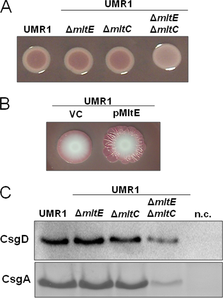
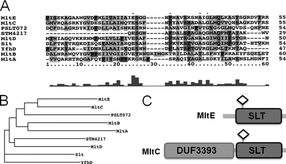
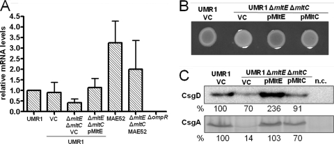
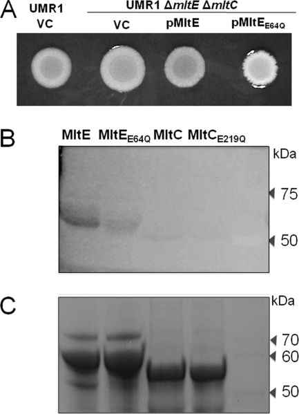
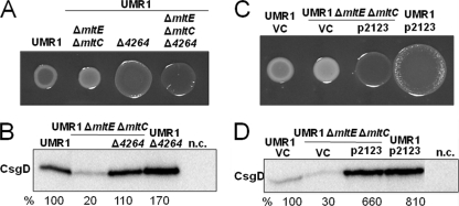
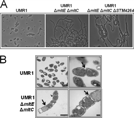
In Salmonella enterica serovar Typhimurium, a biofilm mode of growth known as the rdar morphotype is regulated by several networks which sense multiple environmental signals. The transcriptional regulator CsgD is the major target for these regulatory pathways. In this study, we show that two lytic transglycosylases of family I, MltE and MltC, in combination increase CsgD expression and rdar morphotype. MltE and MltC, which share a highly similar transglycosylase SLT domain, work redundantly to regulate CsgD at the transcriptional and posttranscriptional levels. The effect of MltE and MltC on CsgD levels was independent of the known regulatory pathways that sense cell envelope stress. These findings reveal, for the first time, a specific function of lytic transglycosylases in S. Typhimurium and suggest the existence of a new signaling pathway that links cell wall turnover to biofilm formation.


Figures

Fig. 1.
Fig. 1.
MltE in combination with MltC affects the expression of the rdar morphotype through CsgD. (A) Wild-type S. Typhimurium UMR1 and mutants of lytic transglycosylases MltE and MltC grown on Congo red agar plates at 28°C for 24 h. The mltE and mltC single mutants do not show any difference in rdar morphotype expression compared to that of wild-type UMR1, while the mltE mltC double mutant shows a visible downregulation of rdar morphotype expression. (B) MltE overexpression enhanced the rdar morphotype expression of UMR1 compared to that of the strain containing the vector control. Spot colonies were grown on Congo red agar plates at 28°C for 7 days. VC, vector control pBAD30. (C) CsgD and CsgA expression in S. Typhimurium UMR1 and its mltE and mltC mutants. Compared to wild-type UMR1, a significant reduction of CsgD and CsgA expression is observed in the mltE mltC double mutant. No change in CsgD and CsgA levels is detected in the mltE and mltC single mutants. The strains UMR1 ΔcsgD and UMR1 ΔcsgA were used as negative controls (n.c.) for CsgD and CsgA detection, respectively. Strains were grown on LB without NaCl agar at 28°C for 17 h.
Fig. 2.
Fig. 2.
Phylogenetic analysis of the transglycosylase SLT domains of the lytic transglycosylases of S. Typhimurium. (A) Amino acid alignment of transglycosylase SLT domains of S. Typhimurium. The catalytic E-S motif is a characteristic of SLT family I domains. (B) Phylogenetic tree indicating the relatedness of SLT domains of lytic transglycosylases. MltE and MltC lytic transglycosylase SLT domains are highly related. The tree was constructed using Dendroscope. (C) Domain architecture of MltE and MltC. MltE contains only a transglycosylase SLT domain. MltC is composed of an N-terminal DUF3393 domain and a C-terminal transglycosylase SLT domain. The diamond indicates the catalytic residues of MltE and MltC (E64 and E219, respectively).
Fig. 3.
Fig. 3.
Complementation of the S. Typhimurium UMR1 mltE mltC double mutant by MltE. (A) Expression of csgD in the wild-type S. Typhimurium UMR1 and the mltE mltC double mutant was determined by quantitative real-time RT-PCR. csgD mRNA levels decreased in the mltE mltC double mutant compared to those of wild-type UMR1. The expression of MltE from a plasmid restored csgD mRNA to wild-type UMR1 levels. VC, vector control pBad30. A decrease in csgD mRNA levels in the MAE52 mltE mltC double mutant also was observed. The strain UMR1 ΔompR was used as a negative control (45). Bacteria were grown on LB agar plates without salt at 28°C for 17 h. The experiment was performed with three biological replicates, using the mean expression values from four technical replicates. (B) Rdar morphotype expression in the double mutant UMR1 ΔmltE ΔmltC is complemented with a plasmid expressing MltE but not MltC. Strains were grown on Congo red agar at 28°C for 24 h. (C) CsgD and CsgA expression levels in the double mutant UMR1 ΔmltE ΔmltC complemented with pMltE and pMltC. The expression of MltE strongly increases the amount of CsgD and CsgA in the double mutant, while MltC expression leads to the minor upregulation of CsgD and CsgA. The relative protein levels compared to that of S. Typhimurium UMR1, which was set at 100%, are indicated. VC, vector control pBad30. The strains UMR1 ΔcsgD and UMR1 ΔcsgA were used as negative controls (n.c.) for CsgD and CsgA detection.
Fig. 4.
Fig. 4.
Catalytic activity of the transglycosylase SLT domain of MltE is required for rdar morphotype expression. (A) Morphotypes of UMR1 ΔmltE ΔmltC overexpressing pMltE or pMltEE64Q. MltE expressed from a plasmid leads to an upregulation of the morphotype compared to that of strain UMR1 ΔmltE ΔmltC, while the expression of MltEE64Q showed a white and mucoid colony. VC, vector control pBad30. Strains were grown on Congo red agar plates for 24 h at 28°C. (B) Cell wall hydrolytic activity of lytic transglycosylases MltE and MltC. SLT domains from MltE and MltC were partially purified and resolved on SDS-PAGE gel containing M. luteus cells as substrates for lytic transglycosylase activity. The lytic transglycosylase activity of MltE was higher than the activity of MltC. The MltE and MltC proteins containing mutations in the conserved glutamyl residues show only residual hydrolytic activity. (C) An SDS-PAGE gel stained with Coomassie blue as a loading control. Lanes: 1, MltE; 2, MltEE64Q; 3, MltC; 4, MltCE219Q; and 5, molecular mass standard (in kDa).
Fig. 5.
Fig. 5.
Effect of c-di-GMP signaling on the restoration of rdar morphotype expression in the UMR1 ΔmltE ΔmltC double mutant. (A) Deletion of STM4264, encoding a c-di-GMP-dependent phosphodiesterase, in the mltE mltC double mutant upregulated rdar morphotype expression compared to that of the mltE mltC double mutant. As previously shown (45), the STM4264 mutant displayed an upregulated rdar morphotype compared to that of wild-type UMR1. (B) CsgD expression in wild-type UMR1 and mutants. The mltE mltC STM4264 triple mutant shows an increase in CsgD levels compared to that of UMR1 ΔmltE ΔmltC, as CsgD expression increased in S. Typhimurium UMR1 compared to that of its STM4264 mutant. The relative protein levels compared to that of S. Typhimurium UMR1, which was set at 100%, are indicated. n.c., negative-control MAE50 ΔcogD. (C) UMR1 and the mltE mltC double mutant harboring p2123 showed upregulated rdar morphotype expression compared to that of the vector control (VC) pBAD30. (D) CsgD expression in UMR1 and UMR1 ΔmltE ΔmltC containing p2123. CsgD expression increased upon the expression of STM2123 in the wild-type UMR1 and the UMR1 ΔmltE ΔmltC mutant. n.c., negative-control MAE50 ΔcogD. For phenotype observation (A and C), strains were grown on Congo red agar at 28°C for 24 h. To detect CsgD expression (B and D), wild-type UMR1 and mutants were grown on LB without NaCl agar at 28°C for 17 h.
Fig. 6.
Fig. 6.
Morphological characterization of mltE and mltC mutants. (A) Light microscopy of the mltE mltC and mltE mltC STM4264 mutants. The mltE mltC double mutant and the mltE mltC STM4264 triple mutant display the formation of long chains when grown at 28°C in LB without salt medium. The wild-type UMR1 shows rod-shaped cells of standard length. Magnification, ×630. (B) Transmission electron microscopy (TEM) of the ΔmltE ΔmltC double mutant. Long chains of cells are observed in the ΔmltE ΔmltC double mutant due to an impairment of the cleavage of the PG septum (arrow). The wild-type UMR1 shows rod-shaped cells of 0.8 to 1 μm standard length without impairment in the cleavage of the PG septum. Bars indicate 1 (left panel) or 0.2 μm (right panel).

Similar articles

Cited by

References

    1. Ahn S. J., Burne R. A. 2006. The atlA operon of Streptococcus mutans: role in autolysin maturation and cell surface biogenesis. J. Bacteriol. 188:6877–6888 - PMC - PubMed
    1. Artola-Recolons C., et al. 2011. High-resolution crystal structure of MltE, an outer membrane-anchored endolytic peptidoglycan lytic transglycosylase from Escherichia coli. Biochemistry 50:2384–2386 - PMC - PubMed
    1. Bernadsky G., Beveridge T. J., Clarke A. J. 1994. Analysis of the sodium dodecyl sulfate-stable peptidoglycan autolysins of select gram-negative pathogens by using renaturing polyacrylamide gel electrophoresis. J. Bacteriol. 176:5225–5232 - PMC - PubMed
    1. Blackburn N. T., Clarke A. J. 2001. Identification of four families of peptidoglycan lytic transglycosylases. J. Mol. Evol. 52:78–84 - PubMed
    1. Boehm A., et al. 2010. Second messenger-mediated adjustment of bacterial swimming velocity. Cell 141:107–116 - PubMed

Publication types

MeSH terms

LinkOut - more resources