. 2011 Nov;138(21):4627-38.
doi: 10.1242/dev.071464. Epub 2011 Sep 28.

JAK/Stat signaling regulates heart precursor diversification in Drosophila

Affiliations

JAK/Stat signaling regulates heart precursor diversification in Drosophila

Aaron N Johnson et al. Development. 2011 Nov.

Abstract

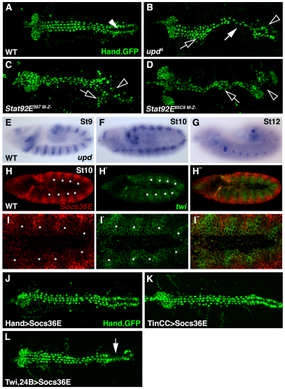
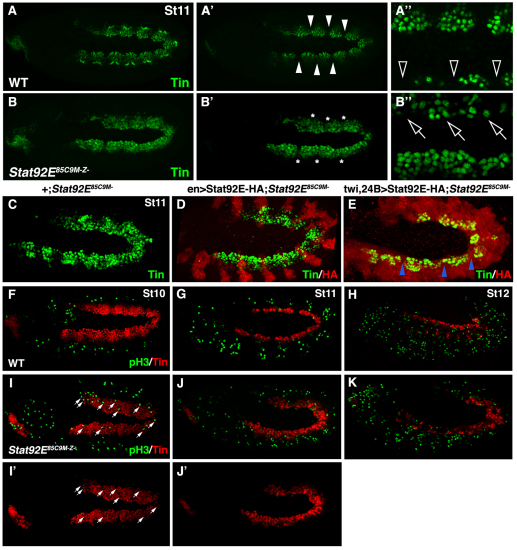
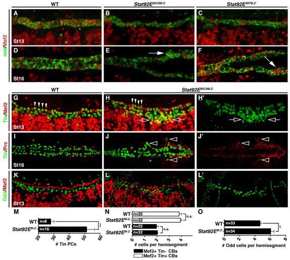
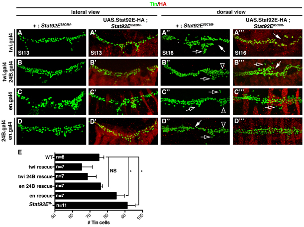
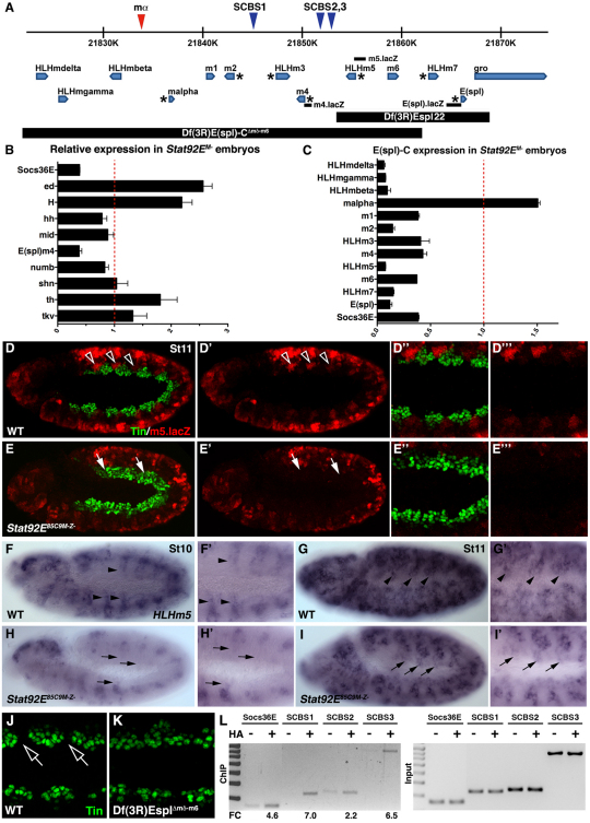
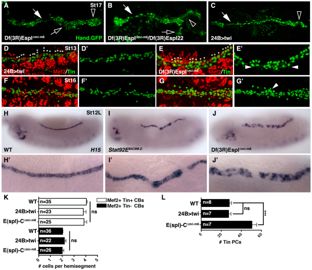
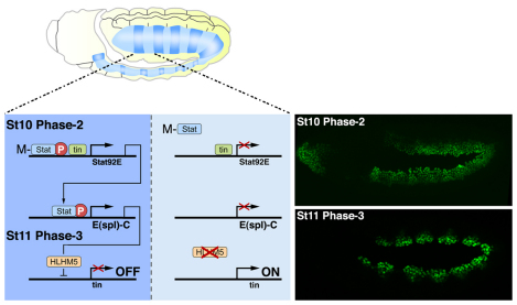
Intercellular signal transduction pathways regulate the NK-2 family of transcription factors in a conserved gene regulatory network that directs cardiogenesis in both flies and mammals. The Drosophila NK-2 protein Tinman (Tin) was recently shown to regulate Stat92E, the Janus kinase (JAK) and Signal transducer and activator of transcription (Stat) pathway effector, in the developing mesoderm. To understand whether the JAK/Stat pathway also regulates cardiogenesis, we performed a systematic characterization of JAK/Stat signaling during mesoderm development. Drosophila embryos with mutations in the JAK/Stat ligand upd or in Stat92E have non-functional hearts with luminal defects and inappropriate cell aggregations. Using strong Stat92E loss-of-function alleles, we show that the JAK/Stat pathway regulates tin expression prior to heart precursor cell diversification. tin expression can be subdivided into four phases and, in Stat92E mutant embryos, the broad phase 2 expression pattern in the dorsal mesoderm does not restrict to the constrained phase 3 pattern. These embryos also have an expanded pericardial cell domain. We show the E(spl)-C gene HLHm5 is expressed in a pattern complementary to tin during phase 3 and that this expression is JAK/Stat dependent. In addition, E(spl)-C mutant embryos phenocopy the cardiac defects of Stat92E embryos. Mechanistically, JAK/Stat signals activate E(spl)-C genes to restrict Tin expression and the subsequent expression of the T-box transcription factor H15 to direct heart precursor diversification. This study is the first to characterize a role for the JAK/Stat pathway during cardiogenesis and identifies an autoregulatory circuit in which tin limits its own expression domain.


Figures

Fig. 1.
Fig. 1.
The Jak/Stat pathway regulates cardiac morphology and is active in the dorsal mesoderm during cardiac precursor specification. (A) WT Hand.GFP embryos express nuclear-localized GFP throughout the dorsal vessel. The contractile ‘heart’ (arrowhead) positioned at the posterior of the dorsal vessel pumps hemolymph through the anterior aorta. (B) Zygotic upd4 embryos lose cardioblast cell-cell adhesion (white arrow) causing inappropriate cell aggregations (open arrow) and incomplete heart lumen formation (open arrowhead). (C,D) Embryos maternal and zygotic mutant for Stat92E397 (C) or Stat92E85C9 (D) phenocopy the dorsal vessel defects of upd embryos. (E-G) upd mRNA expression in WT embryos. upd expression in the ventral ectoderm is segmented during St9 (E). upd expression is restricted to the tracheal system and hindgut in St10 (F) and St12 (G) embryos. (H-I′) St10 WT embryo double labeled for Socs36E (red) and twi (green) mRNA. Socs36E and twi expression is complementary in the dorsal mesoderm. Regions of the mesoderm segment expressing high levels of twi (asterisks) express low levels of Socs36E. (J,K) St17 embryos expressing Hand.GFP. Expressing Socs36E in the heart after cardioblast precursor specification using Hand.gal4 (J) or TinCC.gal4 (K) does not affect heart morphology. (L) Expressing Socs36E in the heart prior to cardioblast precursor specification using Twi.gal4, 24B.gal4 induces mild cardioblast cell-cell adhesion defects. A-G show dorsal views of Hand.GFP expression in live St17 embryos. Embryos are oriented with anterior to the left.
Fig. 2.
Fig. 2.
JAK/Stat signaling restricts Tin expression in the dorsal mesoderm. (A-B′) Oblique views of St11 embryos labeled for Tin. The confocal micrographs in A and B include the entire Tin expression domain whereas the micrographs in A′, A′, B′ and B′ include only the dorsal mesoderm. (A,A′) Tin expression is restricted to the cardiac and visceral muscle progenitors in WT embryos. Note that Tin is not expressed in a subset of dorsal mesoderm cells (arrowheads). (B,B′) In Stat92EM–Z– St11 embryos, Tin expression does not restrict (asterisks). (A′,B′) High magnification micrographs show that the embryos are slightly rotated (oblique) giving a differential projection of the dorsal mesoderm. (A′) This scan captures more medially positioned Tin+ cells in the top of the image compared with the bottom. Note the large gaps between Tin+ cells in the dorsal-most mesoderm of WT embryos (open arrowheads). (B′) This scan captures more medially positioned Tin+ cells in the bottom of the image compared with the top. Tin-expressing cells can be identified throughout the dorsal-most mesoderm of Stat92EM–Z– embryos (open arrows). (C-E) St11 embryos labeled for HA and Tin. St11 Stat92EM– embryos (C) and Stat92EM– embryos that express Stat92E-HA in the ectoderm (D) show inappropriate Tin expression. Stat92EM– embryos that express Stat92E-HA in the mesoderm show restricted Tin expression (E; blue arrowheads). (F-K) Embryos labeled for pH3 and Tin. pH3 is not detectable in Tin+ cells of WT embryos during St10 (F), St11 (G) or St12 (H) or in Stat92EMZ– embryos at the same stages (I-K). pH3+ cells appear to be present in the Tin expression domain of Stat92EMZ– embryos; however, a majority of these nuclei do not co-express Tin and are likely to be in the ectoderm (arrows). Embryos are oriented with anterior to the left.
Fig. 3.
Fig. 3.
JAK/Stat signaling regulates pericardial cell number. (A-F) Lateral views of St13 (A-C) and dorsal views of St16 (D-F) embryos labeled for Mef2 protein and mid mRNA. WT and Stat92EM–Z– embryos express mid in all Mef2+ cardioblasts by St13. mid expression persists to St16 even in the aggregated cardioblasts of Stat92EM–Z– embryos (arrows). (G-H′) Lateral views of St13 embryos labeled for Tin and Mef2. (G) WT St13 embryos express Tin in four cardiomyocytes per heart hemisegment (arrowheads) and in a subset of pericardial cells. Mef2 is not expressed in pericardial cells. (H) Stat92EM–Z– embryos express Tin in four cardiomyocytes in a majority of hemisegments but the number of Tin expressing pericardial cells is significantly increased (open arrows). (I-J′) Dorsal views of St16 embryos labeled for Tin and Prc. (I) Prc localizes to the extracellular matrix between cardiomyocytes and pericardial cells in WT embryos. (J) Ectopic Tin cells positioned lateral to the cardioblasts (open arrowheads) co-express Prc in Stat92EM–Z– embryos. (K-L′) Lateral views of St13 embryos labeled for Odd and Mef2. (K) WT embryos express Odd in four pericardial cells per hemisegment. (L) The number of Odd+ pericardial cells is significantly increased in Stat92EM–Z– embryos. (M) Quantification of Tin+ pericardial cells (PCs) in one half of the mesoderm at St13. Tin+ pericardial cells were identified by lack of Mef2 expression. (N,O) Segmental quantification of Tin+ and Tin–cardioblasts (CBs) at St13 (N) and Odd+ pericardial cells at St13 (O). **P<0.005, ***P<0.001, unpaired t-test. n.s., non-significant. Error bars represent s.e.m. Embryos are oriented with anterior to the left.
Fig. 4.
Fig. 4.
JAK/Stat signaling in the mesoderm restricts pericardial cell number. Embryos labeled for HA and Tin. The crossing scheme is detailed in the text. (A-D) St13 embryos lacking the maternal contribution of Stat92E (Stat92EM–) have an increased number of Tin+ cells compared with WT embryos. (A′,B′,D′) Stat92EM embryos that express Stat92E-HA in the mesoderm show WT levels of Tin+ cells. (C′) Stat92EM– embryos that express Stat92E-HA in the ectoderm show a significant increase in the number of Tin+ cells compared with WT embryos. (A′-D′) St16 Stat92EM– embryos show a loss of cardioblast cell-cell adhesion (white arrows), collapsed ‘heart’ (open arrowheads) and inappropriate cell aggregations (open arrows). (A′′-C′′) Stat92EM– embryos that express Stat92E-HA in either the mesoderm or the ectoderm also show these three cardiac phenotypes, although they are less severe. (D′′) Stat92EM– embryos that express Stat92E-HA in the ectoderm and the mesoderm show mostly normal heart morphology. (E) Quantification of Tin+ cells in St13 embryos from the genotypes shown in A-D. *P<0.05, unpaired t-test. NS, non-significant. Error bars represent s.e.m. Embryos are oriented with anterior to the left.
Fig. 5.
Fig. 5.
Stat92E regulates HLHm5 expression in the dorsal mesoderm. (A) The E(spl) gene complex. Blue triangles mark the position of conserved Stat consensus binding sites (SCBSs); the red triangle shows a non-conserved SCBS. E(spl)-C genes with previously reported mesoderm expression are marked with an asterisk. The position of the sequences comprising the m4.lacZ, HLHm5.lacZ and E(spl).lacZ reporters are shown. Sequences deleted by E(spl)-C deficiencies used in this study are indicated. (B,C) qPCR of extracts from 4- to 6-hour-old embryos showing relative mRNA expression in Stat92EM– embryos compared with WT. Socs36E and m4 are downregulated in Stat92EM– embryos (B). With the exception of mα, all E(spl)-C genes are downregulated in Stat92EM– embryos (C). (D-E′′) St11 embryos double-labeled for HLHm5.lacZ and Tin. (D-D′′) WT embryos express HLHm5.lacZ in the neuroectoderm, lateral mesoderm and in the Tin-cells of the dorsal mesoderm (open arrowheads). (E-E′′) HLHm5.lacZ expression is dramatically reduced in St11 Stat92EM– embryos. lacZ is undetectable in the dorsal mesoderm of Stat92EM– embryos. (F-I′) HLHm5 mRNA expression. In WT embryos, HLHm5 expression initiates as stripes in the dorsal mesoderm during St10 (black arrowheads; F,F′) and expands to include large regions of the dorsal mesoderm by St11 (G,G′). HLHm5 expression does not initiate in the dorsal mesoderm of Stat92EM– embryos by St10 (black arrows; H,H′) and is absent from the dorsal mesoderm at St11 (I,I′). (J,K) Oblique view of St11 embryos labeled for Tin similar in orientation to embryos in Fig. 2. Tin expression restricts in the dorsal-most mesoderm of WT embryos (J, open arrows) but not Df(3R)EsplΔmδ-m6 embryos (K). (L) Stat92E ChIP in 4- to 6-hour-old embryos. Stat92E-HA was expressed in the mesoderm with twi,24B.gal4. Cross-linked chromatin was immunoprecipitated with anti-HA or beads alone and PCR amplified. Stat92E binds SCBS1 and SCBS3 but not SCBS2 in the mesoderm. Stat92E does not bind a non-conserved consensus sequence between mβ and mα (see Fig. S4 in the supplementary material). Fold change (FC) was calculated using Image J. Socs36E served as a positive control. Error bars represent s.e.m. Embryos are oriented with anterior to the left.
Fig. 6.
Fig. 6.
E(spl)-C regulates cardiac morphology and pericardial cell number. (A-C) Dorsal views of Hand.GFP expression in live St17 embryos. Df(3R)EsplΔmδ-m6 homozygous embryos (A) and Df(3R)EsplΔmδ-m6/Df(3R)Espl22 heterozygous embryos (B) show cardiac phenotypes similar to Stat92EM–Z– embryos including a collapsed ‘heart’ (open arrowheads), loss of cardioblast cell-cell adhesion (white arrows) and cell aggregations (open arrows). 24B>twi embryos (C) show a collapsed heart and loss of cardioblast cell-cell adhesion but not cell aggregations. Notice that Hand+ pericardial cells appear to be missing in 24B>twi embryos. (D-G′) Embryos double-labeled for Tin and Mef2. (F) 24B>twi St13 embryos have four Tin+ cells in a majority of hemisegments (asterisks). The number of Tin+ pericardial cells is comparable to WT. (E) Df(3R)EsplΔmδ-m6 St13 embryos also show a largely normal pattern of Tin+ cardioblasts; however, the number of Tin+ pericardial cells is increased compared with WT. (F) 24B>twi St16 embryos show a dramatic reduction in Tin+ cardioblasts and pericardial cells. (G) Df(3R)EsplΔmδ-m6 St16 embryos have ectopic Tin+ pericardial cells. (H-J′) H15 mRNA expression in St13 embryos. WT embryos express H15 in a single row of cardioblasts (H). H15 expression expands ventrally in Stat9292EM–Z– (I,I′) and Df(3R)EsplΔmδ-m6 (J,J′) embryos. (K) Segmental quantification of Tin+ and Tin–cardioblasts (CBs) at St13. (L) Quantification of Tin+ pericardial cells (PBs) in one half of the mesoderm at St13. Tin+ pericardial cells were identified by lack of Mef2 expression. ***P<0.001, unpaired t-test. ns, non-significant. Error bars represent s.e.m. Embryos are oriented with anterior to the left.
Fig. 7.
Fig. 7.
JAK/Stat signaling regulates phase 3 tin expression. During St10, the JAK/Stat pathway is active in one region of each mesoderm segment (dark blue) and largely inactive in the opposing region (light blue). The Tin-responsive Stat92E mesoderm enhancer contains an SCBS (see Fig. S4 and Table S1 in the supplementary material) and directs segmental expression at St10 (Liu et al., 2009). Upd and Upd2 activate the JAK/Stat pathway and induce phosphorylation of maternally contributed Stat92E (M-Stat). Tin and M-Stat co-activate zygotic expression of Stat92E, which induces segmental expression of the E(spl)-C gene HLHm5. HLHm5 then represses tin to establish the phase 3 expression pattern in the dorsal mesoderm. tin expression is sustained in the region of the segment where JAK/Stat signaling is not active. Images on the right show Phase-2 and Phase-3 Tin expression. Embryos are oriented with anterior to the left.

Similar articles

Cited by

References

    1. Bach E. A., Vincent S., Zeidler M. P., Perrimon N. (2003). A sensitized genetic screen to identify novel regulators and components of the Drosophila janus kinase/signal transducer and activator of transcription pathway. Genetics 165, 1149-1166 - PMC - PubMed
    1. Baeg G. H., Zhou R., Perrimon N. (2005). Genome-wide RNAi analysis of JAK/STAT signaling components in Drosophila. Genes Dev. 19, 1861-1870 - PMC - PubMed
    1. Bardin A. J., Perdigoto C. N., Southall T. D., Brand A. H., Schweisguth F. (2010). Transcriptional control of stem cell maintenance in the Drosophila intestine. Development 137, 705-714 - PMC - PubMed
    1. Bertet C., Rauzi M., Lecuit T. (2009). Repression of Wasp by JAK/STAT signalling inhibits medial actomyosin network assembly and apical cell constriction in intercalating epithelial cells. Development 136, 4199-4212 - PubMed
    1. Callus B. A., Mathey-Prevot B. (2002). SOCS36E, a novel Drosophila SOCS protein, suppresses JAK/STAT and EGF-R signalling in the imaginal wing disc. Oncogene 21, 4812-4821 - PubMed

Publication types

MeSH terms