. 2011 Dec 2;286(48):41669-41679.
doi: 10.1074/jbc.M111.301796. Epub 2011 Sep 30.

Qualitative and quantitative cellular glycomics of glycosphingolipids based on rhodococcal endoglycosylceramidase-assisted glycan cleavage, glycoblotting-assisted sample preparation, and matrix-assisted laser desorption ionization tandem time-of-flight mass spectrometry analysis

Affiliations

Qualitative and quantitative cellular glycomics of glycosphingolipids based on rhodococcal endoglycosylceramidase-assisted glycan cleavage, glycoblotting-assisted sample preparation, and matrix-assisted laser desorption ionization tandem time-of-flight mass spectrometry analysis

Naoki Fujitani et al. J Biol Chem. .

Abstract

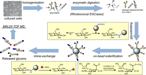
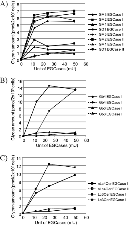
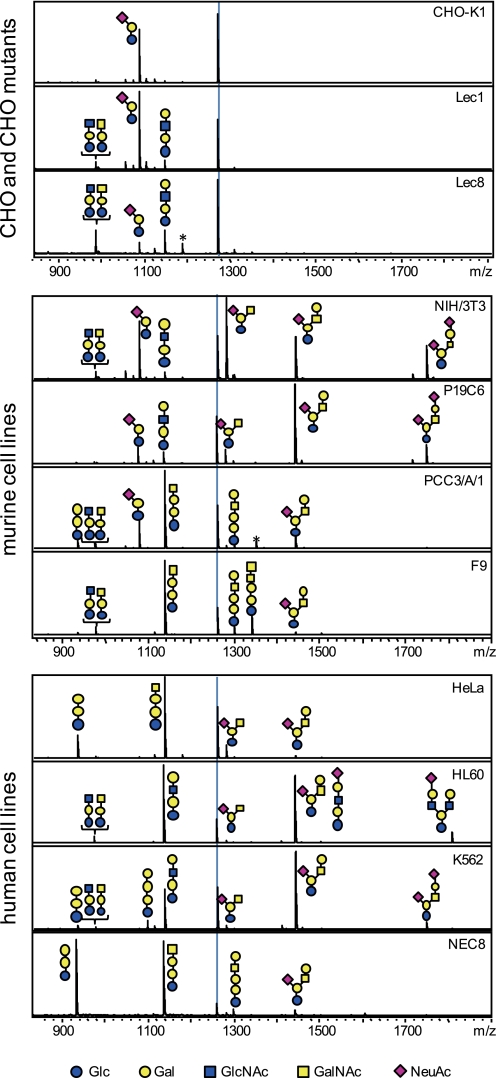
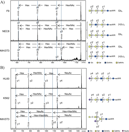
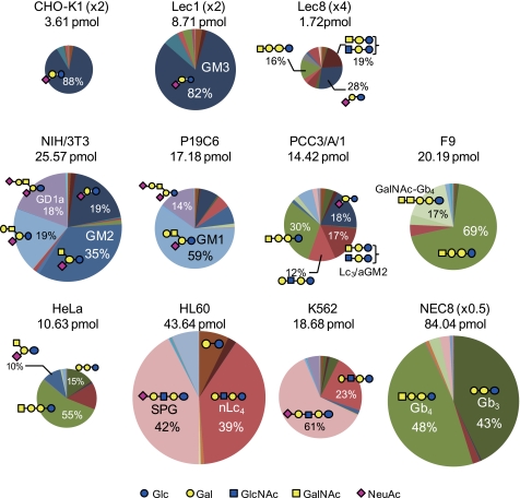
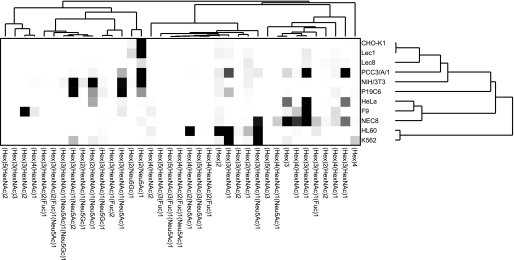
Glycosphingolipids (GSLs) are crucially important components of the cellular membrane, where they comprise microdomains with many critical biological functions. Despite this fact, qualitative and quantitative techniques for the analysis of GSLs still lag behind the needs of researchers. In this study, a reliable procedure for the elucidation of cellular GSL-glycomes was established based on (a) enzymatic glycan cleavage by endoglycosylceramidases derived from Rhodococcus sp. in combination with (b) glycoblotting-assisted sample preparation. The mixture of endoglycosylceramidase I and II was employed to maximize the release of glycan moieties from the major classes of GSLs (i.e. ganglio-, (neo)lacto- and globo-series GSLs). The glycoblotting technique enabled the quantitative detection of GSL-glycans using as few as 2 × 10(5) cells. Thirty-seven different kinds of cellular GSL glycans were successfully observed in 11 kinds of cells, including Chinese hamster ovary cells and their lectin-resistant mutants as well as murine and human embryonic carcinoma cells. Furthermore, in-depth structural clarification in terms of discrimination of isomers was achieved by MALDI-TOF/TOF mass spectrometry analysis and/or linkage-specific glycosidase digestion. These novel analytical techniques were shown to be capable of delineating cell-specific GSL-glycomes. Thus, they are anticipated to have a broad range of applications for the characterization, description, and comparison of various cellular/tissue samples in the fields of drug discovery and regenerative medicine.


Figures

FIGURE 1.
FIGURE 1.
Schematic diagram of the established procedure for quantitative cellular GSL-glycomics. Total lipids were extracted by ultrasonic homogenization in a chloroform and methanol solvent, and glycan moieties were released in the presence of a mixture of EGCase I and II (25 milliunits of each). Because of the high purification capability of glycoblotting, no further purifications were required. GSL-derived glycans were recovered as derivatives of aoWR and subjected to MALDI-TOF MS analysis.
FIGURE 2.
FIGURE 2.
Optimization of EGCase I and II enzymatic reactions. The optimization of cellular GSL-glycomics was performed using cellular GSLs as enzymatic substrates. Total lipids extracted from 2 × 10⁵ NIH/3T3 (A), F9 (B), or HL60 (C) cells were directly digested using various amounts of EGCase I or II in the presence of 0.1% sodium cholate. Ganglio-series GSLs (i.e. GM3, GM2, GM1, and GD1a) in NIH/3T3 cells (A), globo-series GSLs (i.e. Gb3 and Gb4) in F9 cells (B), and (neo)lacto-series GSLs (i.e. nLc4 and Lc3) in HL60 cells (C) were used as cellular GSL substrates. mU, milliunits.
FIGURE 3.
FIGURE 3.
Representative MALDI-TOF MS spectra of GSL-derived glycans derived from 11 kinds of cells. All signals were detected as aoWR-glycan derivatives. The dotted line on each panel indicates the internal standard, GN4 (5 pmol). Estimated structures are shown. Blue circles, yellow circles, blue squares, yellow squares, and purple diamonds represent Glc, Gal, GlcNAc, GalNAc, and Neu5Ac, respectively. Peaks labeled by asterisks stem from contaminating free oligosaccharides.
FIGURE 4.
FIGURE 4.
Representative MALDI-TOF/TOF spectra for the structural determination of glycan isoforms. The isoforms of (Hex)3(HexNAc)1 obtained from F9, NEC8, and NIH/3T3 cells (A) and (Hex)3(HexNAc)1(NeuAc)1 observed in HL60, K562, and NIH/3T3 cells (B) could be distinguished by TOF/TOF analysis. In the case of (Hex)3(HexNAc)1, this glycan corresponded to only Gb4 in the globo-series in F9 cells. On the other hand, TOF/TOF analyses for the glycan in NEC8 and NIH/3T3 cells showed a mixture consisting of Gb4 and (n)Lc4. In similar manner the glycan (Hex)3(HexNAc)1(NeuAc)1 possessed a linear structure (SPG) in HL60 and K562 cells as opposed to a branched structure with a (Hex)1(HexNAc)1 unit at the non-reducing terminal in NIH 3T3 cells, characteristic of GM1.
FIGURE 5.
FIGURE 5.
Cellular delineation/characterization through quantitative GSL-glycomic analyses. Both the absolute quantity of GSL-glycans (i.e. the size of the glycome) and the relative GSL-glycan composition are represented for each cell type. The areas of the circles represent the amount of total GSL-glycans per 2 × 10⁵ cells. The circle size of CHO-K1 and Lec1 cells are magnified 2-fold, that of Lec8 cells is magnified 4-fold, and that of NEC8 is reduced to one-half size to facilitate visualization.
FIGURE 6.
FIGURE 6.
Hierarchical clustering analysis of GSL-glycan expression profiles. Graphic representation of hierarchical clustering results based on expression profiles of 37 GSL-glycans in 11 cell types is shown. Rows, cell types; columns, GSL-glycans. Color intensity from white to red indicates the magnitude of differential expression.

References

    1. Hakomori S. I. (2008) Biochim. Biophys. Acta 1780, 325–346 - PMC - PubMed
    1. Hakomori S. I. (2010) FEBS Lett. 584, 1901–1906 - PMC - PubMed
    1. Lanctot P. M., Gage F. H., Varki A. P. (2007) Curr. Opin. Chem. Biol. 11, 373–380 - PMC - PubMed
    1. Kannagi R., Cochran N. A., Ishigami F., Hakomori S., Andrews P. W., Knowles B. B., Solter D. (1983) EMBO J. 2, 2355–2361 - PMC - PubMed
    1. Kannagi R., Levery S. B., Ishigami F., Hakomori S., Shevinsky L. H., Knowles B. B., Solter D. (1983) J. Biol. Chem. 258, 8934–8942 - PubMed

Publication types

LinkOut - more resources