. 2011 Dec 2;286(48):41758-41766.
doi: 10.1074/jbc.M111.271080. Epub 2011 Sep 29.

hSWS1·SWSAP1 is an evolutionarily conserved complex required for efficient homologous recombination repair

Affiliations

hSWS1·SWSAP1 is an evolutionarily conserved complex required for efficient homologous recombination repair

Ting Liu et al. J Biol Chem. .

Abstract

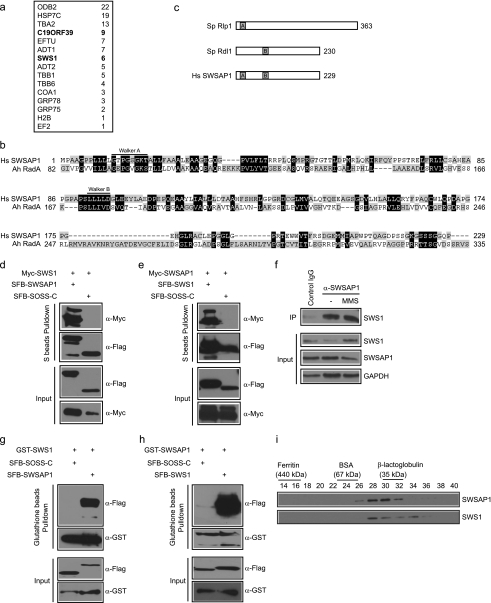
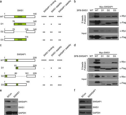
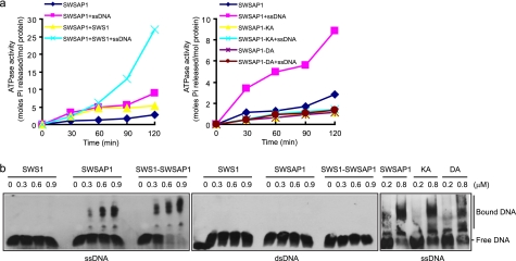
The Shu complex in yeast plays an important role in the homologous recombination pathway, which is critical for the maintenance of genomic integrity. The identification of human SWS1 (hSWS1) as the homolog of budding yeast Shu2 implicated that the Shu complex is evolutionarily conserved. However, the human counterparts of other components in this complex have not yet been identified and characterized. Here we describe the characterization of a novel human component of this complex, SWSAP1 (hSWS1-associated protein 1)/C19orf39. We show that hSWS1 and SWSAP1 form a stable complex in vivo and in vitro. hSWS1 and SWSAP1 are mutually interdependent for their stability. We further demonstrate that the purified hSWS1·SWSAP1 complex possesses single-stranded DNA-binding activity and DNA-stimulated ATPase activity. Moreover, SWSAP1 interacts with RAD51 and RAD51 paralogs, and depletion of SWSAP1 causes defects in homologous recombination repair. Thus, our results suggest that the human Shu complex (hSWS1·SWSAP1) has an evolutionarily conserved function in homologous recombination.


Figures

FIGURE 1.
FIGURE 1.
Identification of SWSAP1 as hSWS1-binding protein. a, 293T cells stably expressing SFB-tagged (S-tag, FLAG epitope tag, and streptavidin-binding peptide tag) hSWS1 was used for tandem affinity purification of protein complexes isolated from chromatin fractions. Tables are summaries of proteins identified by mass spectrometry analysis. Letters in bold indicate the bait proteins. b, alignment of human SWSAP1 with Arcanobacterium hemolyticum RadA. The conserved Walker A and Walker B motifs are indicated. c, schematic representation of fission yeast Rlp1, Rdl1, and human SWSAP1. d and e, the interaction between hSWS1 and SWSAP1 was confirmed by co-immunoprecipitation (IP) experiments. 293T cells were transfected with plasmids encoding Myc-tagged hSWS1 or SWSAP1 together with plasmids encoding SFB-tagged SWSAP1 or hSWS1 as indicated. Cells were collected 24 h after transfection. Precipitation reactions were performed using S beads, and immunoblotting was carried out using antibodies as indicated. f, endogenous hSWS1 and SWSAP1 form a complex in vivo. HeLa cells were mock treated or treated with MMS. Control or anti-SWSAP1 immunoprecipitates were immunoblotted with anti-hSWS1 antibody (top). The expression levels of endogenous proteins were revealed by immunoblotting using anti-hSWS1 and anti-SWSAP1 antibodies (bottom). g and h, hSWS1 and SWSAP1 directly bind to each other. SF9 cells were co-infected with baculoviruses expressing GST-tagged hSWS1 or SWSAP1 together with those expressing SFB-tagged SWSAP1 or hSWS1. Pulldown experiments and immunoblotting were carried out as indicated. i, heterodimeric complex formation was studied by gel filtration chromatography as described under “Experimental Procedures.” Aliquots from peak fractions were analyzed on 12.5% SDS-PAGE and confirmed by Western blot analysis.
FIGURE 2.
FIGURE 2.
hSWS1 and SWSAP1 are interdependent for their stability. a–c, schematic representation of human SWS1 and SWSAP1 and their deletion mutants used in this study. b–d, mapping of the corresponding regions required for hSWS1-SWSAP1 interaction. Precipitation reactions were performed using S beads, and immunoblotting was carried out using antibodies as indicated. e and f, depletion of hSWS1 or SWSAP1 leads to the loss of both proteins in the cell. HeLa cells were transfected with control siRNA or siRNAs specifically targeting hSWS1 or SWSAP1. Lysates were analyzed by immunoblotting using antibodies recognizing hSWS1, SWSAP1, or GAPDH.
FIGURE 3.
FIGURE 3.
SWSAP1 is a DNA-stimulated ATPase and preferentially binds Single-stranded DNA. a, time course for ATPase activity of SWSAP1 and hSWS1·SWSAP1 complex. SWSAP1, hSWS1·SWSAP1 complex, or SWSAP1 ATPase-inactivating mutants was incubated with or without ssDNA as described under “Experimental Procedures.” b, increasing concentrations of hSWS1, SWSAP1, hSWS1·SWSAP1 complex and SWSAP1 ATPase-inactivating mutants were incubated with 5′-biotin-labeled ssDNA or dsDNA as described under “Experimental Procedures.” Protein·DNA complexes and unbound DNA are indicated on the right.
FIGURE 4.
FIGURE 4.
SWSAP1 interacts with RAD51 and RAD51 paralogs and is involved in homologous recombination repair. a, cells with hSWS1 or SWSAP1 down-regulation display increased MMS but not CPT sensitivity. Cell survival assays were performed as described under “Experimental Procedures.” Data are presented as means ± S.D. (error bars) from three different experiments. b, schematic representation of HR assay. The DR-GFP construct consists of direct repeats of two mutated GFP genes, SceGFP and the truncated iGFP. When a single double strand DNA break generated by I-Sce1 is repaired via gene conversion with iGFP, expression of GFP is restored and can be measured by FACS analysis. c, decreasing hSWS1 or SWSAP1 expression impairs HR repair. U2OS DR-GFP cells were electroporated with pCBASce plasmids (see “Experimental Procedures” for details). The percentage of GFP-positive cells was determined by flow cytometry 48 h after electroporation. The data were normalized to those obtained from cells transfected with control siRNA (set as 1.0). Means ± S.E. (error bars) shown are obtained from three independent experiments. d and e, down-regulation of hSWS1 or SWSAP1 impairs MMS-induced RAD51 foci formation. Immunostaining was performed 6 h after MMS treatment using the indicated antibodies. Representative RAD51 foci are shown in d. Data were presented as means ± S.D. (error bars) from three different experiments, more than 100 cells were counted in each experiment (e). f, knockdown efficiency was confirmed by immunoblotting. g, SWSAP1 interacts with RAD51 and several RAD51 paralogs, but not with XRCC2 in vivo. 293T cells were transfected with plasmids encoding SFB-tagged SWSAP1 together with plasmids encoding Myc-tagged RAD51 or RAD51 paralogs. Cells were collected 24 h after transfection. Precipitation reactions were performed using S beads, and immunoblotting was carried out using antibodies as indicated. h, SWSAP1 interacts with RAD51 and several RAD51 paralogs, but not with XRCC2 in vitro. SF9 cells were co-infected with baculoviruses expressing GST-tagged RAD51 or RAD51 paralogs together with those expressing SFB-tagged SWSAP1. Pulldown experiments and immunoblotting were carried out as indicated. i, the ATPase activity of SWSAP1 is required for restoring cellular resistance to MMS. HeLa-derivative cell lines stably expressing siRNA-resistant HA-FLAG-tagged WT (SWSAP1SiR-WT), K18A mutant (SWSAP1SiR-KA), and D96A mutant (SWSAP1SiR-DA) of SWSAP1 were generated. Cell survival assays were performed as described under “Experimental Procedures.” Data are presented as means ± S.D. (error bars) from three different experiments. j, the ATPase activity of SWSAP1 is required for its function in promoting RAD51 foci formation. Immunostaining was performed 6 h after MMS treatment using indicated antibodies. Data are presented as means ± S.D. (error bars) from three different experiments, more than 100 cells were counted in each experiment. k, SWSAP1 expression was confirmed by immunoblotting with the use of FLAG antibody, and extracts were prepared from cells transfected with SWSAP1 siRNA#2.

Similar articles

Cited by

References

    1. van Gent D. C., Hoeijmakers J. H., Kanaar R. (2001) Nat. Rev. Genet. 2, 196–206 - PubMed
    1. Branzei D., Foiani M. (2008) Nat. Rev. Mol. Cell Biol. 9, 297–308 - PubMed
    1. Moynahan M. E., Jasin M. (2010) Nat. Rev. Mol. Cell Biol. 11, 196–207 - PMC - PubMed
    1. Weinstock D. M., Richardson C. A., Elliott B., Jasin M. (2006) DNA Repair (Amst.) 5, 1065–1074 - PubMed
    1. Lukas J., Bartek J. (2004) Cell 118, 666–668 - PubMed

Publication types

MeSH terms