. 2011 Nov 25;286(47):40671-80.
doi: 10.1074/jbc.M111.271189. Epub 2011 Sep 29.

Special AT-rich binding protein-2 (SATB2) differentially affects disease-causing p63 mutant proteins

Affiliations

Special AT-rich binding protein-2 (SATB2) differentially affects disease-causing p63 mutant proteins

Jacky Chung et al. J Biol Chem. .

Abstract

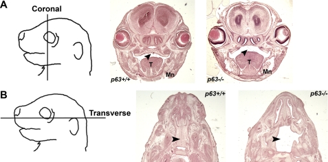
p63, a p53 family member, is critical for proper skin and limb development and directly regulates gene expression in the ectoderm. Mice lacking p63 exhibit skin and craniofacial defects including cleft palate. In humans p63 mutations are associated with several distinct developmental syndromes. p63 sterile-α-motif domain, AEC (ankyloblepharon-ectodermal dysplasia-clefting)-associated mutations are associated with a high prevalence of orofacial clefting disorders, which are less common in EEC (ectrodactyly-ectodermal dysplasia-clefting) patients with DNA binding domain p63 mutations. However, the mechanisms by which these mutations differentially influence p63 function remain unclear, and interactions with other proteins implicated in craniofacial development have not been identified. Here, we show that AEC p63 mutations affect the ability of the p63 protein to interact with special AT-rich binding protein-2 (SATB2), which has recently also been implicated in the development of cleft palate. p63 and SATB2 are co-expressed early in development in the ectoderm of the first and second branchial arches, two essential sites where signaling is required for craniofacial patterning. SATB2 attenuates p63-mediated gene expression of perp (p53 apoptosis effector related to PMP-22), a critical downstream target gene during development, and specifically decreases p63 perp promoter binding. Interestingly, AEC but not EEC p63 mutations affect the ability of p63 to interact with SATB2 and the inhibitory effects of SATB2 on p63 transactivation of perp are most pronounced for AEC-associated p63 mutations. Our findings reveal a novel gain-of-function property of AEC-causing p63 mutations and identify SATB2 as the first p63 binding partner that differentially influences AEC and EEC p63 mutant proteins.


Figures

FIGURE 1.
FIGURE 1.
Cleft palate is observed in p63-knock-out mice. Heads of E17.5 p63+/+ or p63−/− embryos were dissected, and coronal (A) or transverse (B) sections were cut and stained with hematoxylin and eosin. T, tongue; Mn, mandible. Black arrowheads denote the secondary palate.
FIGURE 2.
FIGURE 2.
p63α and SATB2 are co-expressed during development. A, whole mount in situ hybridization of wild-type embryos at E9, E10, E11, and E12. The red arrows and arrowheads at E10 denote the branchial arches and the limb buds, respectively. At E11, the red arrowheads highlight the neural staining. B–D, transverse sections of E10 embryos across the branchial arches (B; red arrowheads) stained with anti-p63 (C) or anti-p63α (D) antibodies and analyzed by confocal immunofluorescence. E, similar sections co-stained with anti-pan-p63 and anti-SATB2-CT antibodies. F, NIH 3T3 cells transfected with TA or ΔNp63α. ChIP (IP) analysis was performed on the perp promoter (perp-1). The β-actin gene promoter was used as a negative control. G, murine newborn P3 brains subjected to ChIP analysis using the indicated antibodies. PCR was performed interrogating three sites of the perp promoter. H, co-localization of SATB2 and p63α onto the perp promoter (perp-1 site) analyzed by ChIP/re-ChIP using the indicated antibodies. Scale bars, 10 μm.
FIGURE 3.
FIGURE 3.
SATB2 inhibits p63α-mediated perp activation. A, NIH 3T3 cells were transiently transfected with the indicated plasmids and quantitative real-time ChIP analysis was performed on the perp promoter (perp-1 site). Values were normalized to the amount of input DNA that was present. Real-time ChIP was also performed on mock-transfected cells as a control. B, a luciferase reporter assay using the murine perp promoter was performed in H1299 cells transfected with TA or ΔNp63α. A mutant reporter (MutD) with a point mutation in the perp promoter was used as a negative control. C, increasing amounts of T7-SATB2 were co-transfected with TAp63α into H1299 cells, and a perp luciferase reporter assay was performed. D, SCC9 cells were infected with lentivirus-expressing and shRNA targeting GFP or two different shRNAs directed against SATB2. Total RNA was extracted, and quantitative RT-PCR was performed using primers specific for perp. Values were normalized to gapdh housekeeper. IB, immunoblotting. E, SCC9 cells stably infected with lentivirus-expressing shGFP or shSATB2–2 were lysed, and Perp expression was analyzed by immunoblotting.
FIGURE 4.
FIGURE 4.
AEC-associated, SAM domain, p63α mutations increase binding to SATB2. A, mutations associated with EEC syndrome are found exclusively in the DNA binding domain of p63 whereas those associated with AEC syndrome map to the SAM domain of p63α. A nonsense mutation leading to a premature stop codon upstream of the SAM domain has also been associated with AEC syndrome. B–D, various wild-type or mutant 3×FLAG-ΔNp63α constructs were co-transfected with T7-SATB2, and lysates were immunoprecipitated (IP) with anti-FLAG antibodies. Bound proteins were separated on SDS-PAGE and analyzed by immunoblotting (IB). E, wild-type 3F-ΔNp63α or 3F-ΔNp63α mutants were in vitro translated together with wild-type T7-ΔNp63α and immunoprecipitated with anti-FLAG antibodies. Bound proteins were resolved on SDS-PAGE and immunoblotted with anti-FLAG or anti-T7 antibodies. F and G, HEK293 cells were co-transfected with the indicated expression plasmids. Lysates were immunoprecipitated with control (anti-HA) or anti-FLAG antibodies and blotted with anti-FLAG or anti-T7 antibodies.
FIGURE 5.
FIGURE 5.
SATB2 has a dominant negative effect on AEC-p63α mutants. A, a luciferase reporter assay was performed using the perp promoter with the indicated TAp63α mutants in H1299 cells (n = 3, mean ± S.E. (error bars)). B, NIH3T3 cells were transfected with the indicated expression plasmids, and ChIP analysis was performed on the perp-1 site in the perp promoter. C, perp luciferase assay was performed with the indicated TAp63α expression plasmids in H1299 cells (representative experiment, mean ± S.D.). D and E, T7-SATB2 was co-transfected, at increasing amounts, with an expression plasmid encoding TAp63α G530V (D) or TAp63α Q536L and a perp luciferase reporter assay was performed (E). IB, immunoblot.

References

    1. Melino G., De Laurenzi V., Vousden K. H. (2002) Nat. Rev. Cancer 2, 605–615 - PubMed
    1. Yang A., McKeon F. (2000) Nat. Rev. Mol. Cell Biol. 1, 199–207 - PubMed
    1. Su X., Paris M., Gi Y. J., Tsai K. Y., Cho M. S., Lin Y. L., Biernaskie J. A., Sinha S., Prives C., Pevny L. H., Miller F. D., Flores E. R. (2009) Cell Stem Cell 5, 64–75 - PMC - PubMed
    1. Carroll D. K., Carroll J. S., Leong C. O., Cheng F., Brown M., Mills A. A., Brugge J. S., Ellisen L. W. (2006) Nat. Cell Biol. 8, 551–561 - PubMed
    1. Lopardo T., Lo Iacono N., Marinari B., Giustizieri M. L., Cyr D. G., Merlo G., Crosti F., Costanzo A., Guerrini L. (2008) PloS One 3, e2715 - PMC - PubMed

Publication types

MeSH terms

Supplementary concepts