. 2011 Oct 1;34(10):1411-21.
doi: 10.5665/SLEEP.1290.

Fast and slow spindles during the sleep slow oscillation: disparate coalescence and engagement in memory processing

Affiliations

Fast and slow spindles during the sleep slow oscillation: disparate coalescence and engagement in memory processing

Matthias Mölle et al. Sleep. .

Abstract

Study objectives: Thalamo-cortical spindles driven by the up-state of neocortical slow (< 1 Hz) oscillations (SOs) represent a candidate mechanism of memory consolidation during sleep. We examined interactions between SOs and spindles in human slow wave sleep, focusing on the presumed existence of 2 kinds of spindles, i.e., slow frontocortical and fast centro-parietal spindles.

Design: Two experiments were performed in healthy humans (24.5 ± 0.9 y) investigating undisturbed sleep (Experiment I) and the effects of prior learning (word paired associates) vs. non-learning (Experiment II) on multichannel EEG recordings during sleep.

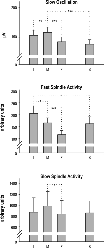
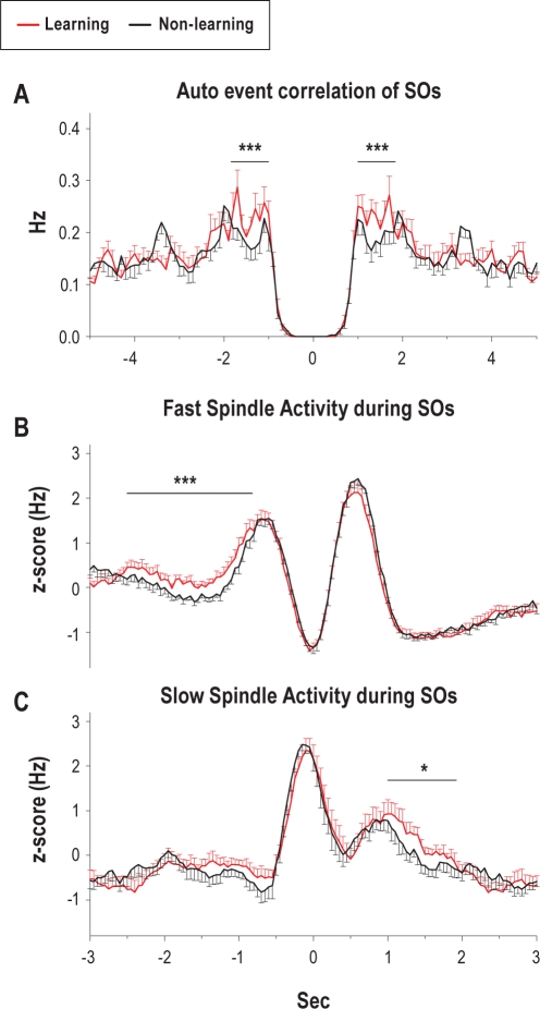
Measurements and results: Only fast spindles (12-15 Hz) were synchronized to the depolarizing SO up-state. Slow spindles (9-12 Hz) occurred preferentially at the transition into the SO down-state, i.e., during waning depolarization. Slow spindles also revealed a higher probability to follow rather than precede fast spindles. For sequences of individual SOs, fast spindle activity was largest for "initial" SOs, whereas SO amplitude and slow spindle activity were largest for succeeding SOs. Prior learning enhanced this pattern.

Conclusions: The finding that fast and slow spindles occur at different times of the SO cycle points to disparate generating mechanisms for the 2 kinds of spindles. The reported temporal relationships during SO sequences suggest that fast spindles, driven by the SO up-state feed back to enhance the likelihood of succeeding SOs together with slow spindles. By enforcing such SO-spindle cycles, particularly after prior learning, fast spindles possibly play a key role in sleep-dependent memory processing.

Keywords: Human; memory; sleep spindles; slow oscillations.


Figures

Figure 1
Figure 1
Averaged fast and slow spindles and averaged slow oscillations. (A) Grand mean averages (± SEM) of original EEG in (top) 12 centro-parietal channels across all detected fast spindles and (bottom) in 12 fronto-central channels across all detected slow spindles. Note, spindles are averaged independently of whether an SO was present. Averaging was performed with reference to the deepest, i.e., most negative, trough in the filtered signal (t = 0). Asterisks (and arrows) indicate a significant (P < 0.001) positive and negative slow potential shift underlying fast and slow spindles, respectively, in the interval between 300 ms before to 300 ms after the spindle peak. (B) Grand mean averages (± SEM) of original EEG in 8 fronto-central channels across all detected slow oscillations. Averaging was performed with reference to the negative half-wave peak of the SO (t = 0). Indicated is also the time interval between 2 succeeding positive-to-negative zero crossings of the EEG signal which was used to identify SOs (see Methods).
Figure 2
Figure 2
Temporal association between fast and slow spindles and SOs. Event correlation histogram (A) of slow spindle activity (i.e., all marked peaks and troughs of all detected spindles) with reference to the peak (most negative trough; t = 0) of fast spindles; (B) of fast spindle activity with reference to the peak of slow spindles; (C) of fast spindle activity with reference to the negative peak (t = 0) of the SOs; and (D) of slow spindle activity with reference to the negative peak of the SOs. Insets show the respective reference spindle and SO. Red lines indicate mean (± SEM) event correlation histograms obtained after randomization of data. Note in (A) and (B), fast spindles are followed (with a 500 ms delay) by an increase in slow spindle activity and, conversely, slow spindles are preceded by an increase in fast spindle activity (500 ms before). Note also in (C), strong increases in fast spindle activity before and after the negative SO down-state coincide with SO up-states and in (D), strong increases in slow spindle activity coincide with the beginning of the downward going negative phase of the SO. Asterisks indicate significant increases and decreases, respectively in spindle activity in the indicated intervals (thin lines) as compared to the 0.5-sec baseline interval (thick line; ***P < 0.001, **P < 0.01).
Figure 3
Figure 3
Time-frequency plots of wavelet-power during slow oscillations. (A) Time-frequency plots of wavelet-power in a time window of ± 1.2 sec and for frequencies of 5-20 Hz around the negative peak of the SO (t = 0) in the frontal (top), central (middle), and parietal (bottom) electrode. Note, strong enhancement in < 12 Hz slow spindle wavelet-power during the transition into SO down-state most pronounced in the frontal electrode. Coloring indicates relative wavelet-power with the average power during the baseline (−1.3 to −1.2 sec) set to “1.” (B) Grand mean averages of original EEG in all 27 recording channels across all detected SOs. Averaging was performed in a time window of ± 1.2 sec with reference to the negative peak of SOs (t = 0).
Figure 4
Figure 4
Analyses of sequences of slow oscillations. Upper panel: Mean (+ SEM) peak-to-peak SO amplitudes (in the −0.2 to 0.7 sec interval) for the 4 types of SOs (across 8 fronto-central channels). Middle and lower panels: Mean (+ SEM) amplitude (peak-to-peak wavelet-power in the −0.3 to 0.8 sec interval) of increase in fast spindle wavelet power (during SO up-state) and slow spindle wavelet power (during transition into SO down-state) for 4 types of SOs (averaged, respectively, across 12 centro-parietal and 12 fronto-central recording sites of interest; n = 11). I, M, F: initial, middle, final SO in a SO sequence, S: single SO. Asterisks indicate significant differences in peak-to-peak SO amplitude and spindle wavelet-power, respectively between the different types of SOs (***P < 0.001, **P < 0.01, *P < 0.05).
Figure 5
Figure 5
Effects of learning on slow oscillations and associated fast and slow spindle activity. (A) Auto-event correlation histogram of all detected SOs after Learning (red) and Non-learning (black). SOs were identified in an average channel across the 6 fronto-central channels of interest. (B) Event correlation histogram of fast spindle activity and (C) of slow spindle activity, both with reference to the negative peak of the SOs, after Learning (red) and Non-learning (black). Spindle activity was defined by all peaks and troughs of all detected spindles (averaged across the 8 channels of interest), and z-transformed for each subject. For all 3 histogram panels, means (± SEMs) are shown. ***P < 0.001, *P < 0.05, for pairwise comparisons between Learning and Non-Learning).
Figure 6
Figure 6
Effects of learning on sequences of slow oscillations. Averages of (A) original EEG and RMS activity for (B) fast and (C) slow spindle frequency range during identified SOs that occurred in sequences for the learning (red) and non-learning (black) condition. Averaging (across identified SO sequences and subjects) for SOs was performed on the original unfiltered signal in the 6 fronto-central channels of interest. Fast and slow spindle RMS activity was averaged, respectively, for the 8 centro-parietal and 8 fronto-central channels of interest. “Initial,” “Middle,” and “Final” indicate position of target SO in a sequence of (3-5) succeeding SOs. Means (± SEMs) are shown. Bottom bars indicate P-values of paired t-tests between Learning and Non-learning condition.

References

    1. Buzsáki G, Draguhn A. Neuronal oscillations in cortical networks. Science. 2004;304:1926–9. - PubMed
    1. Rosanova M, Ulrich D. Pattern-specific associative long-term potentiation induced by a sleep spindle-related spike train. J Neurosci. 2005;25:9398–405. - PMC - PubMed
    1. Czarnecki A, Birtoli B, Ulrich D. Cellular mechanisms of burst firing-mediated long-term depression in rat neocortical pyramidal cells. J Physiol. 2007;578:471–9. - PMC - PubMed
    1. Gais S, Mölle M, Helms K, Born J. Learning-dependent increases in sleep spindle density. J Neurosci. 2002;22:6830–4. - PMC - PubMed
    1. Eschenko O, Mölle M, Born J, Sara SJ. Elevated sleep spindle density after learning or after retrieval in rats. J Neurosci. 2006;26:12914–20. - PMC - PubMed

Publication types