. 2011 Sep;7(9):e1002221.
doi: 10.1371/journal.ppat.1002221. Epub 2011 Sep 22.

APOBEC3A is a specific inhibitor of the early phases of HIV-1 infection in myeloid cells

Affiliations

APOBEC3A is a specific inhibitor of the early phases of HIV-1 infection in myeloid cells

Gregory Berger et al. PLoS Pathog. 2011 Sep.

Abstract

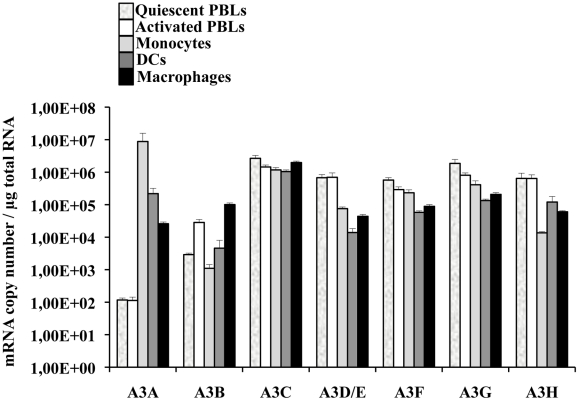
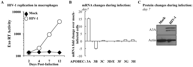
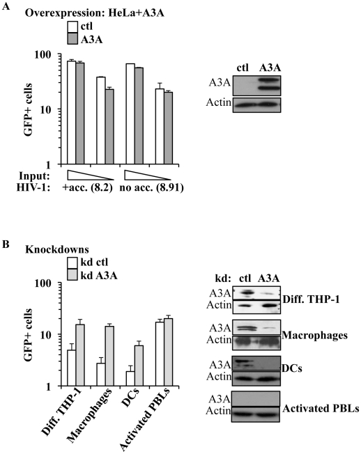
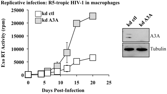
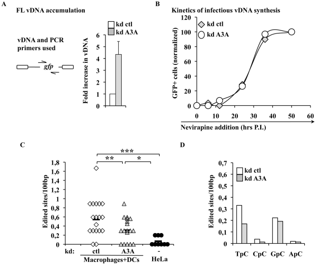
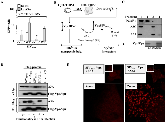
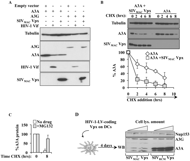
Myeloid cells play numerous roles in HIV-1 pathogenesis serving as a vehicle for viral spread and as a viral reservoir. Yet, cells of this lineage generally resist HIV-1 infection when compared to cells of other lineages, a phenomenon particularly acute during the early phases of infection. Here, we explore the role of APOBEC3A on these steps. APOBEC3A is a member of the APOBEC3 family that is highly expressed in myeloid cells, but so far lacks a known antiviral effect against retroviruses. Using ectopic expression of APOBEC3A in established cell lines and specific silencing in primary macrophages and dendritic cells, we demonstrate that the pool of APOBEC3A in target cells inhibits the early phases of HIV-1 infection and the spread of replication-competent R5-tropic HIV-1, specifically in cells of myeloid origins. In these cells, APOBEC3A affects the amount of vDNA synthesized over the course of infection. The susceptibility to the antiviral effect of APOBEC3A is conserved among primate lentiviruses, although the viral protein Vpx coded by members of the SIV(SM)/HIV-2 lineage provides partial protection from APOBEC3A during infection. Our results indicate that APOBEC3A is a previously unrecognized antiviral factor that targets primate lentiviruses specifically in myeloid cells and that acts during the early phases of infection directly in target cells. The findings presented here open up new venues on the role of APOBEC3A during HIV infection and pathogenesis, on the role of the cellular context in the regulation of the antiviral activities of members of the APOBEC3 family and more generally on the natural functions of APOBEC3A.


Conflict of interest statement

The authors have declared that no competing interests exist.

Figures

Figure 1
Figure 1. A3A is preferentially expressed in myeloid cells.
Primary blood monocytes, monocyte-derived macrophages and DCs were compared to PBLs either quiescent or stimulated for 24 hrs with PHA/IL2 to determine the amount of mRNA of the different members of the APOBEC3 family using primers specific for each member, as already described in . The graph presents results obtained from cells derived from 4 different donors.
Figure 2
Figure 2. The expression of A3A is specifically increased during HIV-1 spread in infected macrophages.
A) M-CSF-derived macrophages were infected with the R5 tropic HIV-1 strain Yu2 at a multiplicity of infection (MOI) of 0,05. Viral spread over time was measured by determining the amount of virion-associated RT activity released in the supernatant (exo-RT). Aliquots of macrophages were lysed at day 7 to determine variations in members of the APOBEC3 family at the mRNA (B) and, in the case of A3A, also at the protein level (C). The panels A and C present one representative experiment out of three, while the panel B presents the average values obtained with cells derived from 3 different donors.
Figure 3
Figure 3. A3A inhibits the early phases of HIV-1 infection specifically in myeloid cells.
A) HeLa cells were transfected with either control or HA-tagged A3A coding constructs and analyzed by WB or challenged 48 hours after transfection with different inputs of VSVg-pseudotyped complete or minimal HIV-1 vectors (i.e. coding or not viral non-structural proteins), carrying a GFP expression cassette in a single-round infectivity assay. The percentage of infected cells was determined 3 days later by flow cytometry. The graph presents averages from 4 different experiments. B) Control or A3A knockdowns were obtained upon LV-mediated transduction of PMA-differentiated THP-1 cells, macrophages, DCs and PHA/IL2-activated PBLs with an equal viral input of miR-shRNAs coding vectors. Four days later, stably silenced cells were analyzed by WB (right panel, presenting representative experiments) or challenged with VSVg-pseudotyped GFP-coding HIV-1 vectors (at MOIs comprised between 3 and 5). The graph presents data obtained from 3 to 6 independent experiments.
Figure 4
Figure 4. A3A silencing increases viral spread of an R5 tropic HIV-1 virus in macrophages.
Macrophages stably silenced as above were infected with an R5 tropic HIV-1 virus (Yu2) at an MOI of 0,05. Viral spread was determined by exo-RT. The graph presents data obtained from 4 independent experiments, while the WB panels present the efficacy of A3A silencing at day 15 in one representative experiment.
Figure 5
Figure 5. A3A impacts the accumulation of vDNA, but not the kinetics of reverse transcription of infectious vDNA and modulates the levels of TC editing of vDNA produced during the infection of myeloid cells.
A) Control and A3A-silenced macrophages were challenged with an equal viral input of GFP-coding HIV-1 virus and cells were lysed at 24 hours post-infection for qPCR analysis with primers specific for the GFP sequence present uniquely on the challenge virus. The scheme presents the relative position of PCR primers on the HIV-1 genome. Control infections were carried out in the presence of RT-inhibitors. Given values are normalized for actin and subtracted for those obtained upon infections carried out in the presence of RT inhibitors (Nevirapine and AZT at 20 µg/ml). B) The kinetics of infectious vDNA accumulation in macrophages silenced or not for A3A were determined as in . Briefly, the accumulation of infectious genomes, defined as those resulting in a GFP-positive cell, is determined upon addition of the RT inhibitor Nevirapine at different times post infection. The percentage of vDNA molecules completed prior to the addition of the inhibitor at each time point is determined 3 days post infection upon normalization to control infections carried out in the absence of RT inhibitor. This assay yields a measure of the speed at which infectious vDNA accumulates, as described in . The graph presents a representative experiment out of 3. C) vDNA produced upon infection of HeLa cells or of primary silenced macrophages was extracted 24 hours post infection, amplified and individual clones, represented by symbols, sequenced. The number of cytidine edited in each sequence with respect to the reference is shown here. Sequences were obtained from 4 independent experiments. Lines represent averages in each sample. Asterisks indicate statistically significant differences after a Student t test (* p<0,05;** p<0,01;*** p<0,001). D) Analysis of the dinucleotide motif of edited cytidine of the sites shown in C.
Figure 6
Figure 6. A3A affects more generally primate lentiviruses. SIVMAC Vpx provides partial protection against A3A and functionally interacts with it.
A) Control and A3A knockdown cells were challenged with an equal amount of SIVMAC vectors carrying or not Vpx and the results of the infection were analyzed 3–4 days after by flow cytometry. B) Scheme used to detect the interaction between Vpx and endogenous A3A expressed in PMA-treated THP-1 cells. Briefly, nichel columns containing purified His-tagged HIV-1 Vpr (non functional during the infection of primary myeloid cells) was used to deplete the cell lysate of non specific interactors prior to its addition to an identical column containing SIVMAC Vpx (functional in the infection of these cells). C) Retained proteins were analyzed by WB with the indicated antibodies, or analyzed by Coomassie staining for the Vpx/Vpr inputs. D) HEK293T cells were transfected with the indicated plasmids coding for Vpx and Vpr proteins whose ability to rescue the infectivity defect of Vpx-deficient SIVMAC vectors we had previously determined [50]). Co-immunoprecipitations were performed using anti-Flag antibody-coupled beads. E) HeLa cells were similarly transfected then fixed and stained for DuoLink immunofluorescence experiments. A positive signal in DuoLink experiments requires the spatial proximity between two proteins (less than 40 nm) for a signal to appear in the form of dots. Representative experiments are shown here.
Figure 7
Figure 7. SIVMAC Vpx degrades A3A but not A3G in both HeLa cells and DCs.
A) HEK293T cells were transfected with the indicated plasmids then lysed for WB analysis 48 hours post transfection. B) As in A, but Cyclohexamide (CHX) was added onto cells at the indicated time prior to cell lysis and WB analysis. C) As in B, with cells incubated in the presence or absence of the proteasome inhibitor MG132 (10 µg/ml) for 8 hours prior to cell lysis. D) Scheme and results used here to determine the degradation of A3A in DCs. HIV-1 vectors coding Vpx proteins derived from SIVRCM and SIVMAC (non-functional and functional during myeloid cells infection, respectively) were produced by transfection of HEK293T cells, normalized by protein content and used to transduce DCs. Vpx-expressing DCs were lysed 4 days post infection and analyzed by WB.

References

    1. Albin JS, Harris RS. Interactions of host APOBEC3 restriction factors with HIV-1 in vivo: implications for therapeutics. Expert Rev Mol Med. 2010;12:e4. - PMC - PubMed
    1. Sheehy AM, Gaddis NC, Choi JD, Malim MH. Isolation of a human gene that inhibits HIV-1 infection and is suppressed by the viral Vif protein. Nature. 2002;418:646–650. - PubMed
    1. Zhang H, Yang B, Pomerantz RJ, Zhang C, Arunachalam SC, et al. The cytidine deaminase CEM15 induces hypermutation in newly synthesized HIV-1 DNA. Nature. 2003;424:94–98. - PMC - PubMed
    1. Mangeat B, Turelli P, Caron G, Friedli M, Perrin L, et al. Broad antiretroviral defence by human APOBEC3G through lethal editing of nascent reverse transcripts. Nature. 2003;424:99–103. - PubMed
    1. Lecossier D, Bouchonnet F, Clavel F, Hance AJ. Hypermutation of HIV-1 DNA in the absence of the Vif protein. Science. 2003;300:1112. - PubMed

Publication types