. 2011;6(9):e25092.
doi: 10.1371/journal.pone.0025092. Epub 2011 Sep 22.

Protection of melanized Cryptococcus neoformans from lethal dose gamma irradiation involves changes in melanin's chemical structure and paramagnetism

Affiliations

Protection of melanized Cryptococcus neoformans from lethal dose gamma irradiation involves changes in melanin's chemical structure and paramagnetism

Abdelahad Khajo et al. PLoS One. 2011.

Abstract

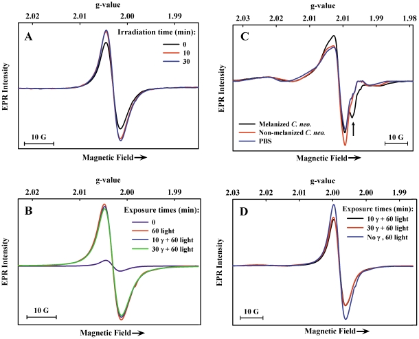
Certain fungi thrive in highly radioactive environments including the defunct Chernobyl nuclear reactor. Cryptococcus neoformans (C. neoformans), which uses L-3,4-dihydroxyphenylalanine (L-DOPA) to produce melanin, was used here to investigate how gamma radiation under aqueous aerobic conditions affects the properties of melanin, with the aim of gaining insight into its radioprotective role. Exposure of melanized fungal cell in aqueous suspensions to doses of γ-radiation capable of killing 50 to 80% of the cells did not lead to a detectable loss of melanin integrity according to EPR spectra of melanin radicals. Moreover, upon UV-visible (Xe-lamp) illumination of melanized cells, the increase in radical population was unchanged after γ-irradiation. Gamma-irradiation of frozen cell suspensions and storage of samples for several days at 77 K however, produced melanin modification noted by a reduced radical population and reduced photoresponse. More direct evidence for structural modification of melanin came from the detection of soluble products with absorbance maxima near 260 nm in supernatants collected after γ-irradiation of cells and cell-free melanin. These products, which include thiobarbituric acid (TBA)-reactive aldehydes, were also generated by Fenton reagent treatment of cells and cell-free melanin. In an assay of melanin integrity based on the metal (Bi(+3)) binding capacity of cells, no detectable loss in binding was detected after γ-irradiation. Our results show that melanin in C. neoformans cells is susceptible to some damage by hydroxyl radical formed in lethal radioactive aqueous environments and serves a protective role in melanized fungi that involves sacrificial breakdown.


Conflict of interest statement

Competing Interests: The authors have declared that no competing interests exist.

Figures

Figure 1
Figure 1. Structure of eumelanin oligomer, adapted from .
R = H, COOH, or other subunits.
Figure 2
Figure 2. EPR spectrum (77 K) of melanized C. neoformans cells (1.9×10⁹ cells/mL).
Figure 3
Figure 3. EPR spectra (77 K) of melanized C. neoformans cells after Xe-lamp illumination.
A) cells were illuminated at room temperature for the time periods indicated and frozen immediately after removal from the light source; B) cells were frozen after collection from cultures and were illuminated at 77 K for the time periods indicated.
Figure 4
Figure 4. EPR spectra (77 K) of γ-irradiated and Xe-lamp illuminated C. neoformans cells.
A) melanized cells γ-irradiated at room temperature (11.94 Gy/min), then frozen in liquid nitrogen; B) γ-irradiated melanized cells from (A) illuminated for the indicated time periods after freezing at 77 K; C) γ-irradiated (11.94 Gy/min) frozen (77 K) melanized cells, non-melanized cells, and PBS; D) γ-irradiated melanized cells from (C) stored frozen for 2 weeks then illuminated at 77 K for the indicated time periods.
Figure 5
Figure 5. Optical spectra of supernatants collected from γ-irradiated or Fenton reagent-treated samples.
A) melanized C. neoformans cells, non-melanized C. neoformans cells, melanized-cell ghosts, and synthetic melanin, irradiated for 30 min at room temperature in PBS; B) as in A, incubated with Fe(II) and H2O2 (Fenton reagent).
Figure 6
Figure 6. Optical spectra of TBA-adducts produced from supernatants collected from γ-irradiated samples.
Spectra are offset for presentation.

Similar articles

Cited by

References

    1. Steinert M, Engelhard H, Flugel M, Wintermeyer E, Hacker J. The Lly protein protects Legionella pneumophila from light but does not directly influence its intracellular survival in Hartmannella vermiformis. Appl Environ Microbiol. 1995;61:2428–2430. - PMC - PubMed
    1. Wang Y, Casadevall A. Decreased susceptibility of melanized Cryptococcus neoformans to UV light. Appl Environ Microbiol. 1994;60:3864–3866. - PMC - PubMed
    1. Zughaier SM, Ryley HC, Jackson SK. A melanin pigment purified from an epidemic strain of Burkholderia cepacia attenuates monocyte respiratory burst activity by scavenging superoxide anion. Infect Immun. 1999;67:908–913. - PMC - PubMed
    1. Turick CE, Caccavo F, Jr, Tisa LS. Electron transfer from Shewanella algae BrY to hydrous ferric oxide is mediated by cell-associated melanin. FEMS Microbiol Lett. 2003;220:99–104. - PubMed
    1. Turick CE, Tisa LS, Caccavo F., Jr Melanin production and use as a soluble electron shuttle for Fe(III) oxide reduction and as a terminal electron acceptor by Shewanella algae BrY. Appl Environ Microbiol. 2002;68:2436–2444. - PMC - PubMed

Publication types

MeSH terms