. 2011;6(9):e25125.
doi: 10.1371/journal.pone.0025125. Epub 2011 Sep 26.

Partial inhibition of estrogen-induced mammary carcinogenesis in rats by tamoxifen: balance between oxidant stress and estrogen responsiveness

Affiliations

Partial inhibition of estrogen-induced mammary carcinogenesis in rats by tamoxifen: balance between oxidant stress and estrogen responsiveness

Bhupendra Singh et al. PLoS One. 2011.

Abstract

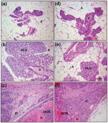
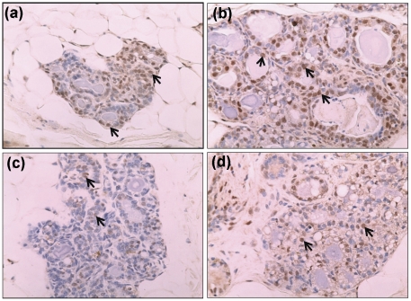
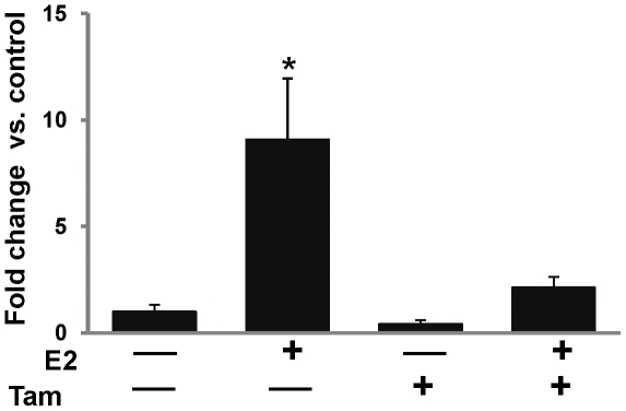
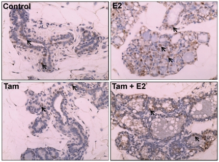
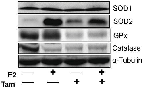
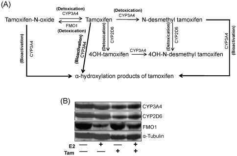
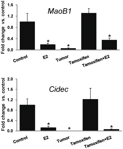
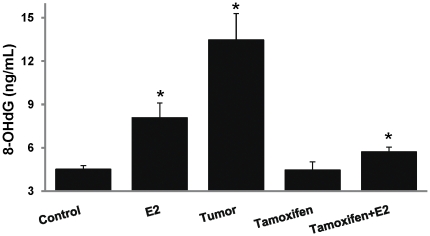
Epidemiological and experimental evidences strongly support the role of estrogens in breast tumor development. Both estrogen receptor (ER)-dependent and ER-independent mechanisms are implicated in estrogen-induced breast carcinogenesis. Tamoxifen, a selective estrogen receptor modulator is widely used as chemoprotectant in human breast cancer. It binds to ERs and interferes with normal binding of estrogen to ERs. In the present study, we examined the effect of long-term tamoxifen treatment in the prevention of estrogen-induced breast cancer. Female ACI rats were treated with 17β-estradiol (E2), tamoxifen or with a combination of E2 and tamoxifen for eight months. Tissue levels of oxidative stress markers 8-iso-Prostane F(2α) (8-isoPGF(2α)), superoxide dismutase (SOD), glutathione peroxidase (GPx), catalase, and oxidative DNA damage marker 8-hydroxydeoxyguanosine (8-OHdG) were quantified in the mammary tissues of all the treatment groups and compared with age-matched controls. Levels of tamoxifen metabolizing enzymes cytochrome P450s as well as estrogen responsive genes were also quantified. At necropsy, breast tumors were detected in 44% of rats co-treated with tamoxifen+E2. No tumors were detected in the sham or tamoxifen only treatment groups whereas in the E2 only treatment group, the tumor incidence was 82%. Co-treatment with tamoxifen decreased GPx and catalase levels; did not completely inhibit E2-mediated oxidative DNA damage and estrogen-responsive genes monoamine oxygenase B1 (MaoB1) and cell death inducing DFF45 like effector C (Cidec) but differentially affected the levels of tamoxifen metabolizing enzymes. In summary, our studies suggest that although tamoxifen treatment inhibits estrogen-induced breast tumor development and increases the latency of tumor development, it does not completely abrogate breast tumor development in a rat model of estrogen-induced breast cancer. The inability of tamoxifen to completely inhibit E2-induced breast carcinogenesis may be because of increased estrogen-mediated oxidant burden.


Conflict of interest statement

Competing Interests: The authors have declared that no competing interests exist.

Figures

Figure 1
Figure 1. Tamoxifen exposure increases the latency of E2-induced breast tumors.
Female ACI rats were treated with E2, Tam or Tam + E2 as described in the Materials and Methods section. Kaplan-Meier survival curves for tumor occurrence were plotted for each treatment group, and the log rank test was used to detect differences in tumor latency curves between groups. Average tumor latency was significantly longer for animals in the Tam + E2 group versus those in the E2 group. Animals in the control or Tam groups did not develop any tumors and are represented by the same line on the graph.
Figure 2
Figure 2. Histopathology of mammary tumor and mammary tissues.
Female ACI rats were treated with E2, Tam or Tam + E2 for 240 days as described in the Materials and Methods section. a) The mammary tissue of a representative control ACI rat shows normal lobular architecture (L) with branched ducts (D) and normal distribution of fat tissue/adipocytes (A); b) E2-treated mammary tissue shows increased proliferation with dilated ducts containing inspissated secretions (D) and increased proliferation and expansion of terminal lobular units (HLU) accompanied by compression of and expansion into the surrounding fat tissue/adipocytes (A); c) Mammary tissue from a rat treated with E2 shows ductal carcinoma in situ (DCIS) containing inspissated secretions in ducts (D) and micro-invasive cancer (IC); d) The mammary gland of Tam only treated rat shows normal lobular architecture (L) with branched ducts (D) and normal distribution of fat tissue (A); e) Mammary tissue from Tam + E2-exposed rat displays increased proliferation compared to control mammary tissue but less than E2-treated mammary tissue. It also shows dilated ducts containing inspissated secretions (D) and increased proliferation and expansion of terminal lobular units (HLU) accompanied by compression of and expansion into the surrounding fat tissue (A); f) Mammary tissue from an animal treated with Tam + E2 shows ductal carcinoma in situ (DCIS) containing inspissated secretions in ducts (D) and micro-invasive cancer (IC). 100× magnification.
Figure 3
Figure 3. Expression of ER-α in mammary tissues.
Female ACI rats were treated with E2, Tam or Tam + E2 for 240 days as described in Materials and Methods section. At the end of the experiment, mammary tissues were collected and used for immunohistochemistry as described in Materials and Methods section. Formalin-fixed/paraffin-embedded mammary sections were immunostained with ER-α antibody. Nuclear expression of ER-α is shown in representative sections (arrow). (a) The mammary tissue of a representative control ACI rat; (b) E2-treated mammary tissue; (c) Tam-treated mammary tissue; and (d) Tam + E2-treated mammary tissue. 100× magnification.
Figure 4
Figure 4. Expression of PR mRNA in mammary tissues.
Female ACI rats were treated with E2, Tam or Tam + E2 for 240 days as described in Materials and Methods section. At the end of the experiment, mammary tissues were collected, total RNA isolated and used for real-time PCR as described in Materials and Methods section. PR mRNA expression was significantly increased in E2-treated mammary tissues and it remained unchanged in Tam- and Tam + E2-treated mammary tissues. mRNA expression data are presented as fold change versus control mammary tissue. These data are reported as an average of values obtained for at least 5 different animals ± SEM. ‘*’ indicates a p value<0.05 compared to controls.
Figure 5
Figure 5. Immunohistochemical detection of proliferation marker PCNA in mammary tissues.
Female ACI rats were treated with E2, Tam or Tam + E2 for 240 days as described in Materials and Methods section. At the end of the experiment, mammary tissues were collected in 10% buffered-formalin. Formalin-fixed/paraffin-embedded mammary tissue sections were immunostained with PCNA antibody. Nuclear expression of PCNA is shown in representative sections (arrows) at 100× magnification.
Figure 6
Figure 6. 8-iso-prostane F (8-isoPGF) formation in rat mammary tissues.
Female ACI rats were treated with E2, Tam or Tam + E2 for 240 days as described in Materials and Methods section. 8-isoPGF levels were measured in mammary tissues from animals in each of these groups. Fold changes were calculated for E2-treated animals relative to age-matched cholesterol-treated controls. Fold changes were calculated for Tam + E2-treated animals relative to age-matched Tam-treated controls. The data are reported as an average of fold change values obtained for at least 8 different animals ± SEM.
Figure 7
Figure 7. Expression of antioxidant enzymes in mammary tissues.
Female ACI rats were treated with E2, Tam or Tam + E2 for 240 days as described in Materials and Methods section. At the end of the experiment, mammary tissues were collected, homogenized and used for western blot analysis. In E2-treated mammary tissue, protein level of CAT is decreased while SOD2 is increased compared to age-matched control. In mammary tissues of rats treated with Tam alone or co-treated with E2, SOD2 is increased, while GPx and CAT are decreased. SOD1 protein levels remain unchanged in all the treatment groups.
Figure 8
Figure 8. Tamoxifen metabolizing enzymes in mammary tissues.
(A) Primary enzymes involved in metabolism of Tam in mammary tissues. Regular arrows indicate the detoxication steps in the pathway, while bold arrows indicate CYP3A4-mediated bioactivation processes. (B) Female ACI rats were treated with E2, Tam or Tam + E2 for 240 days as described in Materials and Methods section. At the end of the experiment, mammary tissues were collected, homogenized and used for western blot analysis. CYP3A4 and CYP2D6 protein levels remain unchanged in all the treatment groups but the FMO1 protein expression is decreased in E2- and Tam + E2-treated mammary tissues.
Figure 9
Figure 9. Expression of estrogen regulated genes MaoB1 and Cidec in mammary tissues.
Female ACI rats were treated with E2, Tam or Tam + E2 for 240 days as described in Materials and Methods section. mRNA expression levels of MaoB1 and Cidec gene in E2-, Tam- and Tam + E2-treated mammary tissues using quantitative real-time PCR are presented as fold change versus control mammary tissues. Expression of each gene was normalized using cyclophilin as internal control. These data are reported as an average of values obtained for at least 5 different animals ± SEM. ‘*’ indicates a p value<0.05 compared to controls.
Figure 10
Figure 10. 8-hydroxydeoxyguanosine (8-OHdG) levels in mammary tissues and mammary tumors.
Female ACI rats were treated with E2, Tam and Tam + E2 for 240 days as described in Materials and Methods section. 8-OHdG levels were measured in mammary tumors and mammary tissues from animals in each of these groups. 8-OHdG levels are significantly increased in E2-treated mammary tissues, mammary tumor tissues and Tam + E2-treated mammary tissues. 8-OHdG levels remain unchanged in Tam-treated mammary tissues compared to control. These data are reported as an average of values obtained for at least 8 different animals ± SEM. ‘*’ indicates a p value<0.05 compared to controls.

References

    1. Bhat HK, Calaf G, Hei TK, Loya T, Vadgama JV. Critical role of oxidative stress in estrogen-induced carcinogenesis. Proc Natl Acad Sci U S A. 2003;100:3913–3918. - PMC - PubMed
    1. Cavalieri E, Chakravarti D, Guttenplan J, Hart E, Ingle J, et al. Catechol estrogen quinones as initiators of breast and other human cancers: implications for biomarkers of susceptibility and cancer prevention. Biochim Biophys Acta. 2006;1766:63–78. - PubMed
    1. IARC. IARC monographs on the evaluation of carcinogenic risks to humans: hormonal contraception and postmenopausal hormone therapy. International Agency for Research on Cancer, Lyon. 1999;72:474–530.
    1. Cavalieri E, Rogan E. Catechol quinones of estrogens in the initiation of breast, prostate, and other human cancers: keynote lecture. Ann N Y Acad Sci. 2006;1089:286–301. - PubMed
    1. Clemons M, Goss P. Estrogen and the risk of breast cancer. N Engl J Med. 2001;344:276–285. - PubMed

Publication types

MeSH terms