. 2011;6(9):e25255.
doi: 10.1371/journal.pone.0025255. Epub 2011 Sep 26.

Isogenic pairs of wild type and mutant induced pluripotent stem cell (iPSC) lines from Rett syndrome patients as in vitro disease model

Affiliations

Isogenic pairs of wild type and mutant induced pluripotent stem cell (iPSC) lines from Rett syndrome patients as in vitro disease model

Gene Ananiev et al. PLoS One. 2011.

Abstract

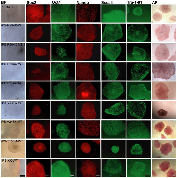
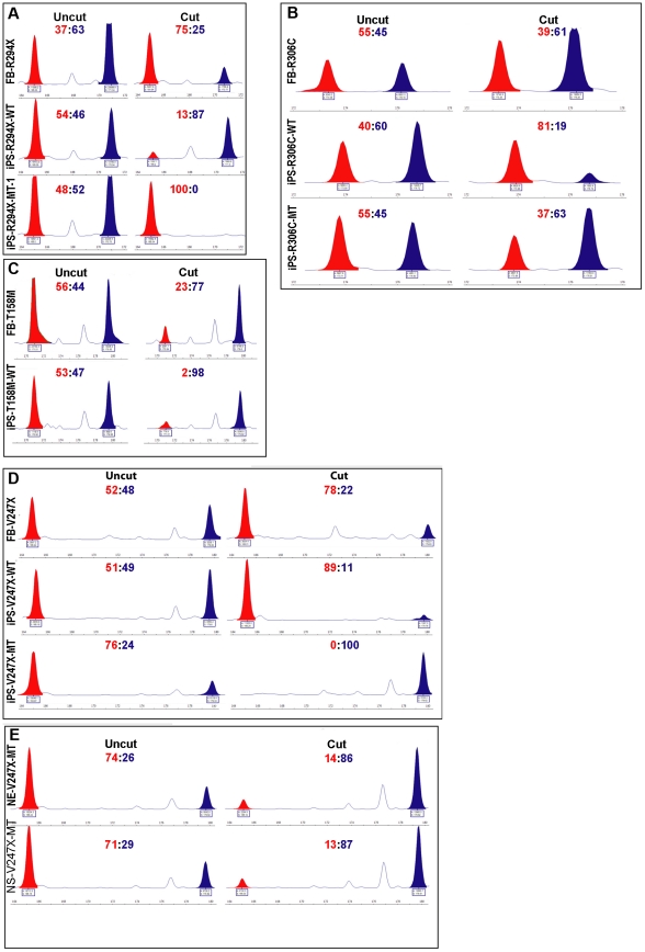
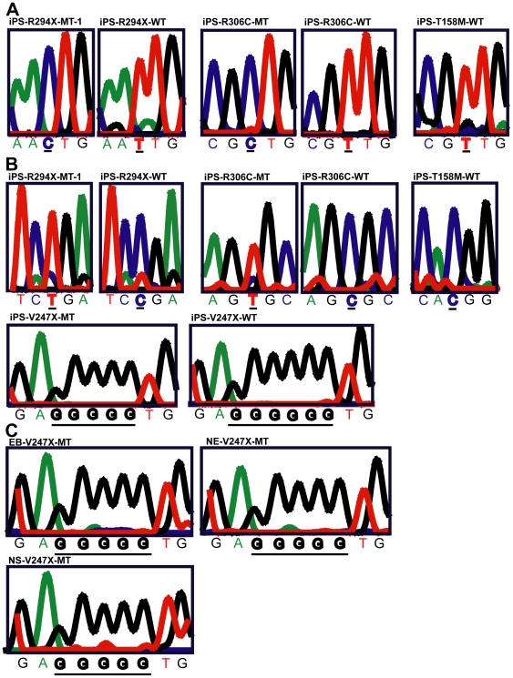
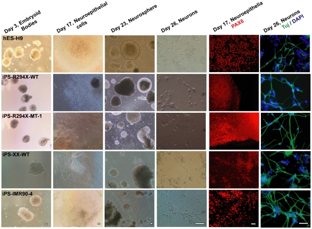
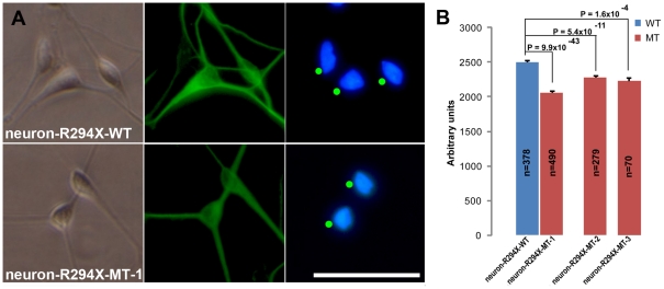
Rett syndrome (RTT) is an autism spectrum developmental disorder caused by mutations in the X-linked methyl-CpG binding protein 2 (MECP2) gene. Excellent RTT mouse models have been created to study the disease mechanisms, leading to many important findings with potential therapeutic implications. These include the identification of many MeCP2 target genes, better understanding of the neurobiological consequences of the loss- or mis-function of MeCP2, and drug testing in RTT mice and clinical trials in human RTT patients. However, because of potential differences in the underlying biology between humans and common research animals, there is a need to establish cell culture-based human models for studying disease mechanisms to validate and expand the knowledge acquired in animal models. Taking advantage of the nonrandom pattern of X chromosome inactivation in female induced pluripotent stem cells (iPSC), we have generated isogenic pairs of wild type and mutant iPSC lines from several female RTT patients with common and rare RTT mutations. R294X (arginine 294 to stop codon) is a common mutation carried by 5-6% of RTT patients. iPSCs carrying the R294X mutation has not been studied. We differentiated three R294X iPSC lines and their isogenic wild type control iPSC into neurons with high efficiency and consistency, and observed characteristic RTT pathology in R294X neurons. These isogenic iPSC lines provide unique resources to the RTT research community for studying disease pathology, screening for novel drugs, and testing toxicology.


Conflict of interest statement

Competing Interests: The authors have declared that no competing interests exist.

Figures

Figure 1
Figure 1. RTT iPSCs express the same set of pluripotency markers as previously characterized hESC and hiPSCs lines.
Representative images of hESC (H9) and hiPSC colonies stained with antibodies against pluripotency markers Sox2, Oct4, Nanog, Ssea4, and Tra-1-81, and processed for alkaline phosphatase reaction. Each row represents colonies from one hESC or hiPSC line. iPS-XX-WT was derived from a healthy female. Each column represents one pluripotency marker. BF stands for bright field. Scale bars = 200 µm.
Figure 2
Figure 2. Nonrandom XCI pattern, as indicated by allele-specific methylation of the AR locus, in female human iPSC lines.
The AR assay was used to examine the XCI pattern in RTT fibroblast and iPSC lines. (A) Results from the R294X fibroblast and iPSC lines. (B) Results from the R306C fibroblast and iPSC lines. (C) Results from the T158M fibroblast and iPSC lines. (D) Results from the V247X fibroblast and iPSC lines. (E) Results from the neuroepithelia (NE-V247X-MT) and neurospheres (NS-V247X-MT) differentiated from iPS-V247X-MT. Uncut traces detected presence of both parental AR alleles: 165 bp and 171 bp for the R294X lines; 173 bp and 176 bp for the R306C lines; 171 bp and 180 bp for the T158M lines; and 165 bp and 180 bp for the V247X lines. Cut traces only detected the hypermethylated AR allele on the inactivated X chromosome, which was resistant to digestion by the methylation-sensitive restriction enzyme HhaI. The source of genomic DNA was labeled to the left of each trace. The ratio between the two AR alleles was at the top of each trace. The molecular weight standard was provided under each trace.
Figure 3
Figure 3. Clonal and allele-specific expression of XIST SNPs and MECP2 in female human iPSC lines.
(A) Representative sequencing traces of SNP analysis to distinguish allele-specific transcription of XIST in iPS-R294X-MT-1, iPS-R294X-WT, iPS-R306C-MT, iPS-R306C-WT, and iPS-T158M-WT. SNP rs1894271 (C or T) was examined in iPS-R294X-MT-1, iPS-R294X-WT. SNP rs16992442 (C or T) was examined in iPS-R306C-MT, iPS-R306C-WT, and iPS-T158M-WT. (B) Representative sequencing traces of regions of the MECP2 gene that contain RTT mutations to distinguish allele-specific transcription of MECP2 in iPS-R294X-MT-1, iPS-R294X-WT, iPS-R306C-MT, iPS-R306C-WT, and iPS-T158M-WT, iPS-V247X-MT, and iPS-V247X-WT. MT denotes expression of the mutant MECP2 allele. WT denotes expression of the wild type MECP2 allele. (C) Representative sequencing traces of the region of the MECP2 gene that contains RTT mutation V247X (705delG) to distinguish allele-specific transcription of MECP2 at the stages of embryoid body, neuroepithelia and neurosphere during directed neural differentiation from iPS-V247X-MT.
Figure 4
Figure 4. Isogenic pairs of wild type and mutant RTT iPSC lines efficiently differentiate into post mitotic neurons.
Left 4 columns: representative transmitted light microscropy images at key stages of the directed neuronal differentiation. Each row represents differentiation from one hESC or hiPSC line. Each column represents one key stage of the differentiation. Right 2 columns: representative fluorescence images of expression of neuroepithelial marker PAX6 and postmitotic neuron marker beta III tubulin (Tuj). Each row represents differentiation from one hESC or hiPSC line. Scale bars = 200 µm.
Figure 5
Figure 5. Analysis of nuclear size in neurons differentiated from isogenic pairs of R294X iPSC lines.
(A) Representative high magnification images of neurons (neuron-R294X-WT and neuron-R294X-MT-1) derived from the isogenic pair of iPS-R294X-WT and iPS-R294X-MT-1 to demonstrate the characteristic neuronal morphology under transmitted light (left), which was used in combination of beta III tubulin immunoreactivity (middle) to identify neurons for measuring DAPI stained nuclear area (right). The green dots in the DAPI images indicate neuronal nuclei identified for size analysis. Scale bar = 200 µm. Scale bar applies to all panels in this figure. (B) Bar graph of neuronal nuclei area measurements using artificial units. Average nuclear area measurements from R294X neurons (red, neuron-R294X-MT-1, -2, and –3 were differentiated from three isogenic iPSC-R294X-MT lines, respectively, and analyzed 3 days after plating) and their isogenic controls (blue, neuron-R294X-WT was differentiated from the isogenic iPSC-R294X-WT line). Error bars represent standard error of the mean (SEM). The total number of nuclei measured in each line was shown on the bar. All p values are from two-tailed Student's t-tests.

References

    1. Hagberg B. Rett's syndrome: prevalence and impact on progressive severe mental retardation in girls. Acta Paediatr Scand. 1985;74:405–408. - PubMed
    1. Hagberg B, Hagberg G. Rett syndrome: epidemiology and geographical variability. Eur Child Adolesc Psychiatry. 1997;6(Suppl 1):5–7. - PubMed
    1. Chahrour M, Zoghbi HY. The story of Rett syndrome: from clinic to neurobiology. Neuron. 2007;56:422–437. - PubMed
    1. Amir RE, van den Veyver IB, Wan M, Tran CQ, Francke U, et al. Rett syndrome is caused by mutations in X-linked MECP2, encoding methyl-CpG-binding protein 2. Nat Genet. 1999;23:185–188. - PubMed
    1. van den Veyver IB, Zoghbi HY. Methyl-CpG-binding protein 2 mutations in Rett syndrome. Curr Opin Genet Dev. 2000;10:275–279. - PubMed

Publication types

MeSH terms

Substances