. 2011;6(9):e25268.
doi: 10.1371/journal.pone.0025268. Epub 2011 Sep 23.

Role of chaperone mediated autophagy (CMA) in the degradation of misfolded N-CoR protein in non-small cell lung cancer (NSCLC) cells

Affiliations

Role of chaperone mediated autophagy (CMA) in the degradation of misfolded N-CoR protein in non-small cell lung cancer (NSCLC) cells

Azhar Bin Ali et al. PLoS One. 2011.

Abstract

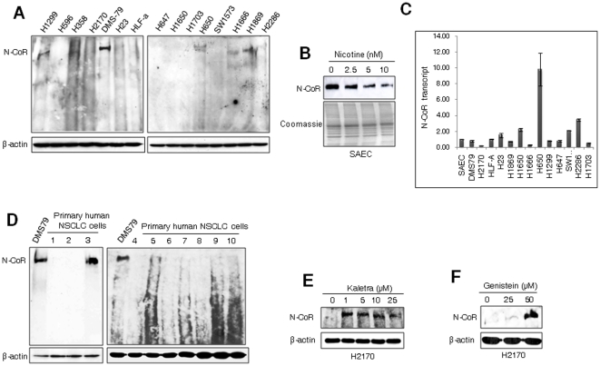
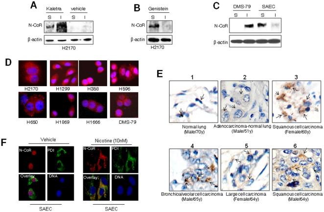
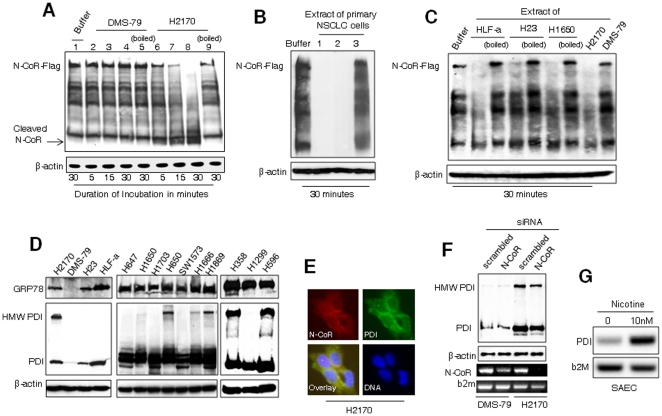
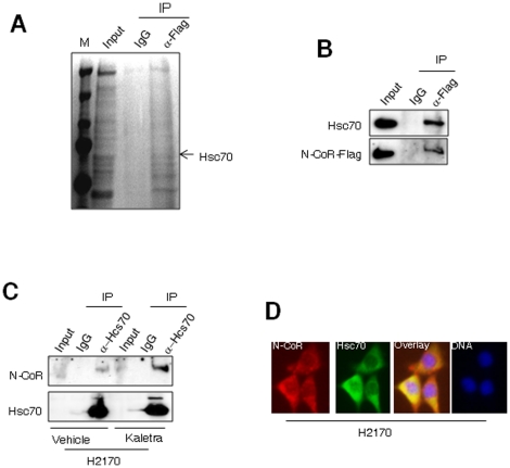
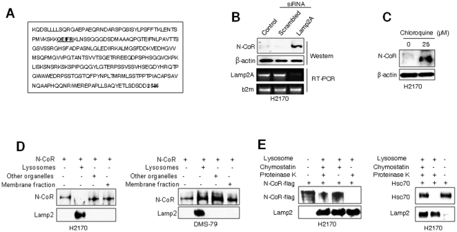
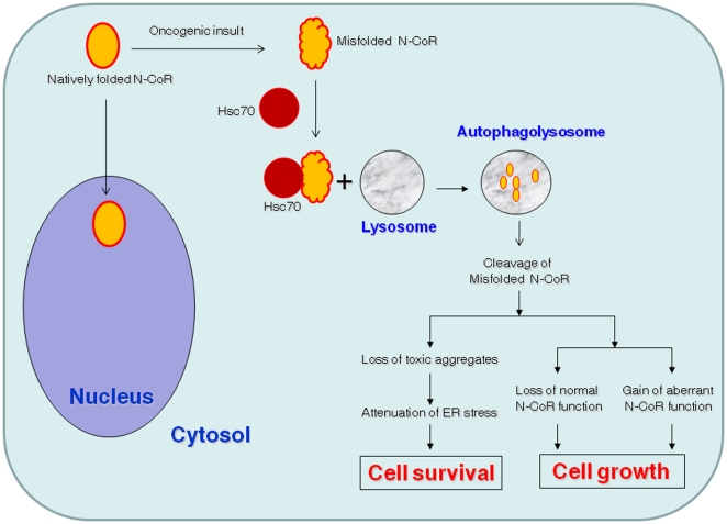
Nuclear receptor co-repressor (N-CoR) plays important role in transcriptional control mediated by several tumor suppressor proteins. Recently, we reported a role of misfolded-conformation dependent loss (MCDL) of N-CoR in the activation of oncogenic survival pathway in acute promyelocytic leukemia (APL). Since N-CoR plays important role in cellular homeostasis in various tissues, therefore, we hypothesized that an APL like MCDL of N-CoR might also be involved in other malignancy. Indeed, our initial screening of N-CoR status in various leukemia and solid tumor cells revealed an APL like MCDL of N-CoR in primary and secondary tumor cells derived from non-small cell lung cancer (NSCLC). The NSCLC cell specific N-CoR loss could be blocked by Kaletra, a clinical grade protease inhibitor and by genistein, an inhibitor of N-CoR misfolding previously characterized by us. The misfolded N-CoR presented in NSCLC cells was linked to the amplification of ER stress and was subjected to degradation by NSCLC cell specific aberrant protease activity. In NSCLC cells, misfolded N-CoR was found to be associated with Hsc70, a molecular chaperone involved in chaperone mediated autophagy (CMA). Genetic and chemical inhibition of Lamp2A, a rate limiting factor of CMA, significantly blocked the loss of N-CoR in NSCLC cells, suggesting a crucial role of CMA in N-CoR degradation. These findings identify an important role of CMA-induced degradation of misfolded N-CoR in the neutralization of ER stress and suggest a possible role of misfolded N-CoR protein in the activation of oncogenic survival pathway in NSCLC cells.


Conflict of interest statement

Competing Interests: The authors have declared that no competing interests exist.

Figures

Figure 1
Figure 1. Selective loss of N-CoR protein in NSCLC cells.
A & B, Level and integrity of full length N-CoR protein in various lung cancer cells was determined through western blotting assay. Level of β-actin was determined as experimental control. DMS-79 is derived from SCLC while rests of the cells are derived from NSCLC. B, Level of full length N-CoR protein in SAEC treated with nicotine for 48 hours in a dose dependent manner was determined. C, Level of N-CoR transcript in cell lines used in Figure 1 A & B was quantified by real time PCR. The identity of cells used in figure 1A & C is presented in Supporting Information S1. D, Level of full length N-CoR protein in ten histologically confirmed human primary NSCLC cells was determined in western blotting assay with N-CoR antibody. The identity of human samples is presented in Supporting Information S1. E and F, Level of intact N-CoR protein in H2170 cells treated with Kaletra (E) or genistein (F) was determined was determined in western blotting assay.
Figure 2
Figure 2. Loss of N-CoR in NSCLC cells is linked to misfolding.
A, B & C, Solubility (S)/insolubility (I) of N-CoR or β-actin in Kaletra (A) or genistein (B) treated H2170 cells, as well as in DMS-79 or SAEC cells (C), was determined by protein solubility assay. D, Subcellular distribution of N-CoR (red signal) in NSCLC (H2170, H1299, H358, H596, H650, H1869 and H1666) and SCLC (DMS-79) cells was determined by immunoflorescence assay performed with N-CoR antibody. DNA (blue signal) was stained with DAPI. E, Subcellular distribution of N-CoR in histologically confirmed human primary NSCLC tissue sections (panel 2–6) and normal human lung tissue section (panel 1) was determined by immunohistochemical (IHC) assay performed with N-CoR antibody. The brownish staining in the cytosol pinpointed by black arrow represents the N-CoR signal (panels 2–6). N-CoR staining in normal lung tissue section is visible as black dots overlapping the nucleus (panel 1). The second panel demonstrates a cross section that contains both cancer tissue (left half) and normal lung tissue (right half) side by side. F, Subcellular distribution of N-CoR and PDI in SAEC cells treated with vehicle or nicotine for 48 hours was determined by confocal microscopy.
Figure 3
Figure 3. NSCLC cell specific N-CoR loss is linked to endoplasmic reticulum (ER) stress.
A, H2170 cells harbor N-CoR cleaving activity. Processing of Flag-tagged N-CoR incubated with the extracts of DMS-79 (lanes 2–5) or H2170 (lanes 6–9) cells for the duration as mentioned was determined by western blotting assay performed with Flag antibody. The extract loaded in lanes labeled as “boiled” was heated at 100°C for 10 minutes prior to incubation. Equal volume of buffer lacking cell extract was used in lane labeled as “buffer”. The arrow marks position of the cleaved 100 kDa N-CoR fragment. B, Processing Flag-tagged N-CoR incubated with the extracts of first three human primary NSCLC cells (Supplementary table 2) was determined by western blotting with Flag antibody. C, N-CoR cleavage assay, using extracts of NSCLC cells HLF-a, H23 or H1650, was performed essentially as described in the legend of Figure 2A. D, Level of ER stress in various lung cancer cells was analyzed by determining the levels of GRP-78 and PDI (basal and HMW: high molecular weight). E, N-CoR localized to ER was visualized by staining H2170 cells with N-CoR (red) and PDI (green) antibodies. DNA was stained with DAPI (blue). F, Level of PDI in H2170 or DMS-79 cells exposed to scramble or N-CoR siRNA was determined by western blotting (upper panel). The N-CoR knock-down efficiency in each subset of cells was confirmed by RT-PCR assay (lower panels). G, Level of PDI transcript in SAEC cells treated with nicotine for 48 hours was determined by RT-PCR analysis.
Figure 4
Figure 4. N-CoR is a substrate of chaperone mediated autophagy (CMA).
A, Column purified cytosolic extracts of H2170 cells was incubated with flag-tagged N-CoR ectopically expressed in 293T cells. N-CoR was immunoprecipitated with anti-flag antibody and proteins co-precipitated with N-CoR were resolved in SDS-PAGE, excised and their identity was determined by MS. B, The association between Flag-tagged N-CoR and Hsc70 was reconfirmed in co-immunoprecipitation assay performed essentially as described in legend of figure 4A. After detection of co-precipitated Hsc70 with Hsc70 antibody (upper panel), the membrane was re-probed with flag antibody to quantify the amount of precipitated N-CoR protein (lower panel). In “input” lanes, an aliquot of whole cell extract was loaded. C, Hsc70 was immunoprecipitated from the extracts of vehicle or 5 µM Kaletra treated H2170 cells and level of co-precipitated N-CoR was determined with N-CoR antibody (upper panel). The membrane was re-probed with Hsc70 antibody (lower panel). In “input” lanes, an aliquot of whole cell extract was loaded. D, N-CoR (red) and Hsc70 (green) distribution in H2170 cells was determined through immunoflorescence analysis using confocal microscopy. The intensity of yellow signals signifies the degree of co-localization of two proteins. DNA was labeled with DAPI (blue).
Figure 5
Figure 5. N-CoR is degraded by chaperone mediated autophagy (CMA).
A, N-CoR contains a consensus lysosomal targeting motif. The lysosomal targeting motif in N-CoR peptide sequence is highlighted in underlined bold. B, Lamp2A ablation blocked N-CoR loss in H2170 cells. N-CoR level in H2170 cells exposed to anti-Lamp2A or control siRNA for 72 hours was determined with N-CoR antibody (upper panel) after confirming Lamp2A ablation by RT-PCR analysis (lower panel). C, Chemical inhibition of lysosomal function stabilized N-CoR. Level of N-CoR in H2170 cells treated for 72 hours with vehicle or 25 µM chloroquine, a selective inhibitor of lysosomal function, was determined with N-CoR antibody. D, Flag-tagged N-CoR was incubated with purified lysosomal or non-lysosomal fractions of H2170 (left) or DMS-79 (right) cells and level of N-CoR degradation was determined with flag antibody. Level of Lamp2, a lysosomal protein, was determined to demonstrate the purity of each fraction. E, N-CoR taken up by chymostatin treated lysosomes was protected from proteinase K digestion. Flag-tagged N-CoR or Hsc70 incubated with lysosomes isolated form H2170 cells pretreated with chymostatin was subjected to proteinase K digestion and relative level of protected N-CoR or Hsc70 was determined with flag or Hsc70 antibody. Lamp2 level was determined to quantify the amount of lysosomes in each sample.
Figure 6
Figure 6. Schematic representation of the role of CMA-induced degradation of misfolded N-CoR in NSCLC cells.
This model illustrates how CMA-induced degradation of misfolded N-CoR could contribute to the survival and growth of NSCLC cells through the loss of normal function and gain of aberrant function mechanism.

Similar articles

Cited by

References

    1. Mallakin A, Sugiyama T, Taneja P, Matise LA, Frazier DP, et al. Mutually exclusive inactivation of DMP1 and ARF/p53 in lung cancer. Cancer Cell. 2007;12:381–394. - PMC - PubMed
    1. Bassères DS, Levantini E, Ji H, Monti S, Elf S, et al. Respiratory failure due to differentiation arrest and expansion of alveolar cells following lung-specific loss of the transcription factor C/EBP in mice. Mol Cell Biol. 2006;26:1109–1123. - PMC - PubMed
    1. Halmos B, Huettner CS, Kocher O, Ferenczi K, Karp DD, et al. Down-regulation and anti-proliferative role of C/EBPalpha in lung cancer. Cancer Res. 2002;62:528–534. - PubMed
    1. Halmos B, Bassères DS, Monti S, D'Aló F, Dayaram T, et al. A transcriptional profiling study of CCAAT/Enhancer binding protein targets identifies hepatocyte nuclear factor 3 as a novel tumor suppressor in lung cancer. Cancer Res. 2004;64:4137–4147. - PubMed
    1. Khan MM, Nomura T, Kim H, Kaul SC, Wadhwa R, et al. PML-RAR alleviates transcriptional repression mediated by tumor suppressor Rb. J Biol Chem. 2001;276:43491–43494. - PubMed

Publication types