. 2011;6(9):e25271.
doi: 10.1371/journal.pone.0025271. Epub 2011 Sep 22.

GW501516, a PPARδ agonist, ameliorates tubulointerstitial inflammation in proteinuric kidney disease via inhibition of TAK1-NFκB pathway in mice

Affiliations

GW501516, a PPARδ agonist, ameliorates tubulointerstitial inflammation in proteinuric kidney disease via inhibition of TAK1-NFκB pathway in mice

Xu Yang et al. PLoS One. 2011.

Abstract

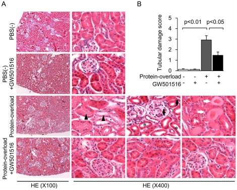
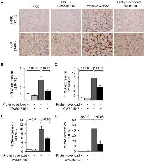
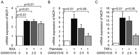
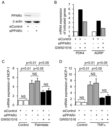
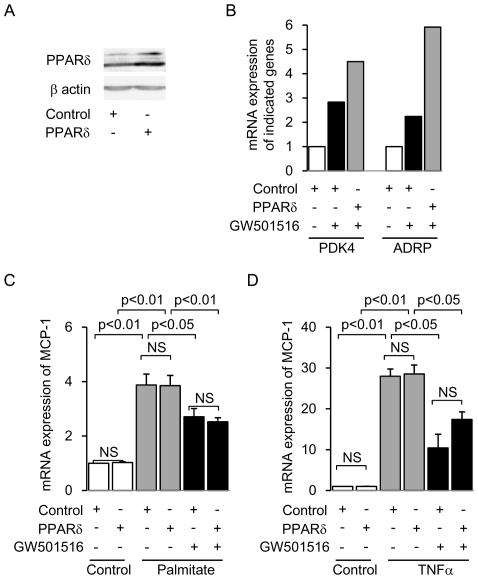
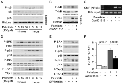
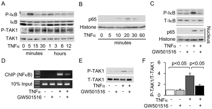
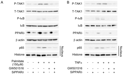
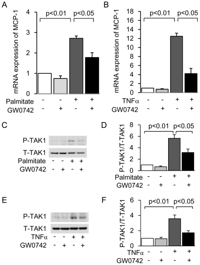
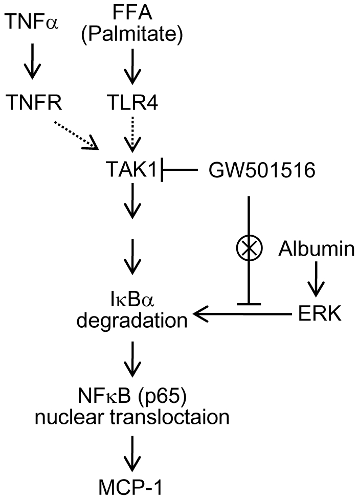
Peroxisome proliferator-activated receptors (PPARs) are a nuclear receptor family of ligand-inducible transcription factors, which have three different isoforms: PPARα, δ and γ. It has been demonstrated that PPARα and γ agonists have renoprotective effects in proteinuric kidney diseases; however, the role of PPARδ agonists in kidney diseases remains unclear. Thus, we examined the renoprotective effect of GW501516, a PPARδ agonist, in a protein-overload mouse nephropathy model and identified its molecular mechanism. Mice fed with a control diet or GW501516-containing diet were intraperitoneally injected with free fatty acid (FFA)-bound albumin or PBS(-). In the control group, protein overload caused tubular damages, macrophage infiltration and increased mRNA expression of MCP-1 and TNFα. These effects were prevented by GW501516 treatment. In proteinuric kidney diseases, excess exposure of proximal tubular cells to albumin, FFA bound to albumin or cytokines such as TNFα is detrimental. In vitro studies using cultured proximal tubular cells showed that GW501516 attenuated both TNFα- and FFA (palmitate)-induced, but not albumin-induced, MCP-1 expression via direct inhibition of the TGF-β activated kinase 1 (TAK1)-NFκB pathway, a common downstream signaling pathway to TNFα receptor and toll-like receptor-4. In conclusion, we demonstrate that GW501516 has an anti-inflammatory effect in renal tubular cells and may serve as a therapeutic candidate to attenuate tubulointerstitial lesions in proteinuric kidney diseases.


Conflict of interest statement

Competing Interests: The authors have declared that no competing interests exist.

Figures

Figure 1
Figure 1. GW501516 attenuates renal proximal tubular cell damage in a protein-overload mouse renal injury model.
(A) Representative HE staining of kidney sections from four groups [PBS(−), PBS(−) + GW501516, Protein-overload and Protein-overload + GW501516]. In the kidney of the protein-overload group, diffused tubular cell vacuolation (black triangles), tubular cell flattening (black arrows), tubular lumen dilation (asterisks) and cast formation (white arrows) were observed (magnification ×100 and ×400). (B) Tubulointerstitial damage scores of kidney sections from four groups. Data are shown as means ± SEM of each individual group.
Figure 2
Figure 2. GW501516 suppresses interstitial inflammation in a protein-overload mouse renal injury model.
(A) Representative photomicrographs of F4/80 immunostaining of the kidney from four groups [PBS(−), PBS(−) + GW501516, Protein-overload and Protein-overload + GW501516] (magnification, upper: ×100, lower: ×400). Renal mRNA expression of F4/80 (B), MCP-1 (C), TNFα (D) and IL-6 (E) in kidney samples from the four groups. Data are shown as means ± SEM. Results are expressed as the fold change relative to the mean value of the mRNA expression level of the PBS(−) group. MCP-1: monocyte chemoattractant protein-1; TNFα: tumor necrotic factor α.
Figure 3
Figure 3. GW501516 inhibits palmitate- and TNFα-induced MCP-1 expression in mouse proximal tubular (mProx) cells.
mRNA expression of MCP-1 determined by real-time PCR in cultured mProx cells stimulated by 30% BSA (A), 150 µM palmitate (B) or 10 nM of TNFα (C) for 12 hours, with or without a 3-hour pretreatment of different concentrations of GW501516 (0, 2.5, 5 µM of GW501516 dissolved in 0.05% DMSO). Results are expressed as fold change relative to control mRNA levels. Data are shown as means ± SEM of at least four independent experiments. MCP-1: monocyte chemoattractant protein-1; BSA: bovine serum albumin; TNFα: tumor necrotic factor α.
Figure 4
Figure 4. Knockdown of PPARδ gene expression does not affect GW501516-mediated inhibition of MCP-1 expression in mouse proximal tubular (mProx) cells treated with palmitate or TNFα.
(A) Representative immunoblot showing protein expression of PPARδ in siRNA-mediated PPARδ-knockdown and control cells. (B) mRNA expression of PPARδ-target genes (PDK4 and ADRP) in PPARδ-knockdown and control cells treated with or without GW501516. Results are expressed as fold change relative to the mRNA from the control group. PDK4: Pyruvate dehydrogenase kinase 4; ADRP: Adipocyte differentiation-related protein. The effect of PPARδ knockdown on GW501516-mediated inhibition of palmitate- (C) and TNFα- (D) induced MCP-1 expression. Results are expressed as fold change relative to the mRNA from the control group. Data are shown as means ± SEM of three independent experiments. MCP-1: monocyte chemoattractant protein-1; TNFα: tumor necrotic factor α.
Figure 5
Figure 5. Overexpression of PPARδ does not affect GW501516-mediated attenuation of MCP-1 expression in mouse proximal tubular (mProx) cells treated with palmitate or TNFα.
(A) Representative immunoblot showing protein expression of PPARδ in retrovirus-mediated PPARδ-overexpressing cells and control cells. (B) mRNA expression of PPARδ-target genes (PDK4 and ADRP) in PPARδ-overexpressing and control cells treated with or without GW501516. Results are expressed as fold change relative to the mRNA from the control group. PDK4: Pyruvate dehydrogenase kinase 4; ADRP: Adipocyte differentiation-related protein. The effect of PPARδ overexpression on GW501516-mediated inhibition of palmitate- (C) and TNFα- (D) induced MCP-1 expression. Results are expressed as fold change relative to the mRNA from the control group. Data are shown as means ± SEM of four independent experiments. MCP-1: monocyte chemoattractant protein-1; TNFα: tumor necrotic factor α.
Figure 6
Figure 6. GW501516 inhibits palmitate-induced NFκB activation via inhibition of TAK1 in mouse proximal tubular (mProx) cells.
(A) Immunoblot analysis showing palmitate-induced phosphorylation of IκB in the total cell lysate and nuclear translocation of the p65 subunit in a time course experiment. (B) Immunoblot analysis showing the effect of GW501516 on palmitate-induced phosphorylation of IκB and nuclear translocation of p65. The pretreatment time with GW501516 was 3 hours and the stimulation time with palmitate was 3 hours for phosphorylation of IκB and nuclear translocation of p65. The images are representative pictures from three independent experiments. (C) Effect of GW501516 on DNA binding of p65 to the MCP-1 promoter determined by chromatin immunoprecipitation (ChIP) analysis in palmitate-treated mProx. (D) Immunoblot analysis showing palmitate-induced phosphorylation of MAPKs (P-ERK1/2, P-p38, P-JNK) and TAK1 in a time course experiment. (E) Immunoblot analysis showing the effect of GW501516 on phosphorylation of MAPKs and TAK1. Pre-incubation time with GW501516 was 3 hours, and stimulation time with palmitate was 1 hour. Images are representative figures from three independent experiments. TAK1: TGF-β activated kinase 1. (F) Quantitative results of the ratio of band density of P-TAK1 and T-TAK1.
Figure 7
Figure 7. GW501516 inhibits TNFα-induced NFκB activation via inhibition of TAK1 in mouse proximal tubular (mProx) cells.
Immunoblot analysis showing TNFα-induced phosphorylation of IκB and TAK1 (A) and nuclear translocation of the p65 subunit (B) in a time course experiment. (C) Immunoblot analysis showing the effect of GW501516 on TNFα-induced phosphorylation of IκB and nuclear translocation of p65. The pretreatment time with GW501516 was 3 hours and the stimulation time with TNFα was 5 minutes for phosphorylation of IκB and 10 minutes for nuclear translocation of p65. Images are representative results from three independent experiments. (D) Effect of GW501516 on DNA binding of p65 to the MCP-1 promoter determined by chromatin immunoprecipitation (ChIP) analysis in TNFα-treated mProx cells. (E) Immunoblot analysis showing the effect of GW501516 on TAK1 phosphorylation. Pre-incubation time with GW501516 was 3 hours and stimulation time with TNFα was 5 minutes. (F) Quantitative result of the ratio of band density of P-TAK1 and T-TAK1. Images are representative figures from three independent experiments. TAK1: TGF-β activated kinase 1; TNFα: tumor necrotic factor α.
Figure 8
Figure 8. Knockdown of PPARδ gene expression does not affect the GW501516-mediated inhibitory effect on the TAK1-NFκB pathway in mouse proximal tubular (mProx) cells treated with palmitate or TNFα.
Immunoblot analysis showing the effect of GW501516 on palmitate- (A) and TNFα- (B) induced phosphorylation of IκB and TAK1 in total cell lysate and nuclear translocation of the p65 subunit in siRNA-mediated PPARδ-knockdown and control cells. Images are representative figures from three independent experiments. TAK1: TGF-β activated kinase 1; TNFα: tumor necrotic factor α.
Figure 9
Figure 9. GW0742 inhibits palmitate- and TNFα-induced MCP-1 expression as well as TAK1 phosphorylation in mouse proximal tubular (mProx) cells.
mRNA expression of MCP-1 determined by real-time PCR in cultured mProx cells stimulated by 150 µM palmitate (A) or 10 nM TNFα (B) for 12 hours, with or without a 3-hour pretreatment with GW0742 (5 µM GW0742 dissolved in 0.05% DMSO). Results are expressed as fold change relative to control mRNA levels. (C) Immunoblot analysis showing palmitate-induced phosphorylation of TAK1. (D) Quantitative result of the ratio of band density of P-TAK1 and T-TAK1. (E) Immunoblot analysis showing TNFα-induced phosphorylation of TAK1. (F) Quantitative result of the ratio of band density of P-TAK1 and T-TAK1. MCP-1: monocyte chemoattractant protein-1. TNFα: tumor necrotic factor α; TAK1: TGF-β activated kinase 1. Data are shown as means ± SEM of at least three independent experiments.
Figure 10
Figure 10. Proposed mechanism underlying the GW501516-mediated anti-inflammatory effect in proximal tubular cells in proteinuric kidney diseases.
Both TNFα and palmitate induce the phosphorylation of TAK1 in a receptor dependent manner. The phosphorylation of TAK1 activates the NFκB pathway via degradation of IκB. The p65 subunit of NFκB translocates into the nucleus, upregulates the transcription of the pro-inflammatory gene, MCP-1, and causes inflammation. GW501516 can inhibit the phosphorylation of TAK1, but not of MAPKs (including ERK), and the activation of the NFκB-associated inflammatory response in proximal tubular cells. TNFα: tumor necrotic factor α; TNFR: TNFα receptor; TLR4: toll-like receptor 4; IKK: IκB Kinase; FFA: free fatty acid; MCP-1: monocyte chemoattractant protein-1.

Similar articles

Cited by

References

    1. Abbate M, Zoja C, Remuzzi G. How does proteinuria cause progressive renal damage? J Am Soc Nephrol. 2006;17:2974–2984. - PubMed
    1. Burton C, Harris KP. The role of proteinuria in the progression of chronic renal failure. Am J Kidney Dis. 1996;27:765–775. - PubMed
    1. Thomas ME, Schreiner GF. Contribution of proteinuria to progressive renal injury: consequences of tubular uptake of fatty acid bearing albumin. Am J Nephrol. 1993;13:385–398. - PubMed
    1. Kamijo A, Kimura K, Sugaya T, Yamanouchi M, Hase H, et al. Urinary free fatty acids bound to albumin aggravate tubulointerstitial damage. Kidney Int. 2002;62:1628–1637. - PubMed
    1. Zoja C, Donadelli R, Colleoni S, Figliuzzi M, Bonazzola S, et al. Protein overload stimulates RANTES production by proximal tubular cells depending on NF-kappa B activation. Kidney Int. 1998;53:1608–1615. - PubMed

Publication types

MeSH terms