. 2011;6(9):e25540.
doi: 10.1371/journal.pone.0025540. Epub 2011 Sep 26.

Basal LAT-diacylglycerol-RasGRP1 signals in T cells maintain TCRα gene expression

Affiliations

Basal LAT-diacylglycerol-RasGRP1 signals in T cells maintain TCRα gene expression

Evan Markegard et al. PLoS One. 2011.

Abstract

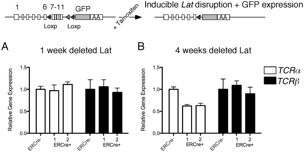
In contrast to the well-characterized T cell receptor (TCR) signaling pathways that induce genes that drive T cell development or polarization of naïve CD4 T cells into the diverse T(H)1, T(H)2, T(H)17 and T(reg) lineages, it is unclear what signals maintain specific gene expression in mature resting T cells. Resting T cells residing in peripheral lymphoid organs exhibit low-level constitutive signaling. Whereas tonic signals in B cells are known to be critical for survival, the roles of tonic signals in peripheral T cells are unknown. Here we demonstrate that constitutive signals in Jurkat T cell lines are transduced via the adapter molecule LAT and the Ras exchange factor RasGRP1 to maintain expression of TCRα mRNA and surface expression of the TCR/CD3 complex. Independent approaches of reducing basal activity through the LAT-diacylglycerol-RasGRP pathway led to reduced constitutive Ras-MEK-ERK signals and decreased TCRα mRNA and surface TCR expression in Jurkat cells. However, loss of TCR expression takes several days in these cell line experiments. In agreement with these in vitro approaches, inducible deletion of Lat in vivo results in reduced TCRα mRNA- and surface TCR-expression in a delayed temporal manner as well. Lastly, we demonstrate that loss of basal LAT-RasGRP signals appears to lead to silencing or repression of TCRα transcription. We postulate that basal LAT-diacylglycerol-RasGRP signals fulfill a regulatory function in peripheral T lymphocytes by maintaining proper gene expression programs.


Conflict of interest statement

Competing Interests: The authors have declared that no competing interests exist.

Figures

Figure 1
Figure 1. Loss of Basal Ras-ERK signaling in Jurkat-PMA-Response-Mutant (JPRM) clones.
(A) RT-PCR analysis for RasGRP-1, -2, -3, -4, and GAPDH gene expression in wild type Jurkat T cells and a panel of mutant JPRM cell lines. Five-fold dilutions of cDNA input were used as template. (B) Western blot analysis of basal levels of phosphorylated ERK-1 and –2 in resting wild type Jurkat T cells, RasGRP1-deficient JPRM441 cells, LAT-deficient J.CaM2 cells, and 4 additional JPRM mutant cell lines. Equal loading is indicated by α-tubulin levels on the same blot. Relative levels of ERK-1 and –2 phosphorylation were calculated by normalizing for α-tubulin and set to 100% in the Jurkat sample. (C) Western blot analysis of RasGRP1 and LAT expression and basal levels of phosphorylated ERK-1 and –2 in the indicated resting cell types. All panels in figure 1 are representative examples of three independent experiments.
Figure 2
Figure 2. Decreased surface TCR/CD3 expression in Jurkat signaling mutant lines and primary T cells deleted for Lat.
(A) Overview of the clonal cell lines used in this study, their identified deficiencies, and surface TCR/CD3 expression characteristics. (B) Surface CD3 expression by FACS analysis in the indicated cell lines. Solid grey histograms represent CD3 expression in wild type Jurkat cells. A representative example of three independent experiments. (C) Schematic of targeting approach that allows tamoxifen-inducible excision of exons 7–11 in Lat. (D, E) Dot plot FACS analyses of surface TCR expression levels and GFP expression against surface CD4 expression on the x-axis. Depicted are representative results of three independent experiments obtained with lymph node cells from ERCre Latf/− and ERCre+ Latf/− mice that either received no tamoxifen or tamoxifen on the first two days and subsequently every week. T cells were analyzed at 1 week or 4 weeks after the initial tamoxifen dose. Figures 2D and 2E are representative of findings with at least three individual mice per genotype.
Figure 3
Figure 3. Loss of TCRα transcripts in mutant Jurkat cell lines with impaired tonic Ras-ERK signaling.
(A, B) Northern blot analysis of transcript levels of TCRα and TCRβ in a panel of resting wild type and mutant Jurkat cell lines. Beta-actin mRNA levels were assessed as a control for loading. (C) Northern blot analysis of transcript levels of the TCRα, TCRβ, and Beta-actin in Jurkat, JPRM441, J.CaM2, and the two mutant lines stably reconstituted with RasGRP1 or LAT cDNA. (D) Western blot analysis of Jurkat and JPRM441 samples stimulated for 10 minutes with the indicated doses of PMA. Levels of ERK-1 and –2 phosphorylation and α-tubulin were determined on the same blot. Northern blot analysis of transcript levels of TCRα and TCRβ in wild type Jurkat and RasGRP1 deficient JPRM441 left unstimulated or stimulated with 2, 10, and 15, or for JPRM441 with 5, 15, and 25 ng/ml PMA for 20 hours, respectively. Percentages of relative expression were determined by normalizing for Beta-actin expression. The unstimulated Jurkat sample was set at 100%. All panels in figure 3 are representative examples of three independent experiments.
Figure 4
Figure 4. LAT-deletion in primary CD4+ T cells results in time-dependent loss of TCRα transcripts.
(A, B) Taqman PCR analysis of TCRα and TCRβ mRNA expression levels in sorted CD4+CD44low T cells from two ERCre+ Latf/− mice and one control ERCre Latf/− mouse. Error bars represent taqman triplicates. The results are a representative example of two independent experiments.
Figure 5
Figure 5. Repression of TCRα expression by limiting DAG-RasGRP1 signals requires several days and can be reversed by TSA or 5-aza inhibitors.
(A) Bargraph representing mean fluorescence signals for surface expression of CD3 on wild type Jurkat cells transfected with vector control, RasGRP1 RNAi, or DGKζ expression constructs, all together with a CD16 expression construct as marker. Surface CD3 expression was determined on CD16 positive cells, at 1, 2, or 3 days following transfection. Expression of CD3 on unmanipulated cells was arbitrarily set at 100%. Means and standard errors of 3 independent experiments are depicted. Western blot analysis of a representative experiment, analyzing unsorted cells for the indicated protein expression 3 days after transfection, indicates the level of FLAG-tagged DGKζ expression and the degree of RasGRP1 knockdown. (B) Northern blot analysis of transcript levels of the indicated TCR components and Western blot analyses of the indicated protein expression levels examining Jurkat T cells 4 days after transient transfection with the indicated RNAi or control construct. (C) Taqman PCR analysis of TCRα and TCRβ mRNA expression levels in sorted CD4+CD44low T cells from wild type mice treated for 24 hours with the indicated inhibitors in the absence of stimuli. Error bars represent taqman triplicates. (D) Northern blot analysis of transcript levels of the indicated TCR components and Western blot analyses of the indicated protein expression levels examining the indicated cell lines treated for 20 hours with Trichostatin A (TSA) or 5-azacytidine (5-aza). Of note, combined treatment of TSA and 5-aza was too toxic to obtain good quality RNA. Panels 5B–5C are representative examples of three independent experiments.

References

    1. Lin J, Weiss A. T cell receptor signalling. J Cell Sci. 2001;114:243–244. - PubMed
    1. Tomlinson MG, Lin J, Weiss A. Lymphocytes with a complex: adapter proteins in antigen receptor signaling. Immunol Today. 2000;21:584–591. - PubMed
    1. Feske S. Calcium signalling in lymphocyte activation and disease. Nat Rev Immunol. 2007;7:690–702. - PubMed
    1. Stone JC. Regulation of Ras in lymphocytes: get a GRP. Biochem Soc Trans. 2006;34:858–861. - PubMed
    1. Lindsten T, June CH, Thompson CB. Transcription of T cell antigen receptor genes is induced by protein kinase C activation. J Immunol. 1988;141:1769–1774. - PubMed

Publication types

MeSH terms