. 2011:1:27.
doi: 10.4103/2156-7514.81294. Epub 2011 May 19.

Diffusion-weighted Magnetic Resonance Imaging: What Makes Water Run Fast or Slow?

Affiliations

Diffusion-weighted Magnetic Resonance Imaging: What Makes Water Run Fast or Slow?

Francesca Fornasa. J Clin Imaging Sci. 2011.

Abstract

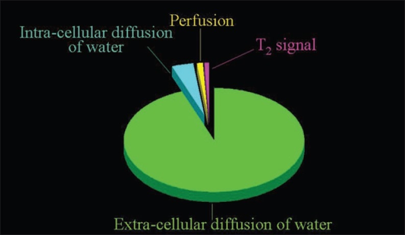
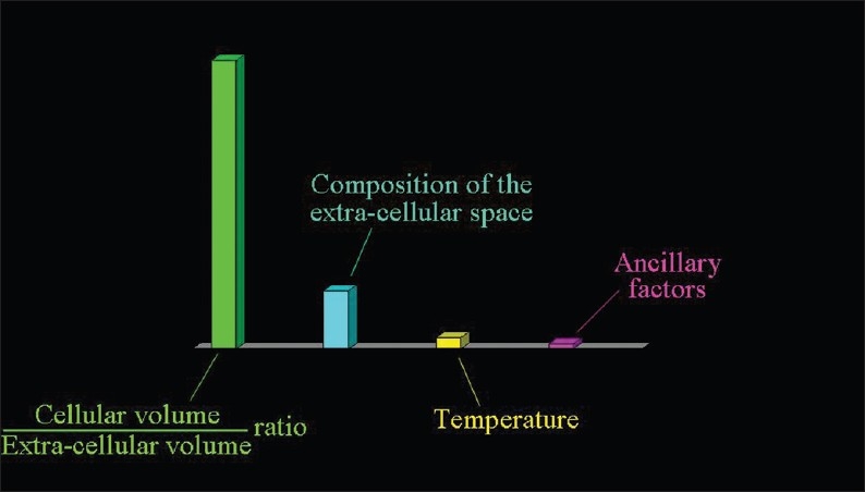
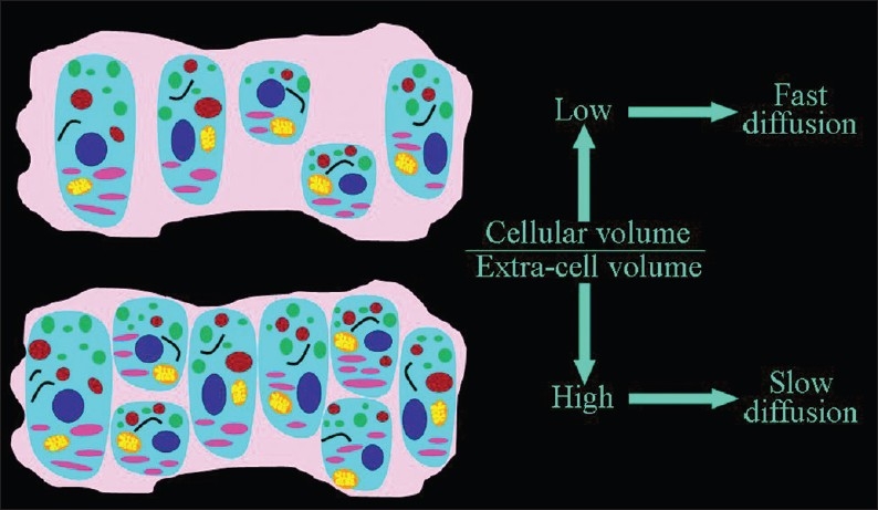
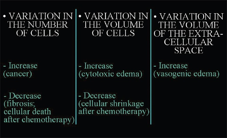
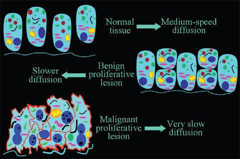
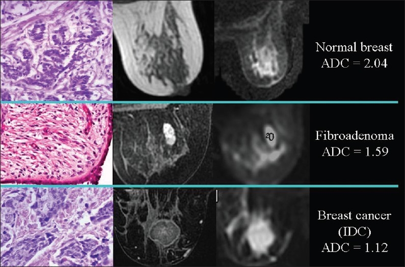
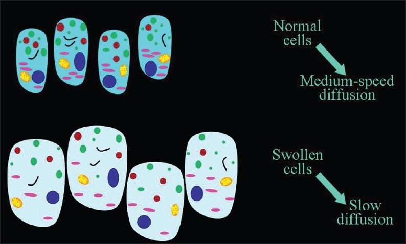
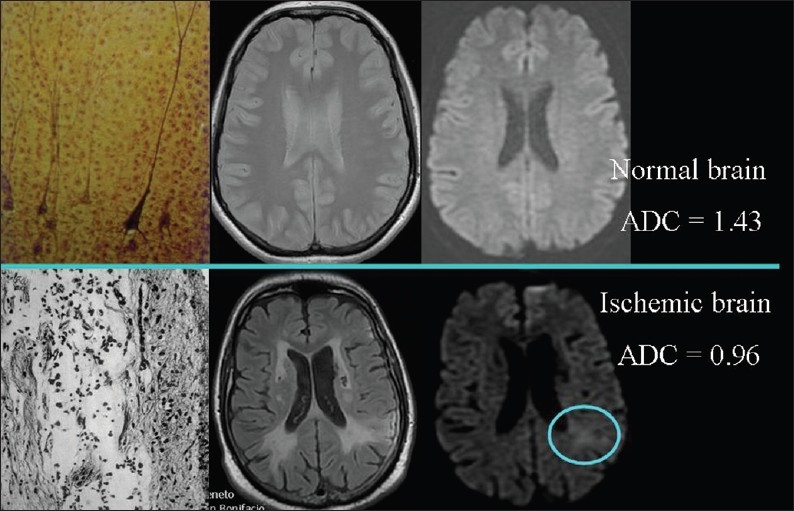
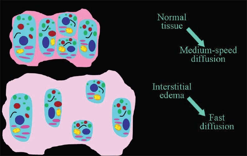
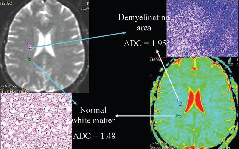
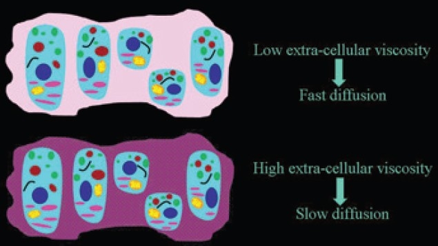
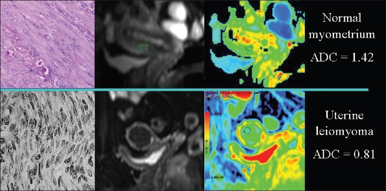
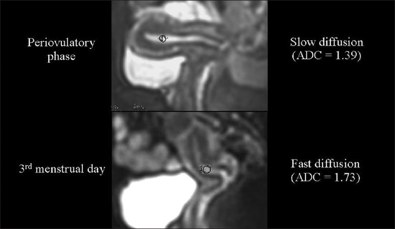
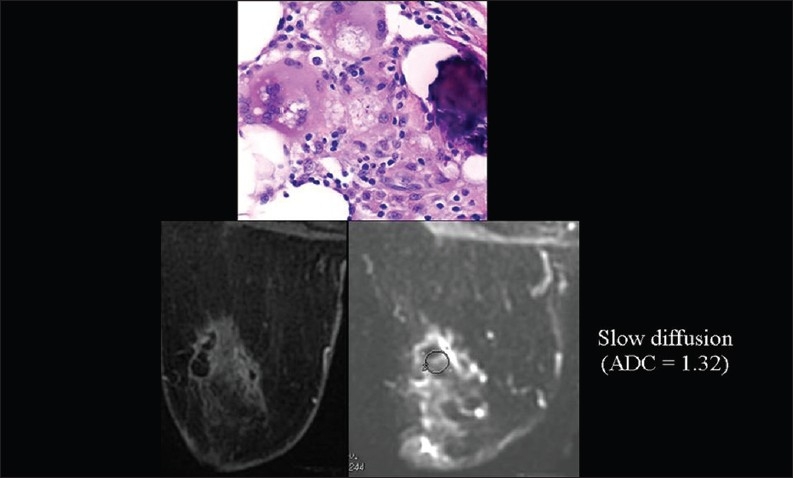
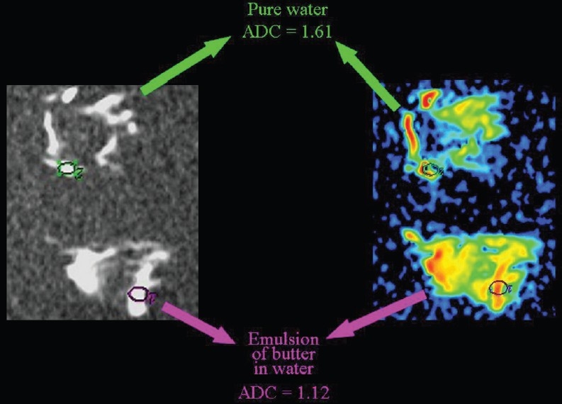
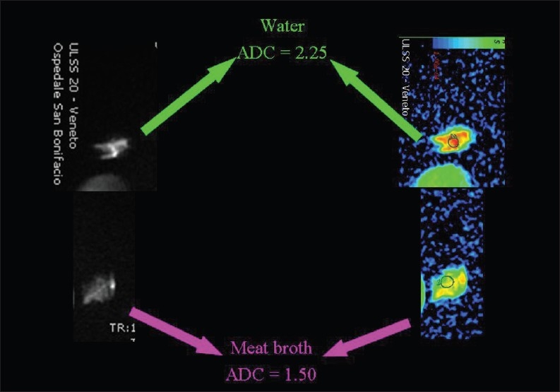
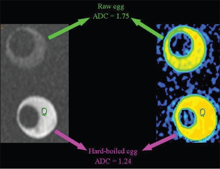
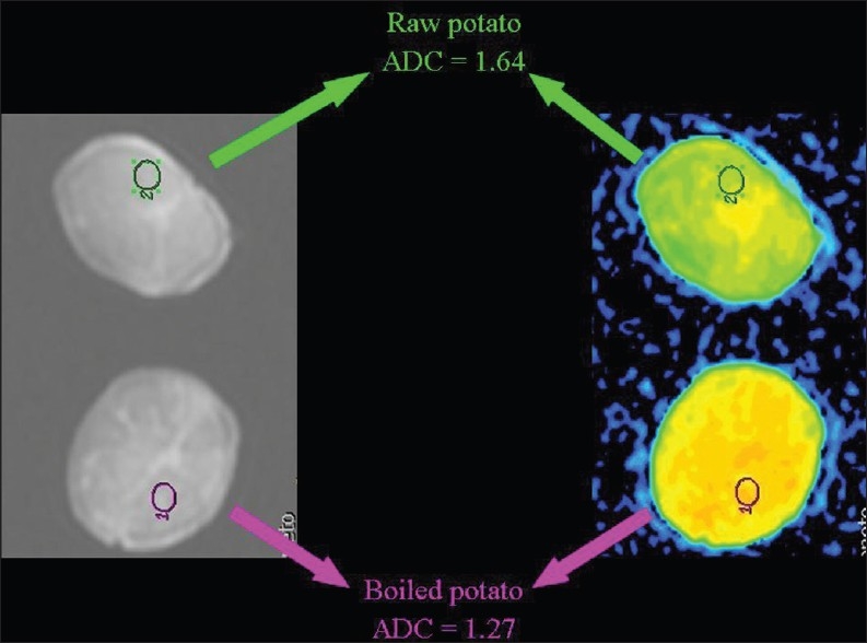
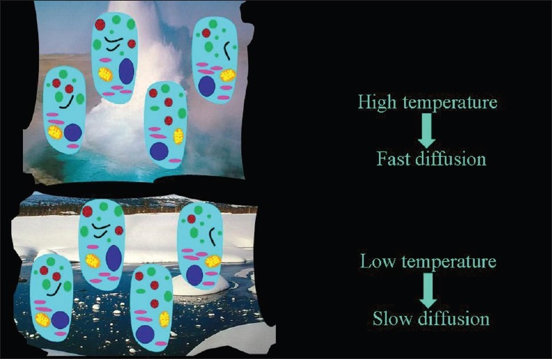
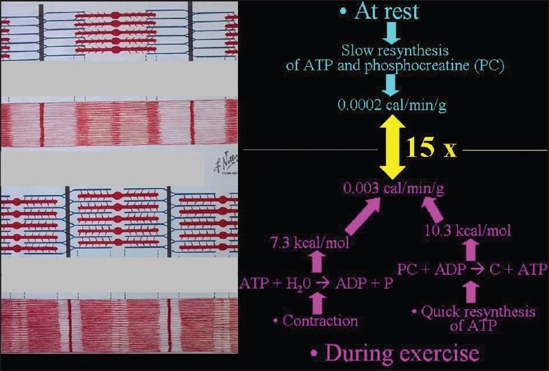
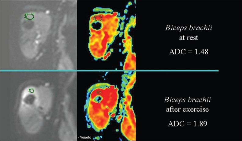
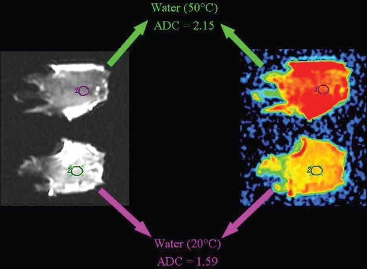
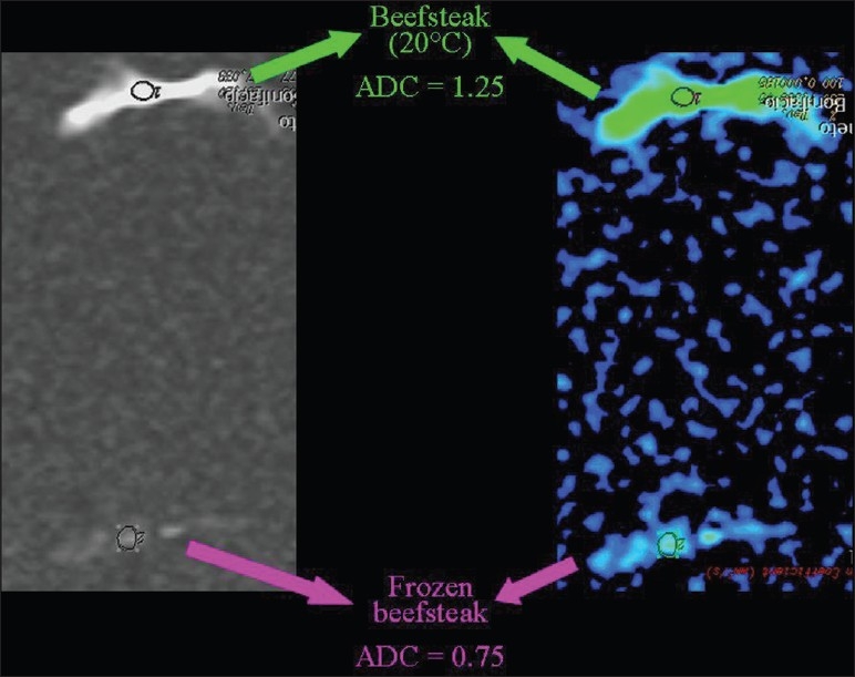
Diffusion-Weighted Magnetic Resonance Imaging (DWI) obtains information useful in diagnosing several diseases through the measurement of random, Brownian diffusion of water molecules in tissues. This pictorial essay illustrates the main factors, i.e., ratio between the volume occupied by cells and the extracellular space, composition of the extracellular space, and temperature, that determine the rate of the water diffusion. The mechanism through which these influencing factors affect water diffusion is explained. Clinical and experimental examples, derived both from physiology and from non-human models, are described.

Keywords: Diffusion weighted imaging; MR imaging; MRI Physics; water.


Conflict of interest statement

Conflict of Interest: None declared.

Figures

Figure 1
Figure 1
The sources of the signal measured with the Diffusion-Weighted Magnetic Resonance Imaging protocols routinely adopted. The overwhelming portion of the signal derives from the extracellular diffusion of water
Figure 2
Figure 2
The factors determining the speed of the extracellular diffusion of water: the ratio between the volume occupied by cells and the extracellular space (the most important), the composition of the extracellular milieu, and the temperature.
Figure 3
Figure 3
The dependence of the speed of the extracellular diffusion on the ratio between the volume occupied by cells and the extracellular space. The higher the percentage of tissue occupied by cells, the slower the extracellular motion of water molecules
Figure 4
Figure 4
The factors that lead to changes in the ratio between the volume occupied by cells and the extracellular space.
Figure 5
Figure 5
The inverse relationship of the speed of diffusion to the number of cells. A slower diffusion can be forecast in malignant than in benign proliferative lesions, and in benign lesions than in normal tissue
Figure 6
Figure 6
The relationship of diffusion to the number of cells. ADC values are lower, because of the higher cellularity, in proliferative lesions than in normal breast tissue; in an invasive ductal carcinoma the diffusion is slower than in a fibroadenoma
Figure 7
Figure 7
The inverse relationship of the speed of diffusion to the volume of cells
Figure 8
Figure 8
The relationship of diffusion to the volume of cells. ADC values are lower, because of cellular swelling, in areas of cerebral ischemia (light blue circle) than in normal brain
Figure 9
Figure 9
The direct relationship of the speed of diffusion to the volume of extracellular space.
Figure 10
Figure 10
The relationship of diffusion to the volume of extra-cellular space. ADC values are higher, because of interstitial edema, in an acute demyelinating plaque than in a normal area of white matter in a patient suffering from multiple sclerosis.
Figure 11
Figure 11
The dependence of the speed of the extracellular diffusion on the composition of the extracellular space. The higher the interstitial viscosity, slower is the extracellular motion of water molecules.
Figure 12
Figure 12
The relationship of diffusion to the composition of the extracellular space. ADC values are lower, because of collagen deposition, in a uterine leiomyoma than in the normal myometrium
Figure 13
Figure 13
The relationship of diffusion to the composition of the extracellular space. Endometrial ADC values are lower (we hypothesize because of glycogen extrusion), during the periovulatory than during the menstrual phase.
Figure 14
Figure 14
The relationship of diffusion to the composition of the extracellular space. ADC values are very low (we hypothesize because of the high amount of extracellular hydrophobic lipid molecules), mimicking cancer, in breast fat necrosis.
Figure 15
Figure 15
The relationship of diffusion to the composition of the extracellular space. ADC values are lower (we hypothesize because of the presence of hydrophobic lipid molecules) within an emulsion of butter in water than in pure water.
Figure 16
Figure 16
The relationship of diffusion to the composition of the extracellular space. ADC values are lower (we hypothesize because of the presence of proteins extruded from muscle tissue) within meat broth than in pure water
Figure 17
Figure 17
The relationship of diffusion to the composition of the extracellular space. ADC values are lower (we hypothesize because of the increased viscosity subsequent to denaturation of albumin molecules) in the white of a hard-boiled than of a raw egg.
Figure 18
Figure 18
The relationship of diffusion to the composition of the extracellular space. ADC values are lower (we hypothesize because of the increased viscosity subsequent to degradation of starch to numerous small saccharide molecules) in a boiled than in a raw potato
Figure 19
Figure 19
The direct relationship of the speed of diffusion to the temperature
Figure 20
Figure 20
The biochemical basis of heat production in muscles during exercise
Figure 21
Figure 21
The relationship of diffusion to the temperature. ADC values are higher, because of heat production, in the arm muscle of a healthy volunteer after exercise than at rest
Figure 22
Figure 22
The relationship of diffusion to the temperature. ADC values are higher in warm than in room temperature pure water
Figure 23
Figure 23
The relationship of diffusion to the temperature. ADC values are lower in a frozen than in a room temperature beefsteak

References

    1. Price WS. Concepts Magn Reson. 1997;9:299.
    1. Clark CA, Le Bihan D. Water diffusion compartmentation and anisotropy at high b values in the human brain. Magn Reson Med. 2000;44:852–9. - PubMed
    1. Harkins KD, Galons JP, Secomb TW, Trouard TP. Assessment of the effects of cellular tissue properties on ADC measurement by numerical simulation of water diffusion. Magn Reson Med. 2009;62:1414–22. - PMC - PubMed
    1. Matsumoto Y, Kuroda M, Matsuya R, Kato H, Shibuya K, Oita M, et al. In vitro experimental study of the relationship between the apparent diffusion coefficient and changes in cellularity and cell morphology. Oncol Rep. 2009;22:641–64. - PubMed
    1. Fornasa F, Pinali L, Gasparini A, Toniolli E, Montemezzi's S. Diffusionweighted magnetic resonance imaging in focal breast lesions: Analysis of 78 cases with pathologic correlation. Radiol Med. 2011;116:264–75. - PubMed