Infrared heat treatment reduces food intake and modifies expressions of TRPV3-POMC in the dorsal medulla of obesity prone rats
- PMID: 21967110
- PMCID: PMC3583363
- DOI: 10.3109/02656736.2011.601283
Infrared heat treatment reduces food intake and modifies expressions of TRPV3-POMC in the dorsal medulla of obesity prone rats
Abstract
Purpose: Infrared heat, a transient receptor potential vanilloid type-3 (TRPV3) sensitive stimulus, may have potential physiological effects beneficial to treating metabolic syndrome.
Materials and methods: Obesity prone (OP) and obesity resistant (OR) rats were fed for seven days on a high-fat diet. Heat treated OP rats were exposed twice daily to infrared light for 20 min each, separated by 80 min of rest. Food intake, blood pressure, blood glucose, and body weight measurements were taken daily and compared between treated OP rats, untreated OP rats, and OR controls. The animals were perfused with 4% paraformaldehyde, and immunohistochemistry was performed on the coronal brainstem sections with polyclonal antibodies against TRPV3 and pro-opiomelanocortin (POMC). The positive-staining cells in the medulla nuclei were quantified using a microscope with reticule grid.
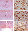
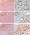
Results: Food intake, body weight, and mean arterial blood pressure (MAP) were higher in OP rats, a diet-induced metabolic syndrome model, accompanied by a reduced expression of POMC, an anorectic agent, in the hypoglossal nucleus (HN) and medial nucleus tractus solitarius (mNTS). Food intake in heat-treated OP rats was significantly decreased. POMC positive neuron count was increased in the HN and mNTS of OP rats following treatment. TRPV3 positive staining neurons were increased in the HN and mNTS of OP control rats and decreased following the heat treatments.
Conclusion: Lowered POMC and heightened TRPV3 expressions in the HN and mNTS are involved in development of hyperphagia and obesity in OP rats. Exposure to infrared heat modifies TRPV3 and POMC expression in the brainstem, reducing food intake.
Figures


Similar articles
-
Effects of electroacupuncture Zusanli (ST36) on food intake and expression of POMC and TRPV1 through afferents-medulla pathway in obese prone rats.Peptides. 2013 Feb;40:188-94. doi: 10.1016/j.peptides.2012.10.009. Epub 2012 Oct 29. Peptides. 2013. PMID: 23116614 Free PMC article.
-
Characterization of the resistance to the anorectic and endocrine effects of leptin in obesity-prone and obesity-resistant rats fed a high-fat diet.J Endocrinol. 2004 Nov;183(2):289-98. doi: 10.1677/joe.1.05819. J Endocrinol. 2004. PMID: 15531717
-
Down-regulation of hypothalamic pro-opiomelanocortin (POMC) expression after weaning is associated with hyperphagia-induced obesity in JCR rats overexpressing neuropeptide Y.Br J Nutr. 2014 Mar 14;111(5):924-32. doi: 10.1017/S0007114513003061. Epub 2013 Oct 7. Br J Nutr. 2014. PMID: 24094067
-
Lean rats with hypothalamic pro-opiomelanocortin overexpression exhibit greater diet-induced obesity and impaired central melanocortin responsiveness.Diabetologia. 2007 Jul;50(7):1490-9. doi: 10.1007/s00125-007-0685-1. Epub 2007 May 16. Diabetologia. 2007. PMID: 17505816
-
Nicotine, food intake, and activation of POMC neurons.Neuropsychopharmacology. 2013 Jan;38(1):245. doi: 10.1038/npp.2012.163. Neuropsychopharmacology. 2013. PMID: 23147487 Free PMC article. Review. No abstract available.
Cited by
-
Stimuli-evoked NOergic molecules and neuropeptides at acupuncture points and the gracile nucleus contribute to signal transduction of propagated sensation along the meridian through the dorsal medulla-thalamic pathways.J Integr Med. 2024 Sep;22(5):515-522. doi: 10.1016/j.joim.2024.07.001. Epub 2024 Jul 5. J Integr Med. 2024. PMID: 39214715 Free PMC article. Review.
-
Involvement of TRP Channels in Adipocyte Thermogenesis: An Update.Front Cell Dev Biol. 2021 Jun 24;9:686173. doi: 10.3389/fcell.2021.686173. eCollection 2021. Front Cell Dev Biol. 2021. PMID: 34249940 Free PMC article. Review.
-
TRPV3 modulates nociceptive signaling through peripheral and supraspinal sites in rats.J Neurophysiol. 2017 Aug 1;118(2):904-916. doi: 10.1152/jn.00104.2017. Epub 2017 May 3. J Neurophysiol. 2017. PMID: 28468993 Free PMC article.
-
MiR-103 inhibiting cardiac hypertrophy through inactivation of myocardial cell autophagy via targeting TRPV3 channel in rat hearts.J Cell Mol Med. 2019 Mar;23(3):1926-1939. doi: 10.1111/jcmm.14095. Epub 2019 Jan 3. J Cell Mol Med. 2019. PMID: 30604587 Free PMC article.
-
Involvement of calcium channels in the regulation of adipogenesis.Adipocyte. 2020 Dec;9(1):132-141. doi: 10.1080/21623945.2020.1738792. Adipocyte. 2020. PMID: 32175809 Free PMC article. Review.
References
-
- Eckel RH, Alberti KG, Grundy SM, Zimmet PZ. The metabolic syndrome. Lancet. 2010;375:181–183. - PubMed
-
- Grundy SM, Brewer HB, Jr, Cleeman JI, Smith SC, Jr, Lenfant C. Definition of metabolic syndrome: Report of the National Heart, Lung, and Blood Institute/American Heart Association conference on scientific issues related to definition. Arterioscler Thromb Vasc Biol. 2004;24:e13–18. - PubMed
-
- Li S, Zhang HY, Hu CC, Lawrence F, Gallagher KE, Surapaneni A, Estrem ST, Calley JN, Varga G, Dow ER, Chen Y. Assessment of diet-induced obese rats as an obesity model by comparative functional genomics. Obesity (Silver Spring) 2008;16:811–818. - PubMed
-
- Tschop M, Heiman ML. Rodent obesity models: An overview. Exp Clin Endocrinol Diabetes. 2001;109:307–319. - PubMed
Publication types
MeSH terms
Substances
Grants and funding
LinkOut - more resources
Full Text Sources
Medical
Molecular Biology Databases
Miscellaneous
