Review
. 2011;22(5):535-49.
doi: 10.1515/RNS.2011.044.

Structural and synaptic plasticity in stress-related disorders

Affiliations
Review

Structural and synaptic plasticity in stress-related disorders

Daniel J Christoffel et al. Rev Neurosci. 2011.

Abstract

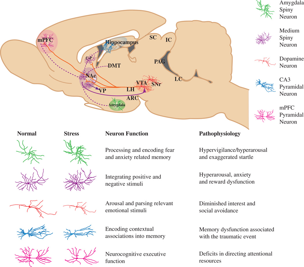
Abstract Stress can have a lasting impact on the structure and function of brain circuitry that results in long-lasting changes in the behavior of an organism. Synaptic plasticity is the mechanism by which information is stored and maintained within individual synapses, neurons, and neuronal circuits to guide the behavior of an organism. Although these mechanisms allow the organism to adapt to its constantly evolving environment, not all of these adaptations are beneficial. Under prolonged bouts of physical or psychological stress, these mechanisms become dysregulated, and the connectivity between brain regions becomes unbalanced, resulting in pathological behaviors. In this review, we highlight the effects of stress on the structure and function of neurons within the mesocorticolimbic brain systems known to regulate mood and motivation. We then discuss the implications of these spine adaptations on neuronal activity and pathological behaviors implicated in mood disorders. Finally, we end by discussing recent brain imaging studies in human depression within the context of these basic findings to provide insight into the underlying mechanisms leading to neural dysfunction in depression.


Figures

Figure 1
Figure 1. A sagittal brain slice showing the mesocorticolimbic reward circuitry of the brain, highlighting the major neuron type of each region
Projections of VTA dopamine neurons (shown in solid red lines) impinge directly on NAc and mPFC neurons, as well as on amygdala and hippocampal neurons (the latter projections are not shown in the figure). The solid purple line represents GABAergic afferents (some direct, some indirect) from the NAc to the VTA, which provide feedback to VTA dopamine neurons. The dotted purple lines represent glutamatergic afferents to the NAc from mPFC, amygdala, and hippocampus. Each structure contains specialized neuronal cell types thought to play an integral role in the complex behavioral phenotypes associated with reward-related behavior. These cell types, color-coded in the key, include amygdala (green) and NAc (purple) spiny neurons, PFC (pink) and hippocampal CA3 (blue) pyramidal neurons, and VTA dopamine neurons (red). Below, cartoon renderings of the affect of stress on neuronal morphology, along with a description of the normal function and pathophysiology of plasticity within each mesocorticolimbic brain region.
Figure 2
Figure 2. Region specific effects of stress on spine plasticity
(A) In the NAc, one study has found a doubling of stubby spines after CSDS. To date, no other studies have examined spine reorganization following stress. (B) In the amygdala, several studies have found a stress-induced increase in dendritic length and spine density; however, there are no studies that have examined changes in spine morphologies. (C) In the PFC, the majority of studies found that stress causes a reduction in dendritic length and spine density. It seems that these changes in density are specific to spine types. Note the increase in thin spines but decrease in mushroom spines. (D) Although there are conflicting reports*, several studies would support the hypothesis that the dendritic atrophy of hippocampal pyramidal neurons is accompanied by spine loss.
Figure 3
Figure 3. Model of interactions between spine type and dopamine and glutamate neurotransmitter systems in the NAc
Spine morphology is an important determinant of synaptic strength. Larger mushroom spines have more AMPA receptors than smaller thin and stubby spines. However, morphology may also have an important impact on responsiveness to different neurotransmitters. Stubby spines, which lack a traditional spine neck, may either receive less or contain very different connections with dopamine terminals extending from the VTA and substantia nigra than thin or mushroom spines. Future studies aimed at characterizing the function of specific terminal populations will further elucidate neurotransmitter specific effects on postsynaptic signaling. (From left to right, stubby spine, thin spine, and mushroom spine.)

Similar articles

Cited by

References

    1. Akram A, Christoffel D, Rocher AB, Bouras C, Kövari E, Perl DP, Morrison JH, Herrmann FR, Haroutunian V, Giannakopoulos P, et al. Stereologic estimates of total spinophilin-immunoreactive spine number in area 9 and the CA1 field: relationship with the progression of Alzheimer’s disease. Neurobiol. Aging. 2008;29:1296–1307. - PMC - PubMed
    1. Andrade C, Rao NS. How antidepressant drugs act: a primer on neuroplasticity as the eventual mediator of antidepressant efficacy. Indian J. Psychiatry. 2010;52:378–386. - PMC - PubMed
    1. Arellano JI, Benavides-Piccione R, DeFelipe J, Yuste R. Ultrastructure of dendritic spines: correlation between synaptic and spine morphologies. Front. Neurosci. 2007;1:131–143. - PMC - PubMed
    1. Autry AE, Adachi M, Nosyreva E, Na ES, Los MF, Cheng P-f, Kavalali ET, Monteggia LM. NMDA receptor blockade at rest triggers rapid behavioural antidepressant responses. Nature. 2011;475:91–95. - PMC - PubMed
    1. Bennur S, Shankaranarayana Rao BS, Pawlak R, Strickland S, McEwen BS, Chattarji S. Stress-induced spine loss in the medial amygdala is mediated by tissue-plasminogen activator. Neuroscience. 2007;144:8–16. - PubMed

Publication types