. 2012 Jan 15;441(2):707-17.
doi: 10.1042/BJ20111379.

Stepwise proteolytic activation of type I procollagen to collagen within the secretory pathway of tendon fibroblasts in situ

Affiliations

Stepwise proteolytic activation of type I procollagen to collagen within the secretory pathway of tendon fibroblasts in situ

Elizabeth G Canty-Laird et al. Biochem J. .

Abstract

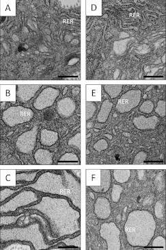
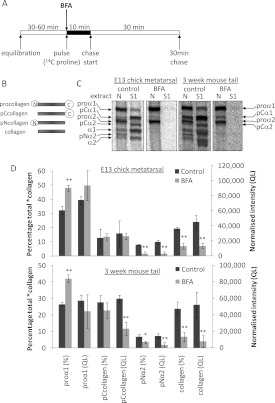
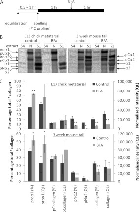
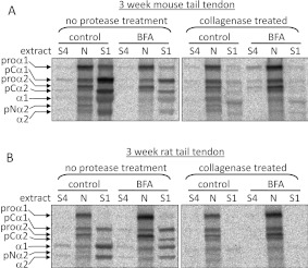
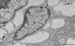
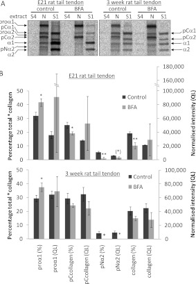
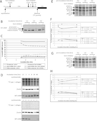
Proteolytic cleavage of procollagen I to collagen I is essential for the formation of collagen fibrils in the extracellular matrix of vertebrate tissues. Procollagen is cleaved by the procollagen N- and C-proteinases, which remove the respective N- and C-propeptides from procollagen. Procollagen processing is initiated within the secretory pathway in tendon fibroblasts, which are adept in assembling an ordered extracellular matrix of collagen fibrils in vivo. It was thought that intracellular processing was restricted to the TGN (trans-Golgi network). In the present study, brefeldin A treatment of tendon explant cultures showed that N-proteinase activity is present in the resulting fused ER (endoplasmic reticulum)-Golgi compartment, but that C-proteinase activity is restricted to the TGN in embryonic chick tendon fibroblasts. In late embryonic and postnatal rat tail and postnatal mouse tail tendon, C-proteinase activity was detected in TGN and pre-TGN compartments. Preventing activation of the procollagen N- and C-proteinases with the furin inhibitor Dec-RVKR-CMK (decanoyl-Arg-Val-Lys-Arg-chloromethylketone) indicated that only a fraction of intracellular procollagen cleavage was mediated by newly activated proteinases. In conclusion, the N-propeptides are removed earlier in the secretory pathway than the C-propeptides. The removal of the C-propeptides in post-Golgi compartments most probably indicates preparation of collagen molecules for fibril formation at the cell-matrix interface.


Figures

Figure 1
Figure 1. BFA treatment protocols
(A) Schematic diagram of the secretory pathway components: RER (rough ER), ERGIC, cis-, medial- and trans-Golgi stacks, TGN, p.m. (plasma membrane) and ECM (extracellular matrix). (B) BFA treatment results in Golgi complex disassembly and formation of a fused ER/Golgi compartment. The TGN becomes connected to the recycling endosomal system. (C) A 10-min pulse results in labelled *collagens in the RER and early secretory pathway. After 30 min of chase, these *collagens would be approximately equally distributed between the secretory pathway and ECM (not shown). (D) A 10-min pulse in BFA-treated samples results in *collagens in the fused ER/Golgi compartment. Retained *collagens would not be secreted during the chase (not shown). (E) Continuous labelling floods the secretory pathway and ECM with labelled *collagens. (F) BFA treatment after continuous labelling results in retrieval of *collagens from the Golgi stacks to the intracellular fused ER/Golgi compartment. *collagens in the TGN at the time of BFA treatment are secreted.
Figure 2
Figure 2. BFA treatment of whole tendon explants results in the expansion and progressive in situ distension of rough ER
Tendon explants were incubated in medium containing BFA for 30 min or 1 h. Samples were fixed, embedded and analysed by TEM as described in the Experimental section. RER denotes regions of the section showing ribosome-studded regions of rough ER. RER distension is most pronounced in samples treated with BFA for 1 h. (AC) Chick metatarsal tendon (E13); (DF) CD1 mouse tail tendon (3-week-old postnatal). (A and D) Control incubation; (B and E) BFA incubation (30 min); (C and F) BFA incubation (1 h). Scale bars, 500 nm.
Figure 3
Figure 3. BFA treatment of embryonic and postnatal tendon explants results in the intracellular retention of newly synthesized procollagen and reduces the extent of intracellular processing at the procollagen C-propeptide
(A) Schematic diagram of the pulse–chase protocol used to label, and then to follow the fate of, newly synthesized type I procollagen in tendon explants. (B) Schematic diagram of the intermediates obtained on removal of the N- and/or C-propeptides from type I procollagen. (C) Embryonic chick metatarsal tendons and postnatal CD1 mouse tail tendons, incubated according to the protocol shown in (A), were subjected to sequential salt and NP40 detergent extractions, and the extracts were analysed by electrophoresis and autoradiography as described in the Experimental section. Single representative images are shown. ‘S1’ denotes the first extracellular salt extract and ‘N’ is the intracellular NP40 detergent extract. Bands corresponding to each of the type I procollagen-processing intermediates are indicated. (D) The relative (percentage total) and absolute [normalized intensity (QL)] amounts of each *collagen were determined as described in the Experimental section (n=4). *P<0.05; **P<0.01 (Student's t test).
Figure 4
Figure 4. Intracellular retention of fully processed collagen is triggered by post-labelling BFA treatment in postnatal CD1 mouse tail tendon, but not in embryonic chick metatarsal tendon
(A) Schematic diagram of the retrieval protocol used to label all *collagens within the secretory pathway of embryonic chick and postnatal mouse tail tendon fibroblasts in situ, before subsequent disruption of the secretory apparatus using BFA. (B) Embryonic chick metatarsal and postnatal mouse tail tendons incubated according to the protocol shown in (A) were subjected to sequential salt and NP40 detergent extractions, and the extracts were analysed by electrophoresis and autoradiography as described in the Experimental section. Single representative images are shown. ‘S1’ denotes the first extracellular salt extract, ‘S4’ represents the fourth and final salt extract, and ‘N’ is the intracellular NP40 detergent extract. Bands corresponding to each of the type I procollagen-processing intermediates are indicated. (C) The relative (% total) and absolute [normalized intensity (QL)] amounts of each *collagen were determined as described in the Experimental section (n=3). *P<0.05; **P<0.01 (Student's t test).
Figure 5
Figure 5. *collagens present in the intracellular ‘N’ extract are protected from collagenase digestion
Postnatal mouse tail tendons (A) and postnatal rat tail tendons (B) were incubated according to the protocol shown in Figure 3(A) and treated with collagenase as described in the Experimental section. Tendons incubated according to the protocol shown in Figure 3(A) were subjected to sequential salt and NP40 detergent extractions, and the extracts were analysed by electrophoresis and autoradiography. Single representative images are shown. ‘S1’ denotes the first extracellular salt extract, ‘S4’ represents the fourth and final salt extract, and ‘N’ is the intracellular NP40 detergent extract. Bands corresponding to each of the type I procollagen-processing intermediates are indicated. Fully processed collagen is present in the intracellular (N) extract following BFA and/or collagenase treatment.
Figure 6
Figure 6. Transmission electron micrograph of embryonic E21 rat tail tendon
The image shows fibricarriers (collagen fibrils within the main body of the cells) (short arrows) and fibripositors (collagen fibrils located within cellular projections) (long arrows), in transverse view. Fibripositors and fibricarriers are characteristic features of embryonic tendon. Scale bar, 1 μm.
Figure 7
Figure 7. Intracellular retention of fully processed collagen in rat tail tendon following post-labelling BFA treatment
(A) Embryonic and postnatal rat tail tendons incubated according to the protocol shown in Figure 3(A) were subjected to sequential salt and NP40 detergent extractions, and the extracts were analysed by electrophoresis and autoradiography as described in the Experimental section. Single representative images are shown. ‘S1’ denotes the first extracellular salt extract, ‘S4’ represents the fourth and final salt extract, and ‘N’ is the intracellular NP40 detergent extract. Bands corresponding to each of the type I procollagen-processing intermediates are indicated. (B) The relative (% total) and absolute [normalized intensity (QL)] amounts of each *collagen were determined as described in the Experimental section (n=3). *P<0.05; **P<0.01 (Student's t test).
Figure 8
Figure 8. Effect of the furin inhibitor Dec-RVKR-CMK on intracellular procollagen processing in embryonic chick metatarsal and postnatal CD1 mouse tail tendon
(A) Schematic diagram of the protocol used to assess the effect of pre-incubation with Dec-RVKR-CMK on the extent of intracellular procollagen processing. (B) Intracellular (N) extracts from tendons treated with Dec-RVKR-CMK were analysed by Western blotting, using an antibody directed against the catalytic domain of MT1-MMP. Furin inhibitor treatment resulted in the appearance of bands corresponding to the proenzyme and intermediate forms of MT1-MMP. (C) The relative amounts of the proenzyme, intermediate and mature forms of MT1-MMP were quantified by densitometry. Furin inhibitor resulted in a time-dependent decrease in the relative amount of the mature enzyme and an increase in the amount of the proenzyme. (D) To determine whether MT1-MMP would be completely turned over during 4 h of incubation, embryonic chick metatarsal tendons were treated with the protein synthesis inhibitor cycloheximide for various durations up to 48 h. Samples were labelled for 1 h before the end of each incubation. The intracellular extracts were analysed by electrophoresis and autoradiography, to confirm the effectiveness of cycloheximide, and also by Western blotting using antibodies directed against MT1-MMP and β-actin. (E and G) Embryonic chick metatarsal (E) or postnatal CD1 mouse tail (G) tendons were incubated according to the protocol shown in (A) were subjected to sequential salt and NP40 detergent extractions, and the extracts were analysed by electrophoresis and autoradiography as described in the Experimental section. The intracellular (N) extracts are shown for each treatment. Bands corresponding to each of the type I procollagen-processing intermediates are indicated. (F and H) The relative amount of procollagen, pCcollagen, pNcollagen and fully processed collagen in each of the intracellular NP40 detergent extracts from embryonic chick metatarsal (F) or postnatal CD1 mouse tail (H) tendon was quantified by densitometry.

Similar articles

Cited by

References

    1. Chapman J. A., Tzaphlidou M., Meek K. M., Kadler K. E. The collagen fibril: a model system for studying the staining and fixation of a protein. Electron Microsc. Rev. 1990;3:143–182. - PubMed
    1. Bellamy G., Bornstein P. Evidence for procollagen, a biosynthetic precursors of collagen. Proc. Natl. Acad. Sci. U.S.A. 1971;68:1138–1142. - PMC - PubMed
    1. Kadler K. E., Hojima Y., Prockop D. J. Assembly of collagen fibrils de novo by cleavage of the type I pC-collagen with procollagen C-proteinase: assay of critical concentration demonstrates that collagen self-assembly is a classical example of an entropy-driven process. J. Biol. Chem. 1987;262:15696–15701. - PubMed
    1. Bonfanti L., Mironov A. A., Martinez-Menarguez J. A., Martella O., Fusella A., Baldassarre M., Buccione R., Geuze H. J., Luini A. Procollagen traverses the Golgi stack without leaving the lumen of cisternae: evidence for cisternal maturation. Cell. 1998;95:993–1003. - PubMed
    1. Polishchuk E. V., Di Pentima A., Luini A., Polishchuk R. S. Mechanism of constitutive export from the golgi: bulk flow via the formation, protrusion, and en bloc cleavage of large trans-Golgi network tubular domains. Mol. Biol. Cell. 2003;14:4470–4485. - PMC - PubMed

Publication types