Review
. 2012 May 15;16(10):1150-80.
doi: 10.1089/ars.2011.4085. Epub 2012 Jan 13.

Mitochondrial regulation of cell cycle and proliferation

Affiliations
Review

Mitochondrial regulation of cell cycle and proliferation

Valeria Gabriela Antico Arciuch et al. Antioxid Redox Signal. .

Abstract

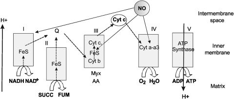
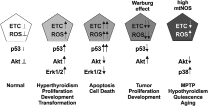
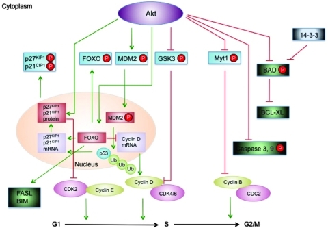
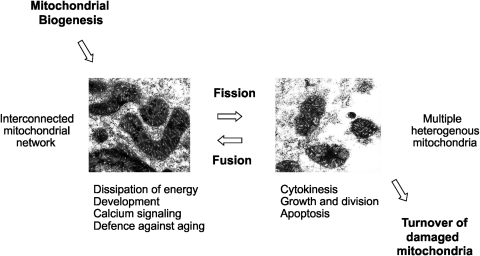
Eukaryotic mitochondria resulted from symbiotic incorporation of α-proteobacteria into ancient archaea species. During evolution, mitochondria lost most of the prokaryotic bacterial genes and only conserved a small fraction including those encoding 13 proteins of the respiratory chain. In this process, many functions were transferred to the host cells, but mitochondria gained a central role in the regulation of cell proliferation and apoptosis, and in the modulation of metabolism; accordingly, defective organelles contribute to cell transformation and cancer, diabetes, and neurodegenerative diseases. Most cell and transcriptional effects of mitochondria depend on the modulation of respiratory rate and on the production of hydrogen peroxide released into the cytosol. The mitochondrial oxidative rate has to remain depressed for cell proliferation; even in the presence of O₂, energy is preferentially obtained from increased glycolysis (Warburg effect). In response to stress signals, traffic of pro- and antiapoptotic mitochondrial proteins in the intermembrane space (B-cell lymphoma-extra large, Bcl-2-associated death promoter, Bcl-2 associated X-protein and cytochrome c) is modulated by the redox condition determined by mitochondrial O₂ utilization and mitochondrial nitric oxide metabolism. In this article, we highlight the traffic of the different canonical signaling pathways to mitochondria and the contributions of organelles to redox regulation of kinases. Finally, we analyze the dynamics of the mitochondrial population in cell cycle and apoptosis.


Figures

FIG. 1.
FIG. 1.
General organization of mitochondrial electron transfer chain and the formation of O2 species are modulated by NO. Electrons from reduced metabolites from the intermediary metabolism and tricarboxylic cycle enter the respiratory chain as NADH (to NADH dehydrogenase at complex I) or from succinate (to succinate dehydrogenase at complex II) and lead to a two-step reduction of reduced ubiquinol (to semiubiquinone, and ubiquinone). This sequential pathway finally reduces O2 to water and, depending on electron entrance, extrudes two or three protons that creates an inner membrane potential and re-enter by ATP synthase with dissipation of energy and formation of ATP. From a low to high concentration, NO progressively inhibits cytochrome oxidase, complex II-III, and complex I. Myx, myxothiazole; AA, antimycin A; FeS, Fe sulfur complex; Cyt, cytochrome; SUCC, succinate; FUM, fumarate.
FIG. 2.
FIG. 2.
Production of superoxide anion and hydrogen peroxide by the inhibition of mitochondrial oxygen uptake. (A) Inverse relationship between oxygen utilization and superoxide formation in mitochondria. The inverse relationship between superoxide production and residual Complex I activity is shown in fibroblasts of patients with isolated Complex I deficiency—Measurement of superoxide production was performed with hydroethydine in an inverted fluorescence microscope. Fluorescence intensity in the indicated compartment (left y-axis) is expressed as percentage of vehicle-treated control (CT). Closed and open symbols represent patient cell lines with a known (13 patients) and hitherto unknown (8 patients) as mutation, respectively. Linear regression analysis reveals an inverse correlation between superoxide production and residual CI activity for the whole cohort of patient cell lines. Reprinted from Verkaart et al. (224). © 2007 by Elsevier. (B) Effects of NO on electron transfer rate and hydrogen peroxide production. The effects of NO result in the inhibition of mitochondrial respiratory rate with an inverse increase of mitochondrial H2O2 yield, the product of dismutation of formula image. The trace was obtained by simultaneous polarographic determinations of O2 utilization and fluorometric detection of H2O2, in rat heart submitochondrial particles (reproduced from Poderoso et al.) (182). © Elsevier, 1996. CI, confidence interval.
FIG. 3.
FIG. 3.
The IMS, a redox compartment with different functions. Many unfolded proteins traverse the IMS to reach the inner membrane or to exit mitochondria. The graphic shows the disulfide bridge relay given by the two complementary intermediaries Mia40 and Erv1 in the IMS. The two components compose a cycle by which an unfolded protein with thiol groups forms a disulfide bridge with oxidized Mia40 (Mia40ox) that allows the protein to be refolded with intermolecular disulfide formation and Mia40 reduction. Mia40red returns to its oxidized state by reduction of Erv1, subsequently recovered by given electrons to Cyt c, and ultimately to cytochrome oxidase. MIA40, mitochondrial intermembrane space assembly machinery; ERV1, endogenous retroviral sequence; Cyt c, cytochrome c; COX, cytochrome oxidase; IMM, inner mitochondrial membrane; IMS, intermembrane space; OMM, outer mitochondrial membrane.
FIG. 4.
FIG. 4.
Interaction between ROS and p53. An equilibrated production of ROS and p53 in normal cells (left) could be disrupted by a modest increase of p53 that increases discretely the ROS production, which increases the activation of pro-proliferative kinases such as Akt and ERK1/2. High p53 arrest cells and increases apoptosis, with low Akt and high respiratory activity and very high ROS. In contrast lack of p53, reduces the activity of cytochrome oxidase, lowers ROS, and provides the platform for transformation. In the end, NO lowers electron transfer rate but differentially with the Warburg condition is not proliferative because it increases ROS and decreases Akt activity; whereas it increases the activity of pro-apoptotic kinases, such as p38 and mitogen-activated protein kinase. Akt, protein kinase B; ERK, extracellular signal-regulated kinase; ETC, electron transfer chain; p53, tumor protein; ROS, reactive oxygen species.
FIG. 5.
FIG. 5.
Electron microscopy of normal and tumor mitochondria. In the upper panel, mitochondria from LM3 mammary tumor cells (right) are compared with those from normal mammary cells of pregnant mice. In the lower panel, a similar comparation is made between mitochondria isolated from lung tumor P07 cell line (right) and organelles from normal lung cells. Bar=0.33 μM. Reproduced according to American Association for Cancer Research (AACR) from Galli et al. (78).
FIG. 6.
FIG. 6.
Hypoxia signaling and HIF. The scheme shows the pathways for HIF degradation and proliferation in accord to oxygen levels. HIF, hypoxia inducible factor.
FIG. 7.
FIG. 7.
Transition from high to low respiratory rate: The Warburg requirement for cell proliferation. Different mechanisms underlie the Warburg effect characterized by low O2 utilization in the presence of enough available oxygen. This effect is given by increased glycolysis associated to high expression and activation of glycolytic enzymes (PK, GAPDH, PFK, HK, and LDH) and reduced mitochondrial respiration. Low respiratory rate depends on several factors such as mitochondrial fission, and defective combined effects of p53, c-myc, and Akt that sustain high oxidative rate or promote adhesion of HK II. GAPDH, glyceraldehide 3-phosphate dehydrogenase; HK, hexokinase; LDH, lactate dehydrogenase; PFK, phosphofructokinase; PK, pyruvate kinase.
FIG. 8.
FIG. 8.
The fate of NIH/3T3 cells depends on the redox status. (A) Cyclin D1 expression increased on low H2O2 stimuli, whereas it decreased at a high H2O2 concentration. (B) High redox status triggered apoptosis by 10-fold as determined by flow cytometry with Annexin V staining (upper panel) and propidium iodide (lower panel). (C) Translocation of Bcl-xL and Cyt c from mitochondria to cytosol was determined 24 and 48 h after H2O2 stimuli. *represents p<0.05 vs control. From Antico et al., according to Creative Commons Attribution License (11). Bcl-xL, B-cell lymphoma-extra large. (To see this illustration in color the reader is referred to the web version of this article at www.liebertonline.com/ars).
FIG. 9.
FIG. 9.
Akt phosphorylates and inactivates the FOXO family of transcription factors. In lower metazoans, FOXO proteins promote the expression of pro-apoptotic genes, such as BIM and FAS. In mammalian cells, FOXO also promotes expression of p27KIP1 and p21CIP1 to inhibit cell cycle entry. The CDK inhibitor p27KIP1 and p21CIP1 proteins can also be phosphorylated by Akt, leading to their accumulation in the cytoplasm. Akt increases the translation of cyclin D mRNA through phosphorylation of GSK3, and Akt phosphorylation of Myt1 drives the cell cycle to M phase. A prominent function of Akt on cell survival is mediated by its direct phosphorylation and negative regulation on caspases 3 and 9 as well as BAD. Phosphorylation of MDM2 by Akt leads to its nuclear translocation and, thus, promoting p53 degradation and finally leading to a reduction in transcription of p21CIP1 mRNA. CDK, cyclin-dependent kinase; FOXO, forkhead O-box; GSK3, glycogen synthase kinase-3; BIM, Bcl-2-like protein 11; MDM2, mouse double minute 2; Myt1, myelin transcription factor; UB, ubiquitin; FAS, TNF receptor superfamily member 6.
FIG. 10.
FIG. 10.
Regulation of mitochondrial biogenesis machinery by calcium and kinases. Extracellular stimuli increase intracellular calcium levels resulting in kinases activation by transcriptional pathways. Some kinases activate PGC-1α coactivator by transcriptional activation of its gene or by direct phosphorylation, thus resulting in the activation of nuclear respiratory factors NRF-1 and NRF-2 that mediate the transcription of several mitochondrial genes that will promote mitochondrial biogenesis. CaMK, calcium/calmodulin-dependent protein kinase; CREB, cAMP response element-binding protein; ER, endoplasmic reticulum; NRF, nuclear respiratory factor; PGC-1, peroxisome proliferator-activated receptor-γ-coactivator-1; TFAM, mitochondrial transcription factor A.
FIG. 11.
FIG. 11.
NO, oxidative stress, antioxidant enzymes, and mitochondrial biogenesis. Increased mitochondrial NO mostly by increased mtNOS activity derives in increased production of formula image by mitochondrial respiratory complex inhibition. NO is an activator of PGC-1α and subsequently of mitochondrial biogenesis, and increased production of ROS activates the nuclear transcription factor NRF2 that regulates antioxidant enzymes genes transcription and mitochondrial biogenesis. SOD, superoxide dismutase; Prx, peroxiredoxin; HO-1, heme oxigenase 1; Trx, thioredoxin; GPx, glutathione peroxidase.
FIG. 12.
FIG. 12.
Mitochondrial dynamics and biological functions. Mitochondrial biogenesis starts the mitochondrial cycle by division of pre-existing organelles, and mitophagy ends it by degradation of impaired mitochondria. In between, mitochondria undergo several cycles of fission and fusion that generate multiple heterogeneous mitochondria or interconnected mitochondrial networks depending on the physiological conditions. Fused mitochondrial networks are important for the dissipation of metabolic energy and for complementation of mtDNA gene products to counteract the decline of respiratory functions in aging in heteroplasmic cells.
FIG. 13.
FIG. 13.
Mitochondrial dynamics and cell cycle. During the cell cycle, rapid changes in mitochondrial morphology and dynamics accompany mitosis, and allow the appropriate segregation of the organelles and mtDNA into daughter cells.

References

    1. Adams JM. Cory S. Bcl-2-regulated apoptosis: mechanism and therapeutic potential. Curr Opin Immunol. 2007;19:488–496. - PMC - PubMed
    1. Affaitati A. Cardone L. Carlucci A. de Cristofaro T. Ginsberg MD. Varrone S. Gottesman ME. Avvedimento VE. Feliciello A. Essential role of A-kinase anchor protein 121 for cAMP signaling to mitochondria. J Biol Chem. 2003;278:4286–4294. - PubMed
    1. Ahmed NN. Grimes HL. Bellacosa A. Chan TO. Tsichlis PN. Transduction of interleukin-2 antiapoptotic and proliferative signals via Akt protein kinase. Proc Natl Acad Sci U S A. 1997;94:3627–3632. - PMC - PubMed
    1. Almeida A. Bolaños JP. Moncada S. E3 ubiquitin ligase APC/C-Cdh1 accounts for the Warburg effect by linking glycolysis to cell proliferation. Proc Natl Acad Sci U S A. 2010;107:738–741. - PMC - PubMed
    1. Alonso M. Melani M. Converso DP. Jaitovich A. Paz C. Carreras MC. Medina JH. Poderoso JJ. Mitochondrial extracellular signal-regulated kinases 1/2 (ERK1/2) are modulated during brain development. J Neurochem. 2004;89:248–256. - PubMed

Publication types

Substances

LinkOut - more resources