Comparative Study
. 2012 Jan;81(1):3-11.
doi: 10.1124/mol.111.073643. Epub 2011 Oct 3.

Development of novel CH223191-based antagonists of the aryl hydrocarbon receptor

Affiliations
Comparative Study

Development of novel CH223191-based antagonists of the aryl hydrocarbon receptor

Eun-Young Choi et al. Mol Pharmacol. 2012 Jan.

Abstract

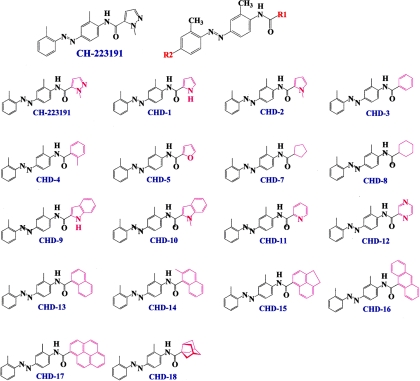
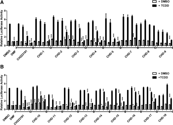
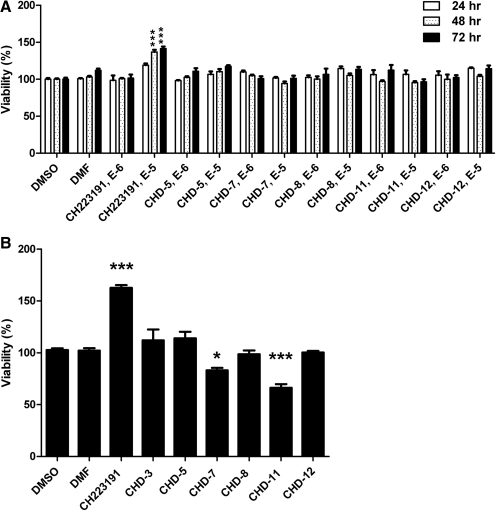
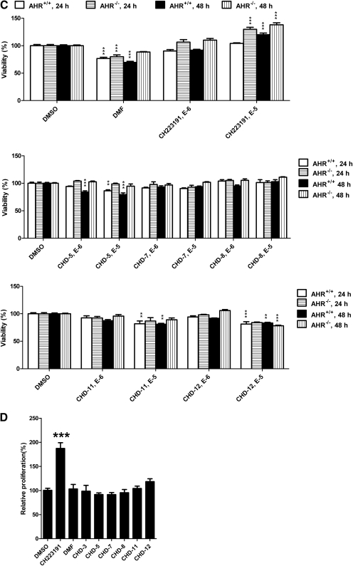
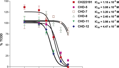
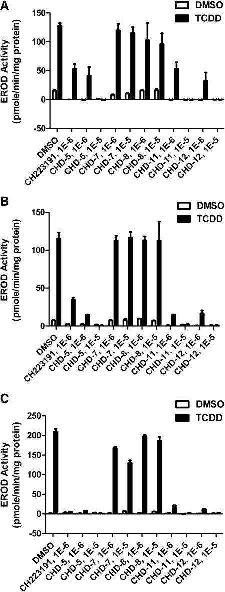
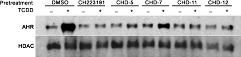
Aryl hydrocarbon receptor (AHR) is a ligand-activated transcription factor that regulates genes involved in drug/xenobiotic metabolism, cell cycle progression, cell fate determination, immune function, and inflammatory response. Increasing evidence that AHR plays a role in the pathophysiology of a number of human disease states is driving the need for improved pharmacological tools to be used for understanding the in vivo impact of AHR modulation. In this study, we have characterized and used structure-activity relationship analyses of a newly synthesized library of derivatives of the potent AHR antagonist 2-methyl-2H-pyrazole-3-carboxylic acid (2-methyl-4-o-tolylazo-phenyl)-amide (CH223191). Initial screening of these compounds revealed that those bearing groups with strong electronegativity at the R1 position (i.e., CHD-5, CHD-11, and CHD-12) versus those that are more electron-poor at this position (i.e., CHD-7 and CHD-8) elicited the most potent AHR antagonistic properties. The ability of these derivatives to inhibit agonist (2,3,7,8-tetrachlorodibenzo-p-dioxin) binding, nuclear translocation of AHR, and agonist-induced enzyme activity also were determined and support the initial findings. Furthermore, CH223191, but not CHD-5, CHD-11, or CHD-12, was found to exhibit AHR-independent proproliferative properties. These results contribute to our understanding of the structural requirements of potent AHR antagonists and the development of effective pharmacological tools to be used for studying the pathophysiological role of AHR.


Figures

Fig. 1.
Fig. 1.
Structures of the CH223191 derivatives. Schematic representation of the CH223191 derivatives that vary with respect to their substituents at either the R1 or R2 (not shown) positions and were synthesized and characterized in this study.
Fig. 2.
Fig. 2.
Reporter analyses of the CH223191 derivatives. A, CHD-1 through CHD-9. B, CHD-10 through CHD-18 HepG2 cells that have been transfected stably with a luciferase reporter regulated by the human CYP1A1 promoter (HepG2-p450luc) were pretreated with either DMSO alone or the indicated compounds (10−5, 10−6, 10−7, or 10−8 M) for 1 h. DMF was used at a concentration of 5 × 10⁻⁶ M. After the addition of TCDD (1 nM), the cells were incubated for an additional 4 h and harvested, and luciferase activities were determined. The mean ratios of triplicate wells (±S.D.) are depicted. The results are representative of at least three independent experiments that were subjected to one-way analysis of variance and Tukey's post hoc test analyses. ***, p < 0.001; **, p < 0.01; *, p < 0.05.
Fig. 3.
Fig. 3.
Impact of the CH223191 derivatives on viability and proliferation. A–C, viability analyses of Hepa1 (A), HepG2 (B), and immortalized murine AHR(+/+) versus AHR(−/−) hepatocytes (C). The cultured cells were incubated with the indicated compounds. After either 24, 48, or 72 h, the cells were harvested, and cell numbers were determined using the WST-1 assay. D, proliferation analyses of HepG2 cells. HepG2 cells were incubated with the indicated compounds for 72 h, and proliferation was addressed using the BrdU incorporation assay. The mean ratios of triplicate wells (±S.D.) are depicted. The results are representative of at least three independent experiments that were subjected to one-way analysis of variance and Tukey's post hoc test analyses. ***, p < 0.001; **, p < 0.01; *, p < 0.05.
Fig. 3.
Fig. 3.
Impact of the CH223191 derivatives on viability and proliferation. A–C, viability analyses of Hepa1 (A), HepG2 (B), and immortalized murine AHR(+/+) versus AHR(−/−) hepatocytes (C). The cultured cells were incubated with the indicated compounds. After either 24, 48, or 72 h, the cells were harvested, and cell numbers were determined using the WST-1 assay. D, proliferation analyses of HepG2 cells. HepG2 cells were incubated with the indicated compounds for 72 h, and proliferation was addressed using the BrdU incorporation assay. The mean ratios of triplicate wells (±S.D.) are depicted. The results are representative of at least three independent experiments that were subjected to one-way analysis of variance and Tukey's post hoc test analyses. ***, p < 0.001; **, p < 0.01; *, p < 0.05.
Fig. 4.
Fig. 4.
CHD-5, CHD-11, and CHD-12, but not CHD-7 and CHD-8, competitively inhibit TCDD binding to AHR. [3H]TCDD, in the absence or presence of varying concentrations of CH223191 and its derivatives (10−10 to 10−5 M) was incubated with protein extracts prepared from Hepa1 cells. After incubation at room temperature for 2 h, nonspecific binding was removed using a 10% dextran-coated charcoal slurry and hydroxyapatite gel. A 200-fold molar excess of 2,3,7,8-tetrachlorodibenzofuran, an analog of TCDD, was used to estimate the nonspecific binding of TCDD. All of the values are expressed as the percentage of the value obtained using TCDD alone. The data are averages ± S.D. of three independent experiments. The IC50 values were determined using GraphPad Prism.
Fig. 5.
Fig. 5.
CHD-5, CHD-11, and CHD-12 inhibit the ability of TCDD to induce CYP1A1 activity in EROD analyses. A–C, either HepG2 (A), HaCaT (B), or T84 (C) cells were aliquoted into 96-well plates. When either 90% (HepG2 and T84) or postconfluent (HaCaT), the indicated chemicals were added. After an additional 16 h of incubation, the cells were harvested, and EROD activities were determined as described under Materials and Methods. The mean ratios of triplicate wells (± S.D.) are depicted. The results are representative of at least three independent experiments. The statistical analyses of these experiments are shown in Table 1.
Fig. 6.
Fig. 6.
CHD-5, CHD-11, and CHD-12, but not CHD-7, inhibit TCDD-induced AHR enrichment in the nuclei. Hepa1 cells were treated with the indicated derivatives (10 μM) for 1 h before the administration of either DMSO (0.1%) or TCDD (1 nM). After incubation for 1 h, the cells were harvested, and the nuclear extracts were prepared and subjected to Western blot analyses. HDAC, histone deacetylase.

References

    1. Beischlag TV, Luis Morales J, Hollingshead BD, Perdew GH. (2008) The aryl hydrocarbon receptor complex and the control of gene expression. Crit Rev Eukaryot Gene Expr 18:207–250 - PMC - PubMed
    1. Bisson WH, Koch DC, O'Donnell EF, Khalil SM, Kerkvliet NI, Tanguay RL, Abagyan R, Kolluri SK. (2009) Modeling of the aryl hydrocarbon receptor (AhR) ligand binding domain and its utility in virtual ligand screening to predict new AhR ligands. J Med Chem 52:5635–5641 - PMC - PubMed
    1. Casado FL, Singh KP, Gasiewicz TA. (2010) The aryl hydrocarbon receptor: regulation of hematopoiesis and involvement in the progression of blood diseases. Blood Cells Mol Dis 44:199–206 - PMC - PubMed
    1. Denison MS, Nagy SR. (2003) Activation of the aryl hydrocarbon receptor by structurally diverse exogenous and endogenous chemicals. Annu Rev Pharmacol Toxicol 43:309–334 - PubMed
    1. Dietrich C, Kaina B. (2010) The aryl hydrocarbon receptor (AhR) in the regulation of cell-cell contact and tumor growth. Carcinogenesis 31:1319–1328 - PMC - PubMed

Publication types

Substances

LinkOut - more resources