. 2011 Oct 24;208(11):2251-62.
doi: 10.1084/jem.20110919. Epub 2011 Oct 3.

Regulation of neutrophils by interferon-γ limits lung inflammation during tuberculosis infection

Affiliations

Regulation of neutrophils by interferon-γ limits lung inflammation during tuberculosis infection

Bisweswar Nandi et al. J Exp Med. .

Abstract

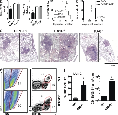
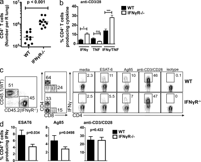
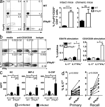
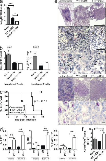
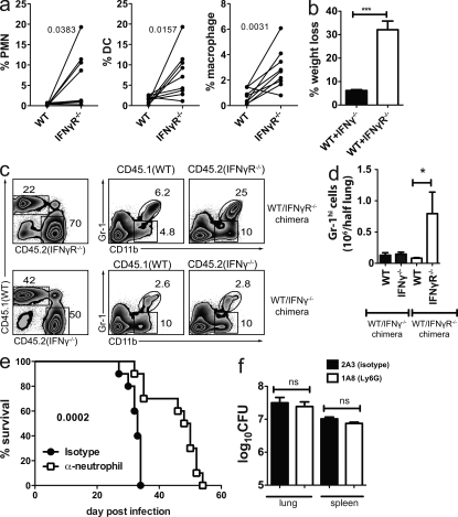
Resistance to Mycobacterium tuberculosis requires the host to restrict bacterial replication while preventing an over-exuberant inflammatory response. Interferon (IFN) γ is crucial for activating macrophages and also regulates tissue inflammation. We dissociate these two functions and show that IFN-γ(-/-) memory CD4(+) T cells retain their antimicrobial activity but are unable to suppress inflammation. IFN-γ inhibits CD4(+) T cell production of IL-17, which regulates neutrophil recruitment. In addition, IFN-γ directly inhibits pathogenic neutrophil accumulation in the infected lung and impairs neutrophil survival. Regulation of neutrophils is important because their accumulation is detrimental to the host. We suggest that neutrophilia during tuberculosis indicates failed Th1 immunity or loss of IFN-γ responsiveness. These results establish an important antiinflammatory role for IFN-γ in host protection against tuberculosis.


Figures

Figure 1.
Figure 1.
IFN-γR−/− mice are more susceptible than RAG2−/− mice to Mtb. (a) Bacterial load (CFU) in the lungs and spleen of WT, IFN-γR−/−, and RAG2−/− mice on day 23 after Mtb infection (n = 5 mice/group). Error bars, SEM. *, P < 0.05. (b) Survival of IFN-γR−/− and RAG2−/− mice after Mtb infection (n = 5 mice/ group). (c) Survival of RAG2−/− and RAG2/IFN-γR−/− mice after Mtb infection (n = 5 mice/ group). (d) Representative lung sections stained with hematoxylin and eosin from the mice represented in a. Two representative lesions are shown for each mouse genotype to demonstrate the size range. (e) Neutrophils (CD11b+Gr1hi) from the lungs of WT (top) or IFN-γR−/− (bottom) mice 4 wk after Mtb infection. Numbers represent percentage of gated cells. (f) Frequency and absolute number of neutrophils (CD11b+Gr1hi) in the lungs of WT and IFN-γR−/− mice (n = 3 mice/group). Error bars, SEM. *, P < 0.05. Data in a and d were from the same experiment. The three-way comparison was performed once; however, pairwise comparisons have been repeated multiple times with similar results. All other data are representative of two independent experiments. Survival analyses were analyzed by the log-rank test; a was analyzed by one way ANOVA; and f was analyzed by Student’s t test.
Figure 2.
Figure 2.
Antigen-specific CD4+ T cells are elicited in the absence of IFN-γ signaling. (a) Absolute number of CD4+ T cells per lung from WT or IFN-γR−/− mice 4–5 wk after Mtb infection. Data represents 11 mice/condition from three independent experiments. Horizontal bars, median. (b) Frequency of WT or IFN-γR−/− CD4+ T cells producing only IFN-γ, only TNF, or both IFN-γ and TNF after stimulation with anti-CD3/CD28, 3.5 wk after Mtb infection. Data are representative of two independent experiments. Error bars, SEM. *, P < 0.05; ***, P < 0.001. (c) Flow cytometric analysis of mixed WT and IFN-γR−/− BM chimeric mice. Lung lymphocytes were gated by size and CD45.1 (WT) or CD45.2 (IFN-γR−/−) cells identified. CD4+ T cells were gated and their production of IFN-γ after stimulation in vitro was determined. (d) Intracellular production of IFN-γ by WT or IFN-γR−/− CD4+ T cells from mixed BM chimera as indicated in c. Error bars, SEM. Data represents the analysis of 16 mice/condition from four independent experiments. Statistical analysis was performed using a Student’s t test.
Figure 3.
Figure 3.
IFN-γ signaling inhibits IL-17A production by CD4+ T cells during Mtb infection. (a) T reg cells in the lung 4 wk after Mtb infection of WT and IFN-γR−/− mice were detected by flow cytometry based on coexpression of CD25 and intracellular FoxP3 (left). T reg cells were enumerated in the lungs of WT and IFN-γR−/− mice (intact, middle) and WT/IFN-γR−/− chimeric mice (right). Each experiment analyzed five mice/group and statistical testing was performed using a Student’s t test. Error bars, SEM. ***, P < 0.001; NS, not significant. (b) Representative flow cytometric analysis (left) and analysis (right) of intracellular IL-17A production by lung CD4+ T cells from WT or IFN-γR−/− mice 4 wk after Mtb (n = 4 mice/group). Data were analyzed using a Student’s t test. Error bars, SEM. *, P < 0.05. (c) Levels of the chemokines KC, MIP-2, and G-CSF were measured in cell-free supernatants of lung homogenates prepared from WT and IFN-γR−/− mice (n = 5 mice/group) 4 wk after Mtb infection. Data was analyzed using a Student’s t test. Error bars, SEM. *, P < 0.05. (d) IL-17A production by lung CD4+ T cells from lungs of mixed WT/IFN-γR−/− BM chimeric mice as described in Fig. 2 c. Mice were analyzed 4–5 wk after Mtb infection (n = 13 mice/condition) or recall response 23–29 d after challenge of memory immune (n = 8 mice/condition). The data are pooled from three independent experiments and analyzed using a paired Student’s t test.
Figure 4.
Figure 4.
IFN-γ is required by CD4+ T cells for suppressing pulmonary inflammation. (a) Lung bacterial burden in recipient RAG2−/− mice 4 wk after Mtb infection and adoptive transfer of no cells, WT, or IFN-γR−/− memory CD4+ T cells. This data was analyzed by one-way ANOVA and is representative of two independent experiments, each with n = 5 mice/group. Error bars, SEM. ***, P < 0.001; *, P < 0.05. (b) Pulmonary bacterial burden 3–4 wk after Mtb infection of RAG2−/− recipient mice that received nothing, WT, or IFN-γ−/− memory CD4+ T cells. Two independent experiments are shown (n = 5 mice/group) analyzed by one-way ANOVA. Error bars, SEM. *, P < 0.05. (c) Survival of RAG2−/− mice that received nothing, WT, or IFN-γ−/− memory CD4+ T cells at the time of Mtb infection (n = 5 mice/group). The figure is representative of two independent experiments analyzed by log-rank test. (d) Cytokine production by WT or IFN-γ−/− memory CD4+ T cells. RAG2−/− mice that received nothing, WT, or IFN-γ−/− memory CD4+ T cells and were infected with Mtb are shown. Lung cells were isolated after 23 d and analyzed after culture alone or stimulation with ESAT61-15. The frequency of CD4+ T cells making IL17A only, TNF only, or both IL17A and TNF was determined (n = 5 mice/condition, from a, Exp1). Error bars, SEM. **, P < 0.01. (e) Lung histology and AFB staining of RAG2−/− mice transferred with nothing, WT, or IFN-γ−/− memory CD4+ T cells (from Exp 1, top; and Exp 2, bottom). Asterisks, neutrophil infiltrates; arrow heads, neutrophils. (f) Neutrophils in the lungs of RAG2−/− mice that received nothing (NoTx), WT, or IFN-γ−/− memory CD4+ T cells (from a, Exp1). Data was analyzed by one-way ANOVA. Error bars, SEM. *, P < 0.05, **, P < 0.01.
Figure 5.
Figure 5.
Neutrophil accumulation is inhibited by IFN-γ and is detrimental to outcome. (a) The percentage of CD45.1 (WT) or CD45.2 (IFN-γR−/−) cells that are neutrophils (PMN), DC, or macrophages in the lungs of mixed BM chimeric mice 4 wk after Mtb infection (n = 9 mice/condition pooled for two independent experiments). (b) Weight loss of [WT+IFN-γR−/−] or [WT+IFN-γ−/−] mixed BM chimeric mice, 4 wk after Mtb infection (n = 5 mice/ group). Error bars, SEM. ***, P < 0.001 by Student’s t test. (c) Flow cytometric analysis of neutrophils in the lungs of mixed [WT+IFN-γR−/−] or [WT+IFN-γ−/−] BM chimeric mice. Numbers indicate the percentage of gated cells. (d) The absolute neutrophil number in the lungs of mixed BM chimeric mice. Error bars, SEM. *, P < 0.05 by Student’s t test. (e) Survival of Mtb-infected IFN-γR−/− mice depleted of neutrophils. The neutrophil-specific antibody anti-Ly6G (clone 1A8) or the isotype control (clone 2A3) was administered to mice and their survival monitored (n = 10 mice/group). Data are analyzed by the log-rank test and are representative of two independent experiments. (f) Bacterial burden in the lungs and spleens of IFN-γR−/− mice treated with 1A8 (neutrophil depleting) or 2A3 (isotype control) monoclonal antibodies, 4 wk after Mtb infection. Data were analyzed by one-way ANOVA and is representative of two independent experiments, each with n = 5 mice/group. Error bars, SEM.
Figure 6.
Figure 6.
IFN-γ regulates survival of neutrophils in vitro and in vivo. (a) In vivo survival of WT or IFN-γR−/− neutrophils. Thioglycollate-elicited peritoneal exudate cells from WT (CFSEhi) or IFN-γR−/− (CFSElo) mice were mixed 1:1 and injected into uninfected or Mtb-infected mice. Cells were isolated from the lungs of recipient mice 4 and 20 h after transfer. After gating the cells for size (first row), the CD11b+CFSE+ cells were identified (second row). The CFSE+ (transferred cells) were regated (third row) to display CD11b+Gr-1+ (neutrophils, top gate) and CD11b+Gr-1lo (monocyte/macrophages, bottom gate) populations. The relative frequency of WT (CFSEhi) or IFN-γR−/− (CFSElo) cells in the CD11b+Gr-1+ (neutrophils, fourth row) and CD11b+Gr-1lo (monocyte/macrophages, fifth row) populations was determined. (b) The relative frequency of surviving WT and IFN-γR−/− monocyte/macrophages (CD11b+Gr-1lo) and neutrophils (CD11b+Gr-1+) at 4 and 20 h after cell transfer. The data are compiled from three mice/condition and are representative of two experiments. (c) Relative ratio of [IFN-γR−/−/WT neutrophils]/[IFN-γR−/−/WT monocyte/macrophages] 4 or 20 h after cell transfer. Results for uninfected and infected mice were pooled from two experiments. Horizontal bars, mean ± SEM. (d) In vitro survival of WT or IFN-γR−/− neutrophils. Peritoneal exudate cells from WT or IFN-γR−/− mice were isolated 4 h after thioglycollate injection. The cells were cultured in the absence or presence of IFN-γ (as indicated in the figure). After 20 h, the cultures were analyzed by flow cytometry and cells with a low (left) or high (right) forward scatter were gated and analyzed separately (middle). Each population was analyzed for the expression of CD11b and Gr-1, and CD11b+Gr-1Hi cells (neutrophils) were analyzed for cell death using a live/dead stain. The numbers indicated the percentage of gated cells. Staurosporine was used to induce cell death as a control. (e) The percentage of dead cells after in vitro treatment with or without IFN-γ. Statistical analysis was done by Student’s t test. Error bars, SEM. *, P < 0.05; **, P < 0.01. The assay was done in triplicate and the data are representative data of two experiments.

References

    1. Aly S., Wagner K., Keller C., Malm S., Malzan A., Brandau S., Bange F.-C., Ehlers S. 2006. Oxygen status of lung granulomas in Mycobacterium tuberculosis-infected mice. J. Pathol. 210:298–305 10.1002/path.2055 - DOI - PubMed
    1. Berry M.P.R., Graham C.M., McNab F.W., Xu Z., Bloch S.A.A., Oni T., Wilkinson K.A., Banchereau R., Skinner J., Wilkinson R.J., et al. 2010. An interferon-inducible neutrophil-driven blood transcriptional signature in human tuberculosis. Nature. 466:973–977 10.1038/nature09247 - DOI - PMC - PubMed
    1. Blanton R.E., Salam E.A., Ehsan A., King C.H., Goddard K.A. 2005. Schistosomal hepatic fibrosis and the interferon gamma receptor: a linkage analysis using single-nucleotide polymorphic markers. Eur. J. Hum. Genet. 13:660–668 10.1038/sj.ejhg.5201388 - DOI - PubMed
    1. Cooper A.M., Dalton D.K., Stewart T.A., Griffin J.P., Russell D.G., Orme I.M. 1993. Disseminated tuberculosis in interferon γ gene-disrupted mice. J. Exp. Med. 178:2243–2247 10.1084/jem.178.6.2243 - DOI - PMC - PubMed
    1. Cooper A.M., Adams L.B., Dalton D.K., Appelberg R., Ehlers S. 2002. IFN-gamma and NO in mycobacterial disease: new jobs for old hands. Trends Microbiol. 10:221–226 10.1016/S0966-842X(02)02344-2 - DOI - PubMed

Publication types

MeSH terms